Causes of heart failure
Physiological factors
Short-term discomfort in the precordial area is possible with severe fear, which is an absolutely normal situation. After eliminating the provoking factor, the unpleasant sensations disappear without a trace. Single extraordinary cardiac contractions, which are clinically manifested by freezing, are observed in 5-15% of absolutely healthy people. Their onset is provoked by nervous or physical stress, or a sudden change in weather.
Extrasystole
This type of arrhythmia most often causes the heart to freeze. Patients describe the sensation as a momentary “cardiac arrest” or talk about missing one heartbeat. Depending on the type of extrasystole, the symptom can be one-time or repeated several times in a row. The most dangerous condition is when freezing is felt in the form of regular episodes that follow every 2-3 heart beats.
The symptom is accompanied by a sharp deterioration of the condition. Patients complain of sudden dizziness, weakness, and shortness of breath. Fading of the heart during extrasystoles is characterized by a feeling of anxiety and lack of air. The skin turns pale or red. Depending on the etiological factor, extrasystole of functional, organic and toxic origin is distinguished.
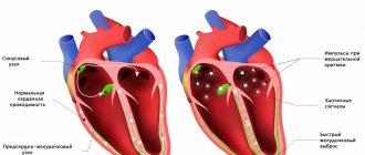
Heart blocks
Heart stopping is felt during atrioventricular blockade, when the conduction of excitation from the atria to the ventricles is disrupted. With 1 degree AV block, such symptoms are observed very rarely. Small pauses in heart contractions are characteristic of 2nd degree blockade. The most pronounced freezing of heart contractions is in stage 3 disease, when the atria and ventricles work in different rhythms.
The symptom is characterized by a combination of constant weakness, decreased performance and physical activity. Freezing is accompanied by severe dizziness and shortness of breath, sometimes the attacks end in fainting. Clinical signs do not depend on the nature, activity and time of day. AV blockades occur with congenital heart defects, organic myocardial lesions, and intoxication.
Bradycardia
Fading of the heart is a common manifestation of bradycardia. The symptom, unlike extrasystoles, is not isolated, but continues for several hours or days. If a person listens to his feelings, he notices that the heart begins to beat much less often. In this case, uniform, long pauses occur between beats, which are interpreted as freezing.
Bradycardia manifests itself in a number of cardiac diseases: coronary heart disease, myocarditis and pericarditis, congenital and acquired defects. A short-term slowdown in rhythm is observed with a reflex effect - a blow to the neck or chest, being in very cold water, squeezing the neck with a tie or tight collar. Bradycardia occurs with hypothyroidism, fulminant hepatitis, and hyperkalemia.
Vegetative-vascular dystonia
With VSD, the symptom develops due to disturbances in the nervous regulation of the cardiovascular system. Freezing is more often observed during excitement, fear, and stress. Vegetative-vascular dystonia is characterized by short-term interruptions in heart function, which are then replaced by tachycardia. Symptoms are complemented by shortness of breath and weakness, dizziness, and increased sweating.
Depression
Complaints of a sinking heart are a typical symptom of a masked form of depression. Symptoms typically appear in the early morning hours. In addition to freezing, patients complain of nagging pain, discomfort, and an unreasonable feeling of fear - the so-called “precardiac melancholy.” During the day and evening, these manifestations are less intense or disappear altogether. With masked depression, other complaints are also disturbing: sleep disturbances, hypochondriacal and panic disorders.
Complications of pharmacotherapy
Many drugs used to treat cardiac patients can cause arrhythmias. Fading of the heart is often provoked by taking beta-blockers, cardiac glycosides, and diuretics. A slowdown in rhythm combined with unpleasant sensations in the heart are observed with incorrect dosage of calcium and potassium solutions, which is accompanied by the development of hyperkalemia.
Fall asleep and not wake up. Why do tragedies happen in dreams?
Cardiovascular diseases are the scourge of modern society. In world statistics, this is the most common cause of death. In Russia alone, about two hundred thousand people a year die from sudden cardiac arrest. The most difficult thing to comprehend is the loss if the person felt well the day before, but did not wake up in the morning.
We talked about the causes of the tragedy and ways to prevent it with cardiologist Tatyana Mikhailovna Kolomeytseva.
— Tatyana Mikhailovna, what can cause a person’s heart to suddenly stop?
As a rule, this is a consequence of existing diseases: coronary heart disease, hereditary cardiomyopathies, congenital heart defects and conduction system. The cause is also thrombosis, spasm of the coronary vessels.
In Russia alone, about two hundred thousand people a year die from sudden cardiac arrest.
— Can a tragedy happen to an absolutely healthy person whose cardiovascular system has never failed?
An absolutely healthy person is not at risk of dying from a heart attack. However, heart disease (in particular, coronary artery disease) can be asymptomatic. In this case, the tragedy becomes the result of an undiagnosed pathology.
“Chronic heart failure (one of the forms of coronary artery disease) is present in 7% of the country’s population.” Quote from the material: “Coronary heart disease: diagnosis and treatment”
— People who are far from medicine associate heart disease with stress or physical activity. But in a dream a person is absolutely calm. Are there any risk factors specific to sleep?
No. The dangers come from the pathologies already listed: both in sleep and while awake.
— Could sleep quality indicate an increased risk of sudden death?
Obstructive sleep apnea is especially dangerous. Its main manifestation is snoring. A person suffering from apnea often does not get enough sleep, does not have time to regain strength during the night, suffers from drowsiness during the daytime, and is also susceptible to increased blood pressure upon awakening.
— Diseases not directly related to the cardiovascular system can become an additional risk factor?
Of course they can. These conditions include diabetes mellitus, obesity, kidney pathology and neurological diseases.
— What age is the most dangerous for cardiac arrest?
The incidence of cardiac disorders increases with age. Therefore, the older the person, the higher the risk.
- When should you be wary? Are there symptoms that allow us to foresee such a dangerous condition in advance?
Warning signs may appear two weeks before cardiac arrest. These are fatigue, shortness of breath, arrhythmia. The most threatening symptom is fainting. They cannot be ignored.
The most threatening symptom is fainting. They can't be ignored
Statistics show that in three out of four cases sudden death was preceded by corresponding symptoms. Therefore, it is important to pay attention to your well-being.
— Not everyone is used to taking care of their health. When a person himself does not talk about his health, can loved ones guess that help is needed? What external manifestations should cause alarm?
From the outside, it is more difficult to predict the impending disaster. But if a person suddenly reduces the pace of activity and cannot cope with his usual workload, this is an alarming symptom. Gait slows down and endurance decreases. It is worth consulting a doctor if your friend or relative does not keep up with you while walking, although you are walking at the speed that is usual for both of you.
Another noticeable symptom is snoring. This is a marker of obstructive apnea.
— Can genetic tests help identify predisposition to heart disease?
Only in the case of hereditary cardiomyopathies. It is not the leading cause of sudden cardiac death. It occupies no more than 20% of the total. To judge the hereditary nature of cardiomyopathy, it is necessary to examine three generations of the same family.
“The main danger is the possibility of sudden cardiac arrest. Therefore, patients with new-onset atrial fibrillation are immediately hospitalized.” Quote from the material “Recognizing the enemy of the heart. What is arrhythmia?
— Recently, fitness bracelets have become widespread. They are worn to control their motor activity. How accurately do they measure the pulse and can they help suspect problems in the cardiovascular system?
It is impossible to talk about fitness bracelets as a full-fledged diagnostic tool. They only measure the pulse, this is not enough to make final conclusions. But they can warn of impending danger. You should be wary if the bracelet detects rhythm disturbances. Simpler models for arrhythmia may simply malfunction - this is also a reason to see a doctor.
“The symmetry of the pulse is affected by a decrease in the lumen of one of the arteries, its incorrect location.” Quote from the material “What should an adult’s pulse be: normal and pathological”
— If cardiac arrest does occur in a dream, can the person be helped somehow?
Most often in this case the person is left without help because there are no witnesses. But if you managed to notice it in time, you need to call an ambulance as quickly as possible.
- What is dangerous for our heart? How to reduce the risk of cardiovascular diseases?
The cardiovascular system suffers from bad habits (in particular, smoking) and chronic emotional overload. Poor nutrition also poses a danger: irregular, with a lot of fat.
To preserve your heart, you need to lead a healthy lifestyle. Give yourself feasible and regular physical activity.
If cardiac pathologies have already been detected, it is important to regularly visit a doctor and follow all recommendations.
“If it was not possible to prevent the tragedy, people can only hope that their loved one at least did not experience pain and fear.” But is there any reason for such hope?
Of course, no research has been done. But it is known that with fatal arrhythmias, consciousness fades very quickly. The parts of the brain responsible for the perception of pain do not function. Therefore, a person does not experience torment.
Other materials on topics:
How to keep your heart healthy?
Serious question: what happens to the heart during an angina attack?
Heart, why don't you want peace? What causes tachycardia?
For reference:
Kolomeytseva Tatyana Mikhailovna
In 2008 she graduated from the Moscow Medical Academy named after. THEM. Sechenov with a degree in medical and preventive care.
In 2009 – professional retraining at the Moscow Medical Academy named after. THEM. Sechenov with a degree in General Medicine. He has professional retraining at the Russian State Medical University of Roszdrav in the specialty “Cardiology”. Completed professional development on the topic “Pulmonary hypertension: clinical workshop” at the European Health Center in Otwock (Poland)
In 2015 – advanced training at the National Medical and Chemical Center named after. N.I. Pirogov “Anticoagulants for atrial fibrillation.” In 2021 – professional development on the topic “Complex issues in the diagnosis and treatment of pulmonary hypertension” on the basis of the RKNPK named after. Myasnikova
In 2017-2018 – advanced training at the First Moscow State Medical University named after. THEM. Sechenov “Modern aspects of diagnosis and treatment of chronic heart failure.”
Member of the Russian Society of Cardiologists.
Has a certificate of a specialist in the specialties “Therapy”, “Cardiology”
Currently working as a cardiologist at Clinic Expert Tula LLC. Receives at the address: st. Boldina, 74
Diagnostics
Even if cardiac arrest is a rare episode, it is a reason to consult a cardiologist. Percussion can determine the expansion of the borders of the heart. During auscultation, muffled heart sounds and the presence of functional or organic noises are heard. To find out the cause of the symptoms, the doctor needs to obtain data from instrumental and laboratory research methods. Typically used:
- ECG.
During blockades, the ECG reveals a prolongation of the PQ interval and periodic loss of the QRS complex. Extrasystole is manifested by an extraordinary ventricular complex, after which a complete or incomplete compensatory pause occurs. Additionally, 24-hour Holter monitoring is prescribed. - EchoCG.
Ultrasound examination is necessary to find organic cardiac pathology that could cause freezing. Sonography can detect thickening of the heart wall, anatomical abnormalities, and a decrease in ejection fraction. Dopplerography is indicated to assess blood flow in the great vessels. - Biochemical tests.
Given the high prevalence of coronary artery disease, it is necessary to examine the blood lipid profile. The content of cholesterol and atherogenic LDL often increases. To exclude rheumatism and myocarditis, acute-phase blood parameters are determined - CRP, sialic acids. - Myocardial markers.
If cardiac arrest is accompanied by a serious condition of the patient and a sharp increase in symptoms, it is necessary to exclude a heart attack. For this purpose, the blood is examined for the level of troponin, myoglobin, AST and CPK enzymes. A slight increase indicates unstable angina, higher concentrations indicate myocardial necrosis. - Additional research.
If organic pathology is suspected, an MRI of the heart is performed. To identify signs of left ventricular heart failure, chest radiography is used. When cardiac arrest progresses to acute coronary syndrome, coronary angiography is performed.
Sudden cardiac arrest associated with early repolarization
METHODS. We reviewed data from 206 patients at 22 centers who were resuscitated from cardiac arrest due to idiopathic ventricular fibrillation and assessed the prevalence of electrocardiographic early repolarization. The latter was defined as an increase in the QRS-ST junction of at least 0.1 mV from baseline in the inferior or lateral lead, manifested as a QRS wander or notch. The control group included 412 patients without heart disease who were matched for age, sex, race, and physical activity level. Follow-up data, which included implantable defibrillator monitoring results, were obtained for all patients.
RESULTS. Early repolarization was more common in subjects with idiopathic ventricular fibrillation than in control subjects (31% vs. 5%, p < 0.001). Among study subjects, those with early repolarization were more likely to be male and have a history of syncope (fainting) or sudden cardiac arrest during sleep than patients without early repolarization. In eight subjects, the origin of ectopia, which initiated ventricular arrhythmias, was located in areas corresponding to the localization of repolarization anomalies. During a mean (±SD) follow-up of 61±50 months, defibrillator monitoring showed a higher rate of recurrent ventricular fibrillation in patients with repolarization abnormality than in patients without such pathology (odds ratio, 2.1, 95% confidence interval, 1. 2 to 3.5, P = 0.008).
CONCLUSIONS. Among patients with a history of idiopathic ventricular fibrillation, there is an increased prevalence of early repolarization.
Sudden cardiac arrest remains . A major public health problem that accounts for approximately 350,000 deaths annually in the United States. Despite advances in emergency medical care, only 3-10% of patients who experience cardiac arrest outside of hospital are successfully resuscitated. Most of these sudden cardiac arrests are caused by ventricular tachyarrhythmias, which occur in 6% to 14% of cases in individuals without structural heart disease. Some of the latter cases are associated with well-recognized electrocardiographic abnormalities that affect ventricular repolarization (eg, long or short QT intervals or Brugada syndrome), while other cases with no signs during sinus rhythm are described as idiopathic ventricular fibrillation. Early repolarization is a common electrocardiographic finding that affects 1 to 5% of people. Although this condition is generally considered benign, its potential arrhythmogenicity has been suggested by experimental studies. However, there is no supporting clinical evidence. We conducted a case-control study of 206 patients with idiopathic ventricular fibrillation to assess the prevalence of early repolarization and assess its potential association with any observed arrhythmias and subsequent outcome monitored by implantable defibrillators.
Methodology.
POPULATION STUDY. Cases of the disease in people under the age of 60 were recorded in 22 territorial centers dealing with arrhythmias. All patients diagnosed with idiopathic ventricular fibrillation in this age group were selected from databases of patients who had received an implantable defibrillator; All patients aged 60 years or older were excluded to minimize the risk of subclinical structural heart disease. Verbal informed consent was obtained from all enrolled patients.
We assessed baseline electrocardiograms for the presence of early repolarization, which was defined as an increase in the QRS-ST junction (J point) in at least two leads during defibrillator implantation. The J-point height amplitude had to be at least 1 mm (0.1 mV) above baseline, either as a QRS blur (a smooth transition from the QRS segment to the ST segment) or a notch (a positive J deflection inscribed on the S wave ) in the inferior lead (II, III and aVF), lateral lead (I, aVL and V4 to V6) or both. Anterior chest leads (V1 to V3) were excluded from the analysis to avoid inclusion of patients with right ventricular dysplasia or Brugada syndrome.
Based on published guidelines, patients were classified as having idiopathic ventricular fibrillation if they had no structural heart disease as demonstrated by normal echocardiographic biventricular dimensions and function, no evidence of ischemic coronary artery disease on coronary angiography or stress testing, and no evidence of any repolarization anomalies. Patients were excluded if they had a heart rate-corrected QT interval (QTc) of less than 340 ms (short QT interval) or more than 440 ms (long QT interval) at baseline and before the arrhythmia. Patients with Brugada syndrome, defined by right branch block and ST segment elevation (>0.2 mV) in precordial leads V1-V3 without intervention or after sodium channel blocker (antiarrhythmic) infusion, were also excluded. In addition, patients with catecholaminergic arrhythmias, defined as arrhythmias during catecholamine infusion or exercise testing, were excluded.
We assessed the prevalence and amplitude of early repolarization in a control group of 412 subjects. This group was composed of health care professionals with normal echocardiographic biventricular dimensions and function and no history of syncope (fainting). The following factors were used for matching (age, gender, race, and physical activity level).
Data collection.
We collected the following clinical data: history of syncope, sudden cardiac arrest, family history of sudden death (age <60 years), physical activity level (>10 hours or ≤10 hours of activity per week), averaged by electrocardiographic signals (both standard amplification and high amplification) and the results of pharmacological testing and invasive electrophysiological testing. Electrocardiographic parameters were measured using automated online software and verified manually. The QTc interval was calculated after heart rate correction using the Bazett formula.
ELECTROPHYSIOLOGICAL STUDY. We performed electrophysiological studies using multielectrode catheters inserted percutaneously through the femoral vessels. Programmed ventricular pacing was performed using a maximum of two or three ventricular extrastimulations from two separate ventricular sites. Ventricular fibrillation was considered inducible if it lasted more than 30 seconds or required electrical cardioversion. No patient had inducible monomorphic ventricular tachycardia.
For subjects with recurrent ventricular fibrillation despite administration of antiarrhythmic drugs, catheter ablation targeting the initiating ventricular ectopy was performed as previously described. Such ectopia was localized by mapping the earliest electrical activity, either Purkinje or myocardial, relative to the onset of the QRS complex. Ablation was performed using radiofrequency energy.
THERAPY AND FOLLOW-UP. All patients with episodes received an implantable defibrillator, which provided accurate information about the recurrence of ventricular fibrillation. Patients were followed regularly every 6–12 months for clinical examination and device testing or, as needed, in the event of symptoms or device battery depletion. In subjects with recurrent arrhythmias, the choice of antiarrhythmic drugs was made by the treating physicians.
STATISTICAL ANALYSIS. Continuous variables were reported as mean ± SD or median (with 25th and 75th percentiles), as appropriate. Comparisons between the two groups were performed with the Student's t test or nonparametric Wilcoxon test, as appropriate, and with the Student t test for paired data. Categorical variables were compared with Fisher's exact test. The prevalence of early repolarization was compared between study subjects and controls using logistic regression analysis (reported as odds ratios with 95% confidence intervals) and adjusted for matching variables. The number of recurrences of ventricular fibrillation was compared using the Wilcokinson test, and the recurrence rate was estimated using actuarial curves. Hazard ratios from Cox proportional hazard models were used to estimate the relative risk associated with early repolarization. All tests were two-sided, and a P value of less than 0.05 was considered statistically significant.
RESULTS.
EARLY REPOLARIZATION.
In the group of patients with idiopathic ventricular fibrillation, there were 123 men and 83 women with a mean age of 36 ± 11 years. The control group included 412 subjects who were matched for age (36 ± 12 years), sex (270 men and 142 women), race (380 whites, 27 Asians, and 5 blacks), and physical activity (44 subjects engaged in more than 10 hours of activity per week ).
Early repolarization occurred in 64 patients (31%) compared with 21 controls (5%, P < 0.001) and was greater in magnitude in study subjects than in control subjects (J-point Height 2.0 ± 0.9 mm vs. 1.2 ± 0.4 mm, P < 0.001). After adjusting for age, sex, race, and physical activity level, the odds ratio for the presence of early repolarization in study subjects compared with control subjects was 10.9 (95% confidence interval [CI], 6.3–18.9).
Cases with early repolarization were more likely to be male, had a history of unexplained syncope or sudden cardiac arrest during sleep, and had a shorter QTc interval than those without early repolarization.
At the initial stage, early repolarization was present in the anterior leads in 28 subjects, in the lateral leads in 6 subjects, and in the inferior and basal leads in 30 patients. This pattern was considered a repolarization rather than a late depolarization because of its slower recording, spontaneous fluctuation in morphologic pattern or amplitude before sustained QRS complexes, and amplitude varying concurrently with the ST segment. The absence of late potentials on high-amplification electrocardiography further supported the repolarization pattern. This pattern occurred in isolation or was accompanied by negative T-wave elevation or discrete ST-segment elevation (horizontal or upward curvature). Electrocardiograms that were obtained several weeks before sudden cardiac arrest were available for 22 subjects and showed a pattern of early repolarization (as described above).
Electrocardiography was performed during the arrhythmic period (including incidence of ventricular ectopy and episodes of ventricular fibrillation) in 18 subjects, all studies showing a consistent increase in early repolarization amplitude compared with baseline. J-point amplitude increased from 2.6 ± 1 mm to 4.1 ± 2 mm (P < 0.001). In most cases, ectopia had a positive QRS morphological pattern in leads V1 to V2, indicating origin from the left ventricle and a short interaction interval initiating ventricular fibrillation (mean, 326 ± 41 ms, range 260 to 400).
Exercise testing or isoproterenol infusion consistently reduced or eliminated early repolarization. Isoproterenol administered to two subjects during repeated episodes of ventricular fibrillation eliminated all arrhythmias when the sinus node heart rate was increased by more than 120 beats per minute. In contrast, beta blockers increased repolarization abnormalities. Their ineffectiveness has led to attempts at catheter ablation of ventricular premature contractions, which resulted in ventricular fibrillation in some subjects.
CORRECTION OF ECTOTIA IN EARLY REPOLLARIZATION. Mapping was performed in eight patients. In two cases, mapping of both ventricles showed no findings during ventricular depolarization that coincided with wide terminal QRS abnormalities, confirming that the latter was associated with repolarization. A total of 26 ectopic patterns were mapped to either ventricular myocardium (16 patterns) or Purkinje tissue (10 patterns). In six patients with early repolarization recorded only in the lower parts, the entire ectopia arose from the lower wall of the ventricle. In two patients with widespread early repolarization, which was recorded by both low and lateral lines, the ectopia arose from multiple regions. Catheter ablation eliminated all ectopia in five subjects and did not eliminate the condition in three subjects.
RELATIONSHIP OF SUBJECTS. Table 2 summarizes results over a mean period of 61 ± 50 months (median, 51 months; interquartile range, 19 to 90) after the initial event, with no subjects losing control. Arrhythmic relapses were more frequent in subjects with early repolarization than in subjects without such repolarization (41% vs. 23%). The hazard ratio for relapse was 2.1 (95% CI, 1.2 to 3.5; P = 0.008), even after adjustment for sex. The three patients with the highest J-point level (>5 mm) had more than 50 episodes of ventricular fibrillation, resulting in death in one case. Quinidine was prescribed to four patients with multiple episodes, which reduced repolarization abnormalities and eliminated recurrent arrhythmias.
DISCUSSION. Sudden cardiac arrest from arrhythmia can occur in individuals who do not have structural heart disease or obvious electrocardiographic abnormalities during sinus rhythm. In our study, such study subjects had a significantly higher prevalence of early repolarization than control subjects, whose prevalence was similar to that of healthy subjects in previously reported studies. In nearly a third of the patients, electrocardiograms obtained before cardiac arrest were available and showed early repolarization, which indicated that this abnormality could not be the result of trauma following sudden cardiac arrest, resuscitation, or the drugs used for resuscitation.
It is unlikely that this anomaly is more common among survivors of cardiac patients than among non-survivors, since the single most important factor determining successful resuscitation is access to rapid defibrillation. This electrocardiographic pattern has also been associated with an increased rate of recurrent ventricular arrhythmias during defibrillator monitoring.
Our results show an association between early repolarization and sudden cardiac arrest, a finding that contradicts the seemingly benign nature of this common phenomenon. First, this finding may be related to the definition of early repolarization, since we specifically included abnormalities in the inferolateral leads, whereas the broad traditional definition of early repolarization included electrocardiographic patterns of varying amplitude, configuration, and extent, most often in the right chest leads. Second, few of the study subjects in our study were from subgroups that have a high prevalence of early repolarization (eg, athletes and blacks), suggesting that cofactors influence the association with sudden cardiac arrest. Third, the beneficial nature of early repolarization is challenged by experimental evidence suggesting that a form of transmural electrical heterogeneity can be dramatically enhanced under certain conditions (use of specific drugs and varying levels of autonomic tone and electrolytes), leading to malignant arrhythmias. Potential arrhythmogenicity thus depends on defective modulation of repolarization, which corresponds to the dynamic changes temporally associated with the arrhythmias that we observed in our case.
The relationship between this electrocardiographic pattern and malignant arrhythmias is supported by increased repolarization before the onset of arrhythmia in study subjects and the occurrence of trigger contractions in the region of early repolarization. Quinidine, which has been shown to restore transmural electrical homogeneity and abolish arrhythmic activity in this condition, reduced the electrocardiographic pattern and eliminated recurrent arrhythmias in four subjects.
Finally, although to our knowledge no multicenter study has examined the association between early repolarization and sudden cardiac arrest, anecdotal reports (mostly from Southeast Asia) have described patients in whom sudden cardiac arrest was associated with abnormal J waves. A repolarization abnormality that is detected in the anterolateral region may be a marker of underlying electrical vulnerability that increases the risk of fatal arrhythmias in the conditions that need to be investigated. These conditions include the presence of genetic defects associated with cardiac ion channels, as evidenced by the fact that 10 of our patients had a family history of sudden cardiac arrest.
These results are potentially important for the evaluation of patients with syncope (fainting) or a family history of sudden death. Arrhythmias associated with repolarization abnormalities may be responsible for a proportion of unexplained deaths, predominantly in young men, as previously reported. Such arrhythmias may also be responsible for some undiagnosed causes of syncope, which have been reported to increase the risk of premature death.
The results of our study, which require confirmation by other researchers, have several limitations. Although the cohort included subjects with well-defined common features, data collection was uneven among centers. In our study population, we did not have subjects with structural heart disease and did not have many athletes or blacks, so the results may not apply to these subgroups. Most importantly, although our results suggest that early repolarization is a marker of malignant arrhythmia disorder, studies predict a favorable course for most of these patients. Further research is needed to identify factors that modulate underlying arrhythmogenicity and predict which patients are at risk.
In conclusion, this multicenter study showed a higher than expected prevalence of early repolarization in patients younger than 60 years of age who had idiopathic ventricular fibrillation that caused syncope and sudden cardiac arrest.
Treatment
Help before diagnosis
When heart palpitations occur, a person needs to immediately sit down and take a comfortable position. In case of rare heart contractions, it is better to lie on your back with your legs slightly elevated. It is necessary to provide access to fresh air by opening windows or doors. Usually the state of health returns to normal within a few minutes. If the patient feels worse, complains of pain in the precardiac region or fainting, emergency medical attention is required.
Conservative therapy
Effective treatment can begin only after the causes of cardiac arrest have been established. Patients with severe illness receive intensive parenteral therapy in the hospital or intensive care unit, but in most cases outpatient treatment is sufficient. To eliminate the root cause of the symptom, normalize heart function and regulate the heart rate, the following is prescribed:
- Antiarrhythmic drugs.
In the arsenal of cardiologists there are 4 classes of drugs that are indicated for arrhythmias. They start with monotherapy, but if high doses of drugs are ineffective or intolerable, the optimal result is shown by a combination of 2-3 drugs. - Beta adrenergic agonists.
The drugs improve conductivity and increase heart rate in patients with atrioventricular block. Medicines are used in short courses. If therapy is ineffective, pacemaker installation is considered. - Anti-inflammatory drugs.
For myocarditis and rheumatic diseases, NSAIDs are recommended, which reduce inflammatory damage to the myocardium. In severe systemic processes, it is advisable to treat with glucocorticoids. - Metabolic agents.
Antihypoxants and antioxidants are widely used, which improve myocardial function in conditions of insufficient blood supply and prevent free radical oxidation. To increase the contractility of the heart muscle, ATP drugs are administered.
Cardiophobia is a neurosis of the heart.
A special form of phobia, along with panic syndrome, is cardiophobia, which should be described especially due to its characteristic clinical picture and significant frequency. It mainly occurs in young people, more often in men, and also in children.
Cardiophobia is not the only form of functional heart disorder. It is necessary to preface it with the following: the function of the heart is autonomous and is not noticed during its normal activity. Only a strong load, as well as emotional stress, lead to increased cardiac activity and tangible reactions of the heart.
Symptoms and course.
Paroxysmal anxiety states, in which patients fear the cessation of heart function and death, can occur without the presence of a somatic disease. At the beginning of the next attack, nausea, dizziness, internal anxiety, and slight compression of the heart appear. However, in many cases, without any warning, a severe attack occurs: a strong heartbeat felt throughout the body, a slight increase in blood pressure, a severe feeling of squeezing and tightness in the heart area, shortness of breath, sweating, dizziness and a feeling of fainting (but not loss of consciousness), trembling throughout the body and elementary fear. The patient believes that his heart will stop in a second and he will drop dead. This is the fear of self-destruction and death. When very excited, patients run around and beg for help. If an attack of fear occurs while traveling in a car, the patient is forced to stop and take a break.
Fear may not be caused by cardiac functional disorders, and cardiac sensations themselves should not be considered as pure consequences of fear. It is more important to establish a psychosomatic connection: cardiac sensations and affect are an expression of a single somatopsychic process - fear. The attack lasts from a quarter of an hour to two hours. When help arrives or is just expected, excitement and fear subside. The appearance of a doctor is more important than sedative medication. In hospitals, where a doctor is always available, cardiophobic attacks are less common than outside the hospital. Pathophysiologically, during attacks of phobias, sympathetic-vasal shifts are found; but not every sympathetic-vasal attack has cardiophobic symptoms.
After the first attack, phobic development occurs. Patients lose their mental balance, live in constant fear, waiting for the next attack or death, experiencing the fear of fear (fear of anticipation, phobia). At the same time, neither the therapist’s message about normal heart function nor persuasion that previous attacks had consequences helps them. The frequency of attacks and the intervals between them are irregular. At intervals, the patient cautiously monitors his cardiac functions, controls the pulse and records its slightest deviations. He perceives random extrasystoles as indisputable signs of a disease with a hopeless outcome. Patients observe with caution other vegetative manifestations, as well as slight fluctuations in their well-being. Patients take care of themselves, hardly dare to walk, strive to eliminate all stress, worries and, first of all, difficult situations in order to prevent an attack (evasive behavior). Instead of the fear of death, in many cases the fear of fear and of fear-inducing situations increasingly appears.
Because of the fear of attacks, the patient is reluctant to remain alone. Many people fear that they will have an attack suddenly in their sleep, and they will not be able to react quickly enough. The consequence of this is persistent sleep disturbances. Others can no longer remain among people - cardiophobia begins to be combined with agoraphobia or claustrophobia. Fear and its avoidance lead to phobic behavior, which causes significant harm to both the professional activity and the private life of the patient.
Conditions of occurrence.
The reason for the first cardiophobic attack is often an acute conflict and overstrain, separation and disappointment, a situation of loneliness and abandonment, as well as anxiety in the event of the cardiac death of someone close. Knowing that cardiac death can always happen, even in the young and healthy, becomes a worrying factor. Intensive consumption of coffee and nicotine can trigger this process.
However, these factors are not enough to explain. The actual occasion may be the final blow after a long neurotic development. The beginning often comes from childhood. Mostly spoiled and dependent children are affected, with a pronounced dependence on the mother, largely with ambivalent attitudes: the expectation of love, on the one hand, and the desire for independence with aggressive impulses, on the other, with contradictory fantasies of attachment and separation. Such attitudes are especially dangerous when connections are broken, separations and disappointments occur. A cardiophobe often lives in fear of separation before he realizes that he wants it and is afraid of it. Joint problems with parents and conflicts with partners regularly arise.
Classification: F45.3 according to ICD 10.
Treatment
If in an acute condition the presence of a doctor and a conversation with him do not cause improvement, tranquilizers or beta blockers are indicated. Like other patients with neuroses with fear, many phobics try to self-medicate with alcohol; but its effect is insufficient, and the danger of becoming dependent on alcohol is great. Pharmacotherapy is only an auxiliary remedy, primarily in acute cases, as well as an initial effective remedy.
Psychotherapy is decisive. The sooner it starts, the better. Studying the causes and conflict situations immediately after the first cardiophobic attacks can stop subsequent phobic development. Later treatment is more difficult and long-term psychotherapy is required. For these and other anxiety disorders, behavioral therapy (arousal confrontation, cognitive therapy, self-confidence training) is especially indicated.
A distinctive feature of anxiety avoidance training is that it works on a desensitization model (to the corresponding conditions of everyday situations), and anxiety management training uses forced immersion in a phobic situation (flood) and the formation of coping strategies. Severe anxiety disorders require clinical treatment using various models of psychotherapy.