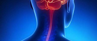
Meningitis is a severe infectious inflammatory disease of the membranes of the brain and/or spinal cord. The causative agent of meningitis can be bacteria, viruses, pathogenic fungi and some protozoa. The disease has a clear clinical picture (high fever, headache), and if it occurs, you should immediately consult a doctor. With modern medical treatments, meningitis is highly treatable.
However, it is very important to seek medical help immediately, as the disease has a high tendency to progress and can lead to serious consequences and death of the patient. When the first signs of meningitis appear, you can seek help at the Yusupov Hospital. Experienced doctors will quickly diagnose and begin treatment. A timely visit to the hospital will save time and avoid serious consequences.
Symptoms
Prognosis for a diagnosis of meningitis depends on the timeliness of diagnosis and initiation of treatment. The first signs of the disease are:
- headache;
- a sharp increase in body temperature;
- attacks of nausea and vomiting. In this case, after vomiting, the patient does not experience relief;
- numbness of the muscles in the neck;
- feeling of weakness and general malaise;
- lethargy, agitation. Sometimes hallucinations may appear;
- lack of appetite;
- diarrhea.
These symptoms include the following:
- feeling of pressure in the eye area;
- pain when pressing in the eyebrow area, under the eye or in the trigeminal nerve area;
- inflammation of the lymph glands;
- Brudzinski's symptom - when tilting the head or pressing on various parts of the body, legs and other parts of the body move reflexively;
- Kernig's symptom - the legs at the knee joint stop extending due to tension in the posterior thigh muscles;
- Bekhterev's symptom - when tapping on the zygomatic arch, the facial muscles contract;
- Mendel's symptom - the occurrence of pain when pressing on the area of the external auditory canal;
- Pulatov’s symptom – when the skull is tapped, the patient experiences pain;
- Lesage's symptom - in young children the large fontanelle is tense and pulsating. If you take the baby under the armpits, he throws his head back and reflexively presses his legs towards his stomach.
Nonspecific symptoms of meningitis can also be identified, including:
- body muscle spasms;
- increased blood pressure;
- hearing loss;
- impaired visual function – possible double vision, strabismus, ptosis, nystagmus;
- runny nose, cough, sore throat;
- bradycardia and tachycardia;
- increased irritability;
- drowsiness.
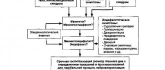
Diagnostics
Before treating meningitis, the doctor must find out the etiology of the disease. For this purpose, additional research is prescribed:
- general blood test - to identify inflammatory changes;
- general urinalysis - severe meningitis and sepsis can lead to kidney damage;
- spinal puncture with examination of cerebrospinal fluid;
- biochemical blood test - to determine the degree of damage to internal organs;
- bacteriological culture of mucus from the nasopharynx to identify meningococcus, pneumococcus;
- bacteriological examination of cerebrospinal fluid and blood - if purulent meningitis is suspected;
- stool tests to detect enteroviruses and polio viruses (PCR);
- A blood test to determine the acid-base balance and coagulation system is prescribed for severe meningitis and infectious-toxic shock.
In addition, the doctor may need the results of other studies:
- electrocardiograms of the heart - in case of serious condition of the patient and symptoms of damage to the heart muscle;
- chest x-ray - if pneumonia is suspected, which often accompanies pneumococcal meningitis.
Make an appointment
Purulent meningitis
MENINGITIS is an inflammation of the membranes of the brain and spinal cord, which is based on an infectious process, which can be caused by a bacterial, viral, fungal infection, occur acutely, subacutely or chronically, primarily affect the membranes, or be secondary (occur against the background of another disease: otitis media, sinusitis, traumatic brain injury).
The clinical picture of meningitis consists of general infectious symptoms (fever, malaise, tachycardia, muscle pain); general cerebral symptoms (intense headache, nausea, vomiting, confusion or depression of consciousness up to coma) and meningeal syndrome.
Meningeal syndrome includes rigidity of the neck muscles, Kernig's sign, Brudzinski's symptoms, decreased threshold to external stimuli, and pain syndromes. Meningeal symptoms can often be detected even in a coma, but at an early stage of the disease, in children and the elderly, they may be absent. In the elderly, meningitis may present as a combination of fever with confusion or increasing depression of consciousness in the absence of meningeal symptoms.
The study of cerebrospinal fluid is of decisive importance in the diagnosis of meningitis. In this regard, lumbar puncture is mandatory at the slightest suspicion of meningitis. A contraindication to lumbar puncture may be: signs of incipient herniation of the brain stem (increasing depression of consciousness, unilateral pupil dilation, respiratory rhythm disturbance, decortication or decerebrate rigidity), detected by CT or MRI, obstructive hydrocephalus.
PURULAR MENINGITIS.
Meningococcal meningitis most often occurs in childhood and adolescence and is usually sporadic, but sometimes small epidemic outbreaks are observed, especially in children's groups. The infection is transmitted by airborne droplets, and its source can be patients with meningococcal nasopharyngitis. In severe cases of meningococcal meningitis, a characteristic hemorrhagic petechial and purple rash occurs, which has the appearance of stars of various sizes and shapes and is localized on the torso and lower extremities (in the area of the buttocks, thighs, legs). Petechiae can also be on the mucous membranes, conjunctiva, and sometimes on the palms and soles. Much less often, a similar rash is observed with meningitis caused by enteroviruses, Haemophilus influenzae, Listeria, pneumococcus, as well as with staphylococcal bacterial endocarditis, rickettsiosis, and vasculitis.
Pneumococcal meningitis is the most common type of meningitis in people over 30 years of age. It often develops as a result of the spread of infection from distant foci (with pneumonia, otitis media, mastoiditis, sinusitis, endocarditis) and is especially severe in patients with reduced reactivity (with alcoholism, diabetes, multiple myeloma, hypogammaglobulinemia, liver cirrhosis, after splenectomy, against the background of corticosteroid therapy, hemodialysis). Pneumococcus is a common causative agent of post-traumatic meningitis in patients with a basal skull fracture and liquorrhea. Pneumococcal meningitis is usually severe, often causes depression of consciousness, persistent focal symptoms and epileptic seizures, often ends in death, and can recur.
Most cases of meningitis caused by Haemophilus influenzae occur in children under 6 years of age, but occasionally it occurs at older ages, usually against the background of sinusitis, epiglottitis, pneumonia, otitis media, head injury, diabetes mellitus, alcoholism, splenectomy, hypogammaglobulinemia, AIDS.
In addition to general cerebral and meningeal symptoms, with purulent meningitis, focal neurological symptoms are sometimes encountered, caused by the involvement of the cranial and spinal nerves, and less often - the brain substance itself. The oculomotor nerves are especially often affected, but, as a rule, movements of the eyeballs are restored within a few days or weeks, while damage to the auditory nerve often leads to permanent deafness. Approximately 40% of patients with meningitis experience epileptic seizures.
Treatment. If meningitis is suspected, the patient should be urgently hospitalized. After taking a sample of cerebrospinal fluid and blood for bacteriological examination, antibiotics are immediately prescribed.
In adults with normal immunity, the causative agent of meningitis is most often pneumococcus or meningococcus, less often streptococcus, Haemophilus influenzae and listeria; The drugs of choice are penicillin or ampicillin, however, taking into account the emergence of strains of pneumococci and meningococci resistant to penicillin, third generation cephalosporins (ceftriaxone or cefotaxime) have been increasingly used in recent years.
After normalization of the temperature, antibacterial therapy should be continued for an average of 10 days. For meningococcal infection, its duration can be reduced to 5-7 days (after the temperature has normalized). For pneumococcal meningitis, the course duration increases to 14 days.
In the presence of sinusitis, otitis or other parameningeal infection, as well as meningitis caused by gram-negative bacteria, listeria, Pseudomonas aeruginosa, the duration of treatment is increased to 3-4 weeks.
Before discontinuing antibiotics, in order to ensure the sanitation of the cerebrospinal fluid, a control lumbar puncture is performed. The condition for discontinuing the antibiotic is that the cerebrospinal fluid is sterile.
If, during antibacterial therapy, fever persists for more than 2-5 days or occurs again, then you should think about the possibility of complications, inadequacy of therapy, phlebitis, metastatic infection (septic arthritis, pericarditis, endocarditis) or the toxic effect of the drug. If a primary source of infection (for example, suppurative otitis media), an abscess or subdural empyema is identified, urgent surgical intervention is indicated.
Treatment
Doctors at the Yusupov Hospital adhere to the principles of treating patients with meningitis, which include preventing further spread of the pathological process and preventing the development of complications. Neurologists take an individual approach to the treatment of each patient. Complex therapy includes:
- etiotropic treatment (destruction of bacteria that cause inflammation of the meninges);
- detoxification therapy;
- measures aimed at reducing intracranial pressure;
- anti-inflammatory therapy with corticosteroid hormonal drugs;
- treatment of intracranial and extracranial complications;
- relief of convulsive syndrome;
- normalization of body temperature.
Antibiotics for meningitis in adults at the prehospital stage are used only in cases where emergency delivery of the patient to the Yusupov Hospital is impossible for some objective reason within 2.5 or 3 hours.
The introduction of antibiotics for meningitis at an earlier time (up to 60 minutes) is justified only if there is a strong suspicion of the meningococcal nature of the disease when the symptoms of meningitis are combined with hemorrhagic rashes that do not disappear with pressure. Antibiotics for bacterial purulent meningitis are administered only parenterally. Antibacterial tablets are not effective for meningitis. If the meningococcal nature of the disease is suspected, antibiotics for meningitis are administered only against the background of established anti-shock therapy in the presence of vascular access due to the high risk of impaired blood supply to tissues and the development of acute adrenal insufficiency against the background of low blood pressure.
At the prehospital stage of treatment of meningitis, third generation cephalosporins are administered. Penicillin for meningitis in this case is less effective, since in Russia there is no mandatory vaccination against Haemophilus influenzae and a hemorrhagic rash can be a manifestation of meningitis caused by Haemophilus influenzae, which is insensitive to penicillin. If there is information about severe allergic reactions in the patient to beta-lactam antibiotics, sodium chloramphenicol succinate (chloramphenicol) is administered. The administration of an antibiotic is not a reason to delay hospitalization of the patient.
For patients with bacterial purulent meningitis, empirical antibacterial therapy begins no later than an hour after admission to the neurology clinic. If the patient has contraindications to immediate lumbar puncture, antibiotics for meningitis begin to be administered immediately after collecting a blood sample for bacteriological examination. If the patient’s condition is stable, there are no contraindications to cerebrospinal puncture, and it is possible to obtain cerebrospinal fluid within one hour of the patient’s admission to the neurology clinic, antibacterial therapy begins after a cystoscopic examination of Gram-stained cerebrospinal fluid smears.
When empirically selecting antibiotics for patients suffering from meningitis, neurologists take into account:
- patient's age;
- conditions preceding the disease (developmental defects, trauma, neurosurgical interventions, immunodeficiency states, cochlear implantation);
- availability of vaccination against meningococcus, Haemophilus influenzae, pneumococcus;
- the possibility of contact with infectious patients;
- staying in countries with a high incidence of bacterial meningitis;
- regional characteristics of pathogens causing neuroinfectious diseases.
Antibiotics for meningitis of unknown cause
In the presence of an unfavorable background preceding meningitis, newborns and children under one month are prescribed the following antibiotics for meningitis: ampicillin and an aminoglycoside (gentamicin) or cefotaxime.
If the pneumococcal nature of the disease is suspected, the use of vancomycin is indicated. In children from 3 months to 18 years of age, meningitis is caused mainly by pneumococci, Haemophilus influenzae, and meningococci. Patients are prescribed antibiotics that belong to the third generation cephalosporins (cefotaxime or ceftriaxone). If the pneumococcal nature of meningitis is suspected, if strains of bacteria resistant to cephalosporins are present in the region, vancomycin or rifampicin is added to the treatment regimen. If a patient develops meningitis due to a skull fracture, the cause of inflammation of the meninges may be pneumococci, Haemophilus influenzae, or β-hemolytic streptococcus. In this case, neurologists at the Yusupov Hospital use a combination of third-generation cephalosporins with vancomycin.
In case of penetrating head trauma, after neurosurgical interventions, the causative agents of meningitis are Staphylococcus aureus, aerobic gram-negative bacteria. They are most sensitive to the combination of vancomycin with cefepime, ceftazidime or meroneme. If meningitis develops after shunt placement, the same combinations of antibiotics are effective.
At the onset of meningitis, similar symptoms (impaired consciousness, convulsions, fever, meningeal symptoms) and the results of cerebrospinal fluid examination (mixed pleocytosis) do not exclude viral encephalitis. Doctors at the Yusupov Hospital prescribe acyclovir intravenously in all doubtful cases, pending the results of the final examination of the patient, along with antibacterial therapy. After laboratory tests and clarification of the nature of meningitis, antibiotic therapy is adjusted.
Antibiotic therapy for established meningitis pathogens
If, when examining the cerebrospinal fluid obtained during the first lumbar puncture, laboratory technicians identify the causative agent of meningitis, doctors at the Yusupov Hospital prescribe antibiotics to which the identified microorganisms are sensitive.
Cefotaxime, ceftriaxone, and penicillin act on meningococci. Alternative antibacterial drugs are the antibiotics meronem and chloramphenicol. With the development of septic shock and multiple organ failure, doctors give preference to cephalosporins. The main antibiotics that doctors prescribe to treat patients with pneumococcal meningitis are cefotaxime and ceftriaxone. Cefepime, meronem and chloramphenicol are used as reserve antibiotics. When penicillin-resistant strains circulate in the region, combination antibiotic therapy with third-generation cephalosporins in combination with vancomycin or rifampicin is prescribed.
Haemophilus influenzae type b is not sensitive to penicillin. If it is the cause of meningitis, neurologists prescribe ampicillin, cefotaxime, ceftriaxone or chloramphenicol. Cefepime or meronem are used as alternative antibiotics. Streptococci serotype B are sensitive to the antibiotic cefotaxime. Neurologists at the Yusupov Hospital individually select doses and frequency of antibiotic administration depending on the patient’s age. For cryptococcal meningitis, 5-flucytosine and amphotericin B are used, and for tuberculosis meningitis, pyrazinamide, rifampicin, isoniazid and ethambutol are used.
The duration of antibacterial therapy is determined individually for each patient, depending on the cause of meningitis, the characteristics of the course of the disease and the presence of complications. The average duration of antibiotic therapy, depending on the causative agent of meningitis with an uncomplicated course of the disease, is as follows:
- meningococcal meningitis – 7 days;
- hemophilic – from 7 to 10 days;
- pneumococcal – from 10 to 14 days;
- listeria – 21 days;
- streptococcal (group B) – 14 days.
For bacterial meningitis caused by gram-negative enterobacteriaceae or Pseudomonas aeruginosa, antibiotics are administered for 21 days. Antibiotics are discontinued when the symptoms of meningitis completely reverse and cerebrospinal fluid levels return to normal. For meningococcal meningitis, antibiotic therapy is stopped if there are no infectious agents in the cerebrospinal fluid and nasopharyngeal swabs.
Complex treatment of meningitis
In addition to antibiotics for meningitis, doctors at the Yusupov Hospital prescribe glucocorticoids to patients.
Dexamethasone is administered before starting antibiotic therapy or simultaneously with the first dose of antibiotic. If there are indications for prescribing dexamethasone after starting antibiotic treatment, it is administered in the first 4 hours after injection of the antibacterial drug. Dexazone is not prescribed later than 12 hours from the start of antibiotic therapy for meningitis. When prescribing glucocorticoids, neurologists take into account that, by reducing the severity of inflammatory changes in the meninges, they help reduce the permeability of the blood-brain barrier to antibacterial drugs and lead to a decrease in the concentration of antibiotics in the subarachnoid space. This fact is especially taken into account when using vancomycin in patients with pneumococcal meningitis. Long-term use of glucocorticoids in patients with meningitis leads to suppression of the immune system and can cause the development of secondary bacterial complications and activation of viral infections. For signs of septic shock and manifestations of acute adrenal insufficiency, glucocorticoids are also prescribed.
Detoxification therapy is prescribed to maintain circulating blood volume and adequate blood supply to brain tissue. When conducting infusion therapy, doctors use isotonic solutions of sodium chloride, glucose or dextrose, Ringer's solution. All patients with meningitis at the Yusupov Hospital are monitored for blood glucose levels at least once a day.
If there are signs of shock, sodium chloride is administered as an emergency intravenous bolus over 5-10 minutes. If symptoms persist or progress, 0.9% sodium chloride solution or 5% human albumin is reintroduced. If symptoms continue to persist after administration of fluid in a volume of 40 ml/kg, a third injection of 0.9% sodium chloride or 5% human albumin is administered over 5-10 minutes. If the measures taken are ineffective, the patient is immediately transferred to artificial ventilation, vasoactive drugs are added, and infusion therapy is continued.
For infusion therapy with an increasing increase in intracranial pressure and the threat of developing cerebral edema, a mannitol solution is used, followed by the administration of furosemide to prevent “recoil” syndrome. If seizures develop, patients are given diazepam or midozal. If convulsive status develops, valproates (Convulex, Depakine) are used. If epileptic activity persists, patients are connected to a ventilator and sodium hydroxybutyrate or sodium thiopental is administered. For the purpose of immunocorrection, intravenous immunoglobulins enriched with IgM - pentaglobin - are used. They are effective on the first day of the disease. If patients develop acute renal failure, plasmapheresis sessions are performed.
You can undergo a course of adequate treatment of meningitis with antibiotics by first making an appointment and calling the Yusupov Hospital. The clinic’s doctors individually select an antibacterial therapy regimen and use antibiotics that have a minimal range of side effects. Timely adequate treatment with antibiotics can prevent complications of meningitis.
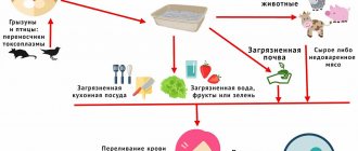
Meningococcal infection
The disease is caused by different strains (varieties) of meningococcus. The source of infection of a child can be a sick person or a “healthy” carrier of meningococcus. The number of such carriers for meningococcal infection is very large: for one case of the generalized form of the disease there are from 2 to 4 thousand healthy carriers of this microbe.
The carriers are usually adults, although they do not know about it, and mostly children get sick.
The pathogen lives in the nasopharynx and is released into the external environment when sneezing or talking. The danger increases when inflammation occurs in the nasopharynx. Fortunately, meningococcus is very unstable in environmental conditions: it survives no more than half an hour.
Infection occurs by airborne droplets with very close (at a distance of up to 50 cm) and prolonged contact. The infection has a pronounced winter-spring seasonality with a peak incidence from February to April.
Periodic increases in the incidence rate are recorded after about 10 years, which is associated with a change in the strain of the pathogen and the lack of immunity to it. Both isolated cases of morbidity in children and widespread cases in the form of outbreaks and epidemics are possible. During the period between epidemics, more young children get sick, and during an epidemic, more older children get sick.
Meningococcus is sensitive to antibiotics and sulfonamide drugs.
When a pathogen enters the mucous membrane of the nasopharynx, it most often does not cause inflammation: this is how a “healthy” carrier state is formed. But sometimes inflammatory changes occur in the nasopharynx, and a localized form of the disease develops: meningococcal nasopharyngitis.
Much less often (in 5% of cases) the microbe penetrates the blood and spreads to various organs. This is how meningococcal sepsis (meningococcemia) develops.
Severe toxic syndrome occurs as a result of the destruction of meningococci (under the influence of produced antibodies or antibiotics) and the release of a significant amount of endotoxin. This may cause the development of infectious-toxic shock.
In addition to internal organs (lungs, joints, adrenal glands, retina, heart), meningococcus can also affect the central nervous system: the membranes and substance of the brain and spinal cord. In these cases, purulent meningitis (or meningoencephalitis) develops.
After an illness and even as a result of carriage of meningococcus, persistent immunity is developed.
Symptoms
The incubation period can last from 2 to 10 days, usually it is short: 2-3 days.
There are localized and generalized clinical forms of meningococcal infection.
Localized:
- asymptomatic meningococcal carriage;
- meningococcal nasopharyngitis.
Generalized:
- meningococcemia (meningococcal sepsis);
- meningitis (inflammation of the membranes of the brain);
- meningoencephalitis (inflammation of both the membranes and substance of the brain);
- mixed form (a combination of meningococcemia and meningitis).
Rare forms include: meningococcal-induced arthritis, pneumonia, iridocyclitis, endocarditis.
Asymptomatic meningococcal carriage is the most common form of the disease (develops in 99.5% of all infected people). More often observed in adults. The condition does not show any signs and the person is unaware of his infection.
Meningococcal nasopharyngitis develops in 80% of patients with meningococcal infection. It manifests itself with the usual symptoms for an inflammatory process in the nasopharynx: acute onset, sore throat, nasal congestion, dry cough, headache. The temperature may rise within 37.5°C. The general condition and well-being suffer little.
Upon examination, redness in the pharynx and swelling of the mucous membrane, sometimes redness of the conjunctiva, and scanty mucopurulent discharge from the nose are revealed. More often the condition is regarded as a manifestation of an acute respiratory disease. The correct diagnosis is made only at the source of infection when examining contact persons.
The duration of the disease is from 2 to 7 days; ends with recovery. But often (about 30% of cases) this form precedes the subsequent development of a generalized form of infection.
Meningococcemia develops acutely, suddenly. Its manifestations increase very quickly. Parents can indicate the exact time of onset of the illness, not just the date. The temperature rises sharply with chills (up to 40°C), which is difficult to reduce with antipyretic drugs. There is repeated vomiting and severe headache and thirst.
But the main and most characteristic sign of meningococcal sepsis is a rash. It appears already on the first day of illness, less often on the second. The earlier the rash appears from the onset of the disease process, the more severe the course and prognosis of the disease.
More often it is localized on the thighs, legs, lower abdomen, and buttocks. The rash spreads quickly, literally “growing before our eyes.” The appearance of rashes on the face indicates the severity of the process. This is an unfavorable prognostic sign.
The size of the rash can vary: from small pinpoint hemorrhages to large irregular (“star-shaped”) elements of a purplish-bluish color. The rash is a hemorrhage into the skin, it does not disappear with pressure, and is located on a pale background of the skin. Pinpoint rashes last 3-4 days, become pigmented and disappear.
In the center of large elements of the rash, necrosis (death) of tissue may develop after a couple of days. The necrotic surface becomes covered with a crust; after it peels off, ulcers form, which scar very slowly (up to 3 weeks or more).
Necrosis can also occur on the tip of the nose, phalanges of the fingers, and ears with the development of dry gangrene.
Clinical symptoms of meningococcemia can grow very rapidly, especially with the fulminant variant of the disease. Hemorrhage into the conjunctiva or sclera of the eyes may appear even earlier than the skin rash. Other manifestations of hemorrhagic syndrome may also occur: bleeding (nasal, gastric, kidney) and hemorrhages in various organs.
Due to impaired blood supply and metabolic processes due to toxicosis, with meningococcemia, children have symptoms of damage to the kidneys, cardiovascular system, lungs, eyes, liver, and joints. All children experience shortness of breath, increased heart rate, and decreased blood pressure.
When the kidneys are involved in the process, changes appear in the urine (protein, red blood cells and white blood cells). Joint damage is characterized by pain in large joints and swelling, and limited range of motion.
In case of hemorrhage in the adrenal glands, acute adrenal insufficiency develops due to hormone deficiency, which can cause death. This complication, just like acute renal failure, is possible with the fulminant form of meningococcemia (hyperacute sepsis).
Clinically, adrenal insufficiency is manifested by a sharp drop in blood pressure, vomiting, the appearance of bluish spots on the skin against the background of severe pallor, frequent weak pulse, severe shortness of breath and subsequent disturbance of the breathing rhythm, and a drop in temperature below normal. In the absence of qualified assistance, death can occur even within a few hours.
A chronic form of meningococcemia with periodic relapses is extremely rare. It may last for several months.
If the meninges are involved in the pathological process, the child’s condition worsens sharply.
Purulent meningococcal meningitis is also characterized by an acute onset. A sharp diffuse headache appears, small children react to it with the appearance of anxiety and piercing crying. The temperature with chills can rise to 40°C and does not decrease after the child takes antipyretic medications.
The headache intensifies in response to any irritant: loud sound, light, even touch: in young children this manifests itself as a symptom of “repulsion of the mother’s hands.” Intensification of the headache is noted with the slightest movement, when turning the head.
No appetite. Repeated vomiting does not bring relief. It is not related to food intake. Diarrhea may also appear, especially at an early age. The child is pale, lethargic, pulse is rapid, blood pressure is reduced.
Muscle tone is increased. The child's position in bed is typical: lying on his side, “curled up,” with his legs pulled to his stomach and his head thrown back.
In small children, there is bulging, tension and pulsation of the large fontanelle. Sometimes there is a divergence of the seams between the bones of the skull. When a small child becomes dehydrated due to vomiting and loose stools, the fontanelle collapses.
Babies may experience reflex constipation and lack of urination.
Sometimes children experience motor restlessness, but there may also be lethargy, drowsiness and lethargy. In young children, you may notice trembling of the chin and hands, which is manifested by symptoms such as impaired consciousness, mental disorders, motor agitation and convulsions.
Upon examination, the doctor identifies focal symptoms: paresis (or paralysis), pathological changes in the cranial nerves (oculomotor disorders, decreased hearing and vision). In severe cases, when cerebral edema occurs, swallowing, speech, cardiac activity and breathing may be impaired.
In the mixed form, both clinical manifestations of meningitis and symptoms of meningococcemia may predominate.
During the course of the generalized form of the disease, rare forms can also develop: damage to the joints, heart, retina and lungs. But if meningococcus enters the lungs directly with air, then meningococcal pneumonia can develop primarily.
.
Diagnostics
During the examination, the doctor assesses the condition of the large fontanel in young children and checks for the presence of meningeal symptoms.
The following methods are used to diagnose meningococcal infection:
- survey of parents and child (if possible by age): allows you to find out the presence of contact with sick people, clarify complaints, the dynamics of the development of the disease and the sequence of symptoms;
- examination of the child by a doctor: assessment of the severity of the condition and identification of a number of clinical signs of the disease (temperature, skin color, rash, meningeal symptoms, condition of the large fontanel in young children, convulsions, etc.);
In the case of generalized forms of the disease, the diagnosis can be made based on clinical manifestations. To confirm the diagnosis, laboratory diagnostic methods are used (it is carried out in a hospital setting after emergency hospitalization of the child):
- clinical examination of blood and urine: in the blood of meningococcal infection there is an increased total number of leukocytes, an increase in the number of band and segmented leukocytes, the absence of eosinophils and an accelerated ESR; Urinalysis allows you to evaluate kidney function;
- clinical examination (bacterioscopy) of a thick drop of blood and cerebrospinal fluid sediment to detect meningococci;
- bacteriological method: culture of mucus from the nasopharynx, culture of cerebrospinal fluid, blood culture to isolate meningococcus and determine its sensitivity to antibiotics;
- a biochemical blood test (coagulogram, liver and kidney complex) allows you to assess the severity of the child’s condition;
- serological blood test (paired sera taken at intervals of 7 days) can detect antibodies to meningococcus and an increase in their titer; a 4-fold increase in titer is diagnostic;
Additional examination methods:
- consultations with a neurologist, ENT doctor and ophthalmologist (fundus examination);
- in some cases, echoencephalography (ultrasound examination of the brain to diagnose complications of the disease) and computed tomography are performed;
- According to indications, ECG and echocardiography may be prescribed.
Treatment
At the slightest suspicion of meningococcal infection, urgent hospitalization is carried out.
At home, it is possible to treat carriers of meningococcus and meningococcal nasopharyngitis (if there are no other children in the family at preschool age).
For the treatment of nasopharyngitis of meningococcal etiology, the following is prescribed:
- antibiotics orally in an age-appropriate dosage;
- gargling with furatsilin solution;
Treatment of generalized forms includes:
- antibacterial therapy;
- hormonal drugs;
- detoxification therapy;
- symptomatic treatment.
In order to influence meningococcus, Penicillin and Levomycetin-succinate are prescribed. The choice of antibiotic, its dosage, and the duration of the course depend on the clinical form of the disease, severity, age and body weight of the child and his other individual characteristics.
When treating meningitis and meningoencephalitis, high doses of antibiotics are used to overcome the blood-brain barrier and create a sufficient concentration of the antibiotic in the brain matter. Penicillin is preferably prescribed.
For meningococcemia, Prednisolone and Levomycetin-succinate are administered at the prehospital stage (in the clinic or by ambulance staff), and not Penicillin, which has a detrimental effect on meningococcus. When the microbe dies, endotoxin is released in large quantities, and infectious-toxic shock can develop. And Levomycetin will only prevent the pathogen from multiplying.
Hormonal drugs (Prednisolone, Hydrocortisone) are used in cases of severe infection in order to suppress the violent reaction of the immune system to the penetration of the pathogen and to maintain blood pressure at the proper level.
In case of developing infectious-toxic shock, treatment is carried out in an intensive care unit.
The following are used as detoxification agents: 10% glucose solution, plasma and plasma substitutes, Ringer's solution, Reopoliglyukin, etc. Plasmapheresis and ultraviolet irradiation of blood can be used.
Symptomatic therapy includes the prescription of anticonvulsants (Sibazon, Relanium, Sodium Oxybutyrate), cardiac drugs (Korglykon, Cordiamin), diuretics (Lasix), vitamins (C, group B), heparin under the control of the blood coagulation system.
To reduce cerebral hypoxia, oxygen therapy and cerebral hypothermia are used (applying an ice pack to the head).
If breathing is impaired, the child is connected to an artificial respiration apparatus.
Prognosis and outcomes of the disease
During the recovery period, weakness and increased intracranial pressure may be noted, which disappear after a few months.
The prognosis is more severe in children under one year of age. In rare cases, they may develop severe consequences in the form of hydrocephalus and epilepsy.
Complications of meningococcal infection are divided into specific and nonspecific. Specific (develops at an early stage of the disease):
- infectious-toxic shock;
- acute cerebral edema;
- bleeding and hemorrhage;
- acute adrenal insufficiency;
- acute heart failure;
- pulmonary edema, etc.
Nonspecific (due to other bacterial flora):
- pneumonia;
- otitis media, etc.
Specific complications are manifestations of the pathological process itself. Any of them can cause the death of a child.
After the illness, residual effects and complications can be detected.
Functional residual effects:
- asthenic syndrome, the manifestation of which at an early age is emotional instability and motor hyperactivity, disinhibition, and at an older age - decreased memory and fatigue;
- vegetative-vascular dystonia during puberty in adolescents.
Organic complications:
- hydrocephalus (increased amount of fluid in the cranial cavity);
- increased intracranial pressure;
- child's lag in psychomotor development;
- decreased or loss of hearing;
- epileptiform (convulsive) syndrome;
- paresis with movement disorders.
Prevention
Preventive measures can be considered:
- early detection and hospitalization of patients;
- measures at the source of infection: identification of carriers of meningococcus and their treatment, 10-day observation of those in contact with the patient and their 2-fold examination (nasopharyngeal swab), admission of contact children to kindergarten only after a negative examination result;
- discharge of a recovered person from the hospital only after a 2-fold negative bacteriological analysis of mucus from the nasopharynx (done 3 days after the course of treatment with an interval of 1 or 2 days);
- limiting contacts;
- during an outbreak of morbidity, exclusion of holding mass events with overcrowding of children;
- treatment of chronic foci of infection;
- hardening;
- vaccination (Meningo A+C vaccine): schoolchildren (if more than 2 cases of meningococcal infection are registered at school) and children before traveling to a region unfavorable for the incidence of this infection. The vaccine can be used in children from 1.5 years of age; immunity is formed by day 10 and lasts for 3-5 years.
Summary
Meningococcal infection is a serious disease, especially for young children. The danger of this infection is not only in the acute period (due to the development of complications and threats to life), but also after recovery (quite serious consequences can remain for life).
Considering the likelihood of a very rapid development of the disease, you should not delay the time to consult a doctor with any disease.
It must be remembered that a spinal puncture (which parents are so afraid of) is a necessary diagnostic procedure that will help the doctor prescribe the correct treatment.
Which doctor should I contact?
If symptoms of inflammation of the nasopharynx appear, you should usually contact a pediatrician, and adults should consult a therapist. If there is a rapid increase in temperature, deterioration of the condition, severe headache and especially the appearance of a skin rash, you should urgently call an ambulance. Treatment is carried out in an infectious diseases hospital.
ANTI-EPIDEMIC MEASURES IN THE SOCIETY OF MENINGOCOCCAL INFECTION
General events. Information about the sick person in the Center for Sensitive Diseases in the form of an Emergency Notification within 12 hours after identifying the patient. Epidemiological examination of the outbreak in order to identify and sanitize carriers and patients with erased forms; determination of the circle of persons subject to mandatory bacteriological examination. Measures regarding the source of the pathogen. Hospitalization of the patient, isolation of carriers.
Discharge from the hospital - with 2 negative bacteriological studies of nasopharyngeal mucus, carried out 3 days after the end of treatment. Measures regarding pathogen transmission factors. Disinfection: daily wet cleaning, ventilation, irradiation with UV rays and bactericidal lamps in the fireplace. Final disinfection is not carried out. Measures regarding contact persons in the outbreak. Medical observation for 10 days from the last visit to the sick team/daily examination of the skin and pharynx with the participation of an ENT doctor, thermometry/. Children, staff of preschool and school institutions, in universities and secondary specialized institutions are subject to bacteriological examination in the 1st year - the entire course where the patient is identified, in senior years - students of the group where the patient or carrier is identified. In kindergartens, biological examinations are carried out 2 times with an interval of 3-7 days. Emergency prevention. Children from 18 months. up to 7 years of age and first-year students, in the first 5 days after contact, active immunization is carried out with a meningococcal polysaccharide vaccine of serogroups A and C. In its absence, normal human immunoglobulin is administered. Previously vaccinated children are not given immunoglobulin.
Rehabilitation
Patients at the Yusupov Hospital who have had meningitis require observation by a neurologist for the next 2 years.
Regular examination in the first year should be carried out once every 3 months, then once every 6 months. Recovery from meningitis is complex, complex and multifaceted. One of the components of the rehabilitation period after meningitis is a diet aimed at restoring the patient’s strength without causing irritation to the gastrointestinal tract. The preferred methods of cooking are boiling, including steaming, stewing, and baking. It is recommended to eat lean meat: rabbit, chicken, veal, lean fish. Vegetables and fruits must be heat-treated to prevent irritation of the sensitive mucous membrane by coarse fiber. Consumption of dairy products is necessary to further enrich the body with protein. Patients are recommended to drink jelly, compotes, and weak tea.
Physiotherapy plays an important role in restoring the body of a patient who has suffered meningitis. Doctors at the Yusupov Hospital prescribe classical massage and various hardware techniques. Electrophoresis of vitamins and medications helps relax or stimulate the desired muscle groups. Patients with coordination and cognitive impairments are prescribed the use of electrosleep, magnetic therapy, and magnetic laser therapy to restore central nervous system functions. The selection of individual physiotherapeutic techniques aimed at treating the consequences of meningitis at the Yusupov Hospital is carried out by qualified physiotherapists, depending on the patient’s condition.
A separate and extensive area of recovery after meningitis is physical therapy. Qualified physical therapy doctors at the Yusupov Hospital develop individual programs for patients to help restore movement skills.
The everyday adaptation of patients is provided with the help of occupational therapy. This complex technique is aimed at restoring the amplitude, strength and coordination of movements.
Cognitive therapy helps restore attention, memory and logical thinking.
Rehabilitation after meningitis is a long and painstaking process, the effectiveness of which requires the experience and knowledge of doctors, which the specialists of the Yusupov Hospital fully possess, as well as the perseverance, consistency and patience of patients.
General recovery regimens are aimed at relieving symptoms, improving function and overall well-being of the patient. In addition, they help improve motor, sensory, cognitive and behavioral function.
The Yusupov Hospital in Moscow is a modern multidisciplinary clinic that offers medical care and care services at the highest level. The Neurology Clinic of the Yusupov Hospital provides comprehensive diagnosis and treatment of meningitis using modern and high-precision equipment, which allows us to identify the disease at an early stage and select the most effective treatment regimen for each patient.
You can make an appointment for diagnosis and treatment with a doctor at the Yusupov Hospital, and find out the cost of the medical services provided by calling or visiting the clinic’s website. The coordinating doctor will answer all your questions.
Make an appointment
Complications
The disease is characterized by an acute course and begins with an increase in body temperature.
Patients complain of severe headaches and vomiting, which does not bring relief. The so-called meningeal syndrome is associated with one of the most obvious symptoms of meningitis - stiffness of the neck muscles. Patients develop drowsiness, photophobia, dizziness, and in some cases, convulsions and even loss of consciousness.
The most common and relatively harmless consequence of meningitis is asthenic syndrome, which manifests itself as causeless malaise, weakness, and low mood. It can last from 3 months to 1 year.
However, according to modern researchers, in 30% of patients who suffered meningitis, the disease caused the development of the following complications:
- intellectual disabilities;
- paresis, paralysis;
- loss of vision;
- hearing loss (sensorineural hearing loss);
- hydrocephalus;
- convulsive syndrome;
- ischemic stroke.
You can make an appointment for diagnosis and treatment with a doctor at the Yusupov Hospital, and find out the cost of the medical services provided by calling or visiting the clinic’s website.
The coordinating doctor will answer all your questions. Make an appointment
HIB-MENINGITIS: clinical picture, diagnosis and treatment
Meningitis caused by Haemophilus influenzae type B, or HIB meningitis, ranks third in frequency in the etiological structure of bacterial meningitis, accounting for 5 to 25% of cases. In the age group up to 4-5 years, this form of meningitis ranks second (from 10 to 50% of cases). In Moscow, in children under 5 years of age, HIB meningitis accounts for 15% of all cases of bacterial meningitis.
The incidence of HIB meningitis in children under 5 years of age before the start of routine vaccination in a number of countries varied from 23 cases per 100,000 children per year in developed European countries to 60 per 100,000 in developing countries. Before the introduction of vaccination against HIB infection, 370,000 cases of HIB meningitis were reported annually worldwide, of which about 100,000 were fatal (mainly in developing countries, which accounted for 97% of deaths). The mortality rate for this form of meningitis varies, according to various authors, from 5% (in developed countries) to 30% (in developing countries); Unfavorable long-term consequences are often observed.
Despite the significant place that HIB meningitis occupies in childhood infectious pathology, there are almost no works devoted to the pathogenic properties of the pathogen, pathogenesis and clinical features of this form of the disease. Publications devoted to the clinical aspects of HIB meningitis are based on a small number of observations and, as a rule, do not provide insight into the clinical and pathogenetic features of HIB meningitis.
Our report is based on an analysis of case histories of 89 patients with HIB meningitis, 71 of whom were treated in Clinical Hospital No. 2, 17 in other hospitals in Moscow. Observations cover the period from 1994 to 2001. Case histories of 150 patients with meningococcal meningitis, 51 with pneumococcal meningitis and 29 patients with meningitis of enteroviral etiology were used as comparison groups.
The diagnosis of HIB meningitis in 22 patients was confirmed after isolating a culture of the pathogen from the CSF or blood; in the other 22 patients, it was confirmed by latex agglutination. In 45 patients, the diagnosis was confirmed both bacteriologically and by latex agglutination.
As can be seen from table. 1, HIB meningitis is a disease of early and young childhood, and more than 11% of cases were children in the first 6 months of life (starting from 9 days), which indicates the absence or insufficiency of innate immunity. On the other hand, the fact that in children over 4 years of age, who belong to the most epidemiologically vulnerable age group, the incidence drops sharply can be explained both by the formation of acquired immunity (in children who have had various forms of HIB infection) and by age-related anatomical and physiological factors. characteristics of children of younger age groups.
Of the four patients over 8 years of age we observed, one child had a burdened premorbid background (exudative diathesis, multiple acute respiratory infections); two children aged 9 years suffered from purulent meningitis, of different etiologies, three to four times, which allows us to suggest the presence of an anatomical defect, most likely Spina bifida; Apparently, an adult woman suffered a skull injury, suffered from nasal liquorrhea and also repeatedly suffered from purulent meningitis of various etiologies.
HIB meningitis was severe in 88.6% of patients; in 12 patients the course of the disease was protracted (more than 2 months) or undulating. 6 patients died (6.7%).
From the data in table. 2 it follows that one of the important causes of mortality is hospitalization in non-core hospitals. This applies not only to patients with HIB meningitis, but also to patients with other neuroinfections.
Another reason is a burdened premorbid background: complicated pregnancy and childbirth, prematurity, organic damage to the central nervous system and other factors.
The third reason is late diagnosis and late hospitalization. The fourth is irrational therapy. Let's look at a typical case.
Patient U. from the Children's Home. Age - 1 year 3 months. He fell ill on February 27, 1997, was treated at the Children's Home for acute respiratory infections, and received ampicillin for the last day. On the 4th day of illness, the patient’s condition worsened and the patient was diagnosed with ARVI and pneumonia and was hospitalized at Children’s Infectious Diseases Hospital No. 12, where he received penicillin. On the 2nd day after hospitalization, the condition worsened, vomiting, muscle tremors, and bulging of the fontanel appeared. During puncture, purulent cerebrospinal fluid was obtained; the patient was transferred to clinical hospital No. 2 on the 6th day of illness. The condition upon admission was extremely serious. Upon repeated lumbar puncture, the diagnosis of purulent meningitis, complicated by cerebral edema, was confirmed. Concomitant disease: Down's disease, 3rd degree malnutrition. Treatment with penicillin was continued and intensive pathogenetic therapy was carried out. The patient's condition stabilized, but on the 3rd day it worsened again. Hyperthermia, convulsions, and loss of consciousness were observed. On the 3rd day of treatment, a culture of HIB was isolated from the CSF and blood, penicillin was replaced with chloramphenicol, but on the 5th day of treatment the patient died. On the day of death, the results of determining the sensitivity of the isolated culture were obtained: resistant to chloramphenicol and penicillin, sensitive to cefotaxime.
Pathological diagnosis: purulent meningitis caused by Haemophilus influenzae, cerebral edema, small-focal pneumonia. Associated complications: grade 3 accidental transformation of the thymus, Down's disease, grade 3 malnutrition, hydrocephalus.
This case illustrates the main causes of mortality: late diagnosis, severe premorbid background, inadequate antibacterial therapy. Nevertheless, thanks to rational pathogenetic therapy, death occurred only after 5 days, while in non-core hospitals all patients died within 24 hours, which, judging by the data of the case histories, is partly due to insufficiently adequate pathogenetic therapy. If we compare the mortality rate for purulent meningitis of various etiologies for the same period of time in the conditions of one medical institution (CIB No. 2), then for HIB meningitis it was 2.8%, for meningococcal meningitis - 6%, and for pneumococcal meningitis - 17%, i.e., in terms of prognosis for life, HIB meningitis is the most benign. At the same time, 8.5% of children were discharged with hearing loss, 13.4% with ataxia and other residual effects.
As can be seen from the above medical history, HIB meningitis often occurs against an unfavorable premorbid background, noted in 72% of patients. In most cases (62%), there were severe organic lesions of the central nervous system with hypertension and convulsive syndromes, developmental delays, paresis, etc. In patients with pneumococcal and meningococcal meningitis, the spectrum of background diseases was completely different, lesions of the central nervous system accounted for 12% and 3% respectively. Moreover, lesions of the central nervous system in pneumococcal meningitis were of traumatic origin. Frequent acute respiratory infections were observed in 52% of patients.
The onset of the disease in patients with HIB meningitis was acute, but the appearance of obvious clinical symptoms of meningitis was often preceded by moderate fever lasting from 1-2 to 5-7 days, accompanied by catarrhal symptoms from the upper respiratory tract. In 51% of patients, ARVI, pneumonia, catarrhal otitis, and sinusitis were initially diagnosed. In 6.8% of patients, fever was accompanied by dyspeptic symptoms, and they were diagnosed with acute intestinal infection.
Respiratory tract involvement is a characteristic manifestation of HIB infection, and it differs from those associated with meningococcal and pneumococcal infections. The absence of signs of meningitis in the early stages of the disease, as well as the underestimation of symptoms such as repeated vomiting, convulsive readiness, lethargy, and drowsiness, became the reason for the relatively late periods of hospitalization of patients.
Thus, in general, the average length of hospitalization was 3.1±0.17 days (for pneumococcal meningitis - 3.5±0.21 days, for meningococcal meningitis - 1.8±0.16).
The length of hospitalization of patients (Table 3) sent directly to Clinical Hospital No. 2 was 2.3 days, that is, they were significantly shorter. Naturally, this applies to children with the most striking clinical picture. More than half of the children were transferred to ICU No. 2 from other hospitals or were treated in them, most of them had problems with diagnosis. If hospitalization during the first two days of illness is conditionally considered timely, then it can be noted that 67.4%, that is, 2/3 of patients, were hospitalized in a specialized hospital during this period, while only 21.8% of patients were hospitalized in other hospitals. Thus, problems with diagnosing HIB meningitis lead to significantly later hospitalization of patients than with meningococcal meningitis. The duration of hospitalization affected not only the outcome of the disease, but also the duration of treatment. In hospitalized patients, in the first two days it was 25±0.3 days, after the 3rd day - 32.5±1.3 days (p<0.001). It should be emphasized that in general the duration of treatment was 27.7±0.9 days and was 8 days longer than for meningococcal meningitis.
What are the clinical features of HIB meningitis?
In addition to the anamnestic data mentioned above, upon admission to the hospital, 58% of patients were diagnosed with acute rhinitis and 87% with acute pharyngitis. The course of the disease was characterized by high fever lasting from 3 to 29 days, on average 11.8±0.9 days (Table 4), which is significantly longer than with meningococcal meningitis (8.4±0.7 days, p<0.001) , and approximately the same as with pneumococcal (13.5±0.9 days).
Moreover, there was not always a connection between the course of meningitis, the duration of fever and the timing of cerebrospinal fluid sanitization. The maximum level of fever varied from 37.8 to 41.3°C, averaging 39.6±0.08°C, that is, it turned out to be significantly higher than with meningococcal (39.2±0.07) and pneumococcal (39 .1±0.11) meningitis. In 53.9% of patients, fever ranged from 39.1 to 40°C, in 13.5% from 40.1 to 41.6°C, and in 4.5% up to 38°C.
In addition to the severity of the febrile reaction, the generalized nature of the infectious process in HIB meningitis is evidenced by the frequent enlargement of the liver (49%) and spleen (36%), as well as (in a large percentage of cases) the release of blood culture (44°C), and in the last two years blood cultures are isolated from almost all patients initially hospitalized in Clinical Hospital No. 2. For comparison: with meningococcal infection, the frequency of blood culture isolation was 16%, and with pneumococcal meningitis - 27%.
The nature and severity of CNS damage also differs compared to other common meningitis of bacterial etiology (Table 5).
Thus, HIB meningitis is characterized by a less severe course of the disease, a lower percentage of cases of cerebral edema, less profound and less prolonged disturbances of consciousness. Generalized convulsions were observed significantly less frequently. At the same time, focal convulsions and focal symptoms were more common, in particular cerebellar ataxia, damage to the cranial nerve, especially the VIII pair.
Meningeal syndrome was observed in almost all patients, but it was often expressed moderately, sometimes not fully. Thus, rigidity of the neck muscles was observed in 95% of patients, and Kernig’s sign and Brudzinski’s symptoms were observed in 74%; on average, meningeal syndrome regressed after 6.8 ± 0.4 days. The picture of the cerebrospinal fluid was similar to that of other purulent meningitis, but had some peculiarities. Pleocytosis above 1000 in μl was in 86% of patients, the content of neutrophils ranged from 70 to 100% (96.3±1.4%). Hyperproteinorachia was moderately pronounced, that is, most patients had a moderate increase in protein content (maximum up to 6.6 g/l), while with meningococcal and especially pneumococcal meningitis in severe cases this figure reached 9-12 g/l and even 30 g/l. On average, the protein level in HIB meningitis was 2.8±0.2 g/l, in meningococcal meningitis - 3.3±0.3 g/l, pneumococcal - 3.8±0.3 g/l.
The glucose content tended to decrease, averaging 1.7±26 g. However, in this case, the average indicator does not reflect the multidirectional nature of the change in this parameter. In more than 45% of patients, the glucose level was sharply reduced (less than 1 g/l) or glucose was not determined at all. This mainly concerned patients admitted late in the course of their illness. Upon early admission, 38% of patients had increased or normal glucose levels, that is, there were inverse relationships between the duration of hospitalization and glucose levels, which reflects the pathogenetic mechanisms on which this indicator depends: on the one hand, as a result of increased permeability of the BBB, the glucose content increases , on the other hand, the consumption of glucose by the pathogen leads to a decrease in its concentration. The same dependence is reflected by the dynamics of this indicator during the treatment process (Table 6).
Concluding the clinical characteristics of HIB meningitis, I would like to dwell on the features of the blood picture taking into account age standards (adults were excluded). The average age of the patients was 2.6±0.2 years. Leukocytosis was on average 13.1±1.17 thousand/μl, with leukopenia observed in 28% of patients, normocytosis in 21%, and normocytosis in half (51%) - leukocytosis. Leukocytosis over 15 thousand per μl was found in only 18.2% of patients, and the average value was only slightly higher than the norm. Even more demonstrative are the data on the number of lymphocytes, which reflects the state of the immune system. It varied from 300 to 5.5 thousand, and absolute lymphopenia was detected in 95% of children. Red blood, although to a lesser extent, indicated an unfavorable premorbid background. The hemoglobin level was also reduced and averaged 115.4±2 g/l. Normal hemoglobin levels upon admission were noted in 40% of patients; In 20% of patients, hemoglobin was below 100 g/l, that is, severe anemia was noted.
Initial therapy in 45 patients was carried out with benzylpenicillin at a dose of 200-400 thousand units/kg per day. After receiving the results of a bacteriological study or, in some cases, data on the sensitivity of the isolated strain to antibiotics, penicillin was replaced with other drugs. In 8 patients, treatment with penicillin was carried out for 4-8 days, after which the drug was replaced due to its ineffectiveness. In 3 patients, treatment was effective; its duration was 11, 14 and 16 days, respectively. Considering that in the younger age group, in addition to HIB, other flora resistant to penicillin (staphylococcus, group B streptococci, gram-negative flora) can also serve as an etiological factor for purulent meningitis, at present penicillin should not be used as a starting drug for purulent meningitis in children under 4-5 years.
In 18 patients, initial therapy was carried out with chloramphenicol-succinate; and in 9 of them it turned out to be ineffective. They replaced it with ceftriaxone. The duration of therapy ranged from 10 to 17 days, the effectiveness of chloramphenicol was 50%. Based on these data, it should be recognized that chloramphenicol is inappropriate to use for initial therapy.
Of the 14 patients treated with ceftriaxone or cefotaxime, 8 had an effect; in 6 cases, they were replaced with chloramphenicol, in one case with meronem, and in another with rifampicin.
When comparing the results of determining the sensitivity of the isolated HIB strains with the clinical effectiveness of treatment with cephalosporins, penicillin, chloramphenicol, it is obvious that the latter is 1.5-2 times lower (Table 7).
In total, of 19 patients who were treated with drugs to which the isolated HIB strain was sensitive, treatment was ineffective in 6 (32%). What are the reasons for this phenomenon? The following factors are likely to be relevant:
- low passage of antibiotics into the subarachnoid space or a rapid decrease in passage due to a decrease in the permeability of the BBB during treatment;
- the presence of zones of biological inaccessibility due to the anatomical age-related characteristics of the subarachnoid space in young children;
- disturbance of liquorodynamics in children with neurological background;
- unreasonable replacement of the drug due to the slow rate of cerebrospinal fluid sanitation.
As is known, β-lactam antibiotics pass through the BBB only in the presence of an inflammatory process, and as it subsides, the passage worsens. This phenomenon was described more than 40 years ago by V.I. Pokrovsky using penicillin as an example. In young children, the volume of the subarachnoid space in relation to body weight is 2-3 times larger, so the exchange of cerebrospinal fluid is slowed down, and accordingly, drug concentrations may be lower. In children with damage to the central nervous system, cerebral hydrops, cerebral hypertension, and partial occlusion of the cerebrospinal fluid pathways are often observed, which accordingly negatively affects the passage of drugs into the subarachnoid space.
Upon receipt of liquorological data indicating the presence of bacterial meningitis, initial therapy in children under 4-5 years of age should be carried out with ceftriaxone. If it is necessary to replace the drug, preference can be given taking into account data on sensitivity to chloramphenicol or benzylpenicillin, the dose of which we can increase to 500 thousand - 1 million / kg of weight, thereby achieving therapeutic concentrations. Preliminary experience also allows us to recommend fluoroquinolones and rifampicins according to individual indications.
To illustrate the above, we present the following case history.
Ganeeva Sarvinoz, 1 year 1 month, was in clinical hospital No. 2 from August 18 to September 15, 2001 with a diagnosis of purulent meningoencephalitis caused by M. inf. Atactic syndrome. Prolonged flow.
She was admitted to the department on the 5th day of the disease, which began gradually on August 14, with the temperature rising to 37.8°C. Became lethargic.
15.08 t = 39.5°C, appetite decreased. Examined by a local doctor. Diagnosis of ARVI.
In the following days, lethargy, drowsiness, and anorexia increased.
On August 18, headache and vomiting began. An emergency doctor admitted her to Children's Hospital No. 6 with a diagnosis of ARVI and meningeal syndrome. In the emergency department, chloramphenicol and prednisolone were administered intramuscularly, and the girl was transferred to clinical hospital No. 2.
Upon admission, the condition was severe, lethargic, loaded. The skin is pale. Muscle tone is reduced. No pathology was detected on the part of the cranial nerve.
Rigidity of the neck muscles, positive upper Brudzinski's sign, Kernig's sign, heart rate - 128 per minute, blood pressure - 90/55, respiratory rate - 24 per minute are pronounced. An LP was performed, which confirmed the presence of purulent meningitis.
19.08 - turbid liquor, C - 1797 liters in 1 mm3, B - 0.8%o. R-tion Pandi ++. The smear is 91% neutral. and 9% lymph. General blood test: Hb - 100, Er. - 3.9, L - 14.2, n - 30, S - 40, L - 27, M - 3, ESR - 48.
Upon admission, antibacterial therapy was carried out with chloramphenicol succinate 100 mg/kg.
On August 20, bacteriological confirmation of the diagnosis was received. HIB, sensitive to chloramphenicol, was isolated from the blood. Taking into account the ongoing fever, intoxication, the development of convulsive syndrome, an increase in blood leukocytosis to 25 thousand, and persistent pathological changes in the cerebrospinal fluid, chloramphenicol was replaced with longocef (100 mg/kg per day). The temperature returned to normal on the 10th day of treatment with Longocef, and cerebral and focal symptoms regressed. The seizures did not return. Lumbar puncture on August 29 and September 8 revealed lymphocytic pleocytosis. Subsequently, the patient’s condition was satisfactory and she was discharged home on September 15.
During the examination on October 16: cytosis - 21, protein - 0.3 g/l, smear - 100% lymphocytes.
This case illustrates the peculiarities of the course of HIB meningitis (late appearance of meningeal symptoms, prolonged fever against the background of effective antibacterial therapy and insufficient effectiveness of treatment with chloramphenicol, despite the sensitivity of the isolated HIB strain to it).
Table 2. Mortality in HIB meningitis
| Hospital | Number of patients | Died | Mortality, % |
| Profile | 79 | 2 | 2,5 |
| Non-core | 10 | 4 | 40,0 |
| Total | 89 | 6 | 6,7 |
back
Table 4. Duration of fever in HIB meningitis
| Number of units | Up to 5 days | 6-10 | 11-15 | 16-20 | More | Total |
| Abs. | 17 | 24 | 28 | 14 | 6 | 89 |
| % | 19,1 | 27,0 | 31,5 | 15,7 | 6,7 | 100 |
back