The development of pain inside the eyeball can be associated with various pathological processes or act as a secondary symptom. In the process of making a diagnosis, the nature of the pain and its duration are of particular importance.
Pain in the eyeball: dangerous causes
Glaucoma attack
The formation of intraocular fluid in a large volume leads to disruption of outflow and increased pressure inside the eye. As the pathology progresses, damage to the vascular system of the retina and optic nerve is observed, which is accompanied by a pronounced clinical picture:
- soreness inside the eyeball;
- scotoma in the field of peripheral vision;
- decreased visual acuity in the evening;
- changes in the perception of colors and shades;
- development of myopia (myopia);
- sensation of pressure and a foreign object in the eye.
In the presence of glaucoma, the main symptoms are increased intraocular pressure and pain inside the eye. These signs require a basic eye examination, including fundus examination and evaluation of peripheral vision.
Causes
The most common reasons:
- Glaucoma. This disease is associated with increased intraocular pressure. The development of pathology is accompanied by pressing pain in the eye. The list of typical symptoms also includes fog before the eyes, a sharp decrease in visual acuity, and sometimes nausea. Glaucoma progresses quickly, so you should consult an ophthalmologist as early as possible.
- Neuritis is inflammation of a nerve. In the case of pain in the eye, the pathological process affects the optic nerve. Discomfort increases with eye movement. More often, neuritis develops against the background of infection.
- Inflammation of the iris. Painful sensations are usually accompanied by photophobia.
- Migraine. The pain is accompanied by loss of part of the visual field. After the attack ends, vision is restored.
- Eye injury.
There are also reasons not related to the function of the eye: for example, sinusitis and increased intracranial pressure.
Sinusitis is an inflammation of the paranasal sinuses. If it affects the upper sinuses, the headache may spread to the eyes. As a rule, the development of the disease is accompanied by an increase in temperature and a deterioration in general well-being.
Increased intracranial pressure can be chronic (due to vascular diseases), as well as acute (occurs due to head injuries or the development of infectious diseases).
In some cases, pressing pain in the eyes may appear due to visual fatigue. This often happens after working at the computer for a long time or with small print, or when working in insufficient lighting. In such cases, it is necessary to ensure working conditions that comply with visual hygiene recommendations. Set up a comfortable computer brightness, color scheme, and ensure adequate lighting of the workplace.
Flickering in the eyes and headache
The main cause of flicker in the eyes and headaches is retinal detachment and rupture. Sharp flashes of light are the result of tension in the eye membrane. After a rupture of the capillaries and retina, a person sees many black dots.
Vitreous detachment also causes tension on the retina. It comes in back and front. The vitreous body is divided into a gel and a liquid part, which passes under the membrane and “disconnects” it from the retina. The main sign of the pathology is “flying flies”. They can be seen when looking at the sun, snow in sunny weather, or blue sky.
Chorioretinitis is inflammation of the vessels of the retina of the eye. The development of the disease is caused by:
- infectious diseases;
- increased levels of radiation;
- allergy;
- intoxication;
- injuries and damage to the organs of vision;
- autoimmune conditions;
- decreased immunity after long-term treatment or in HIV-infected patients.
Chorioretinitis can be congenital. Symptoms of the disease include darkening of the eyes and decreased visual acuity. The patient cannot determine the size of objects; it is difficult for him to navigate in low light. Ophthalmologists at the Yusupov Hospital use the latest diagnostic equipment from leading global manufacturers to examine patients and apply innovative treatment methods.
Intraocular hypertension and glaucoma
Intraocular hypertension is a pathological increase in intraocular pressure. The pressure that the intraocular fluid and vitreous body exert on the membranes of the eyeball. It can only be measured at an appointment with an ophthalmologist.
At CELT you can get advice from an ophthalmologist.
- Initial consultation – 3,900
- Repeated consultation – 2,000
Make an appointment
Temporary increases in blood pressure can be caused by drinking alcohol, smoking, or eye strain. If the sensation of pain does not go away for a long time, then this is a symptom of glaucoma - one of the most common and dangerous diseases. It manifests itself as a decrease in visual acuity and over time can lead to complete blindness.
The insidiousness of glaucoma is that it develops gradually: in the first stages, a slight pressing pain may be the only symptom. The disease develops at any age, but most often in older people.
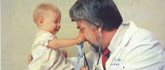
Pain in the eyes of a child
Most often, eye pain in children occurs due to inflammation and enlargement of the adenoids or tonsils. If a child complains of pain in the eyes, you should listen to his nasal breathing - perhaps it is difficult due to the adenoids. In this case, the child may wheeze and snore in his sleep; in severe cases, he may breathe through his mouth rather than his nose. The more enlarged the adenoids are, the stronger and more often the child’s eyes and head hurt. Enlarged tonsils are visible if you ask the child to open his mouth and breathe through his mouth. Treatment of adenoids and chronic tonsillitis in most cases eliminates eye pain in a child. Pain in the eye area in children can also be caused by severe allergic swelling of the nasal mucosa; in this case, the child most often complains of severe pain in the temples and eyes along with impaired nasal breathing. If the inflammatory process is one-sided, then the eye area hurts on the same side where the nose is stuffy.
Treatment
Before prescribing treatment, the ophthalmologist will conduct an examination and make a diagnosis. As a rule, the main stages are measuring intraocular pressure, examining the fundus, and examining with a slit lamp. This set of examinations allows you to identify the main and most common eye diseases.
In the case of glaucoma, special medications will need to be dripped into the eyes to reduce intraocular pressure. In most cases, you will need to prepare for surgery - glaucoma does not respond to conservative treatment.
If the pain is caused by an inflammatory disease, then you will need to identify the pathogen and start taking appropriate medications: antibacterial or antiviral.
Treatment for pain caused by fatigue involves following basic visual hygiene recommendations. The patient will have to be more attentive to his health. This pathology is most often found among office workers, and it has even been called visual fatigue syndrome. An ophthalmologist may prescribe eye exercises and recommend coming for preventive examinations.
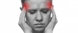
Eyes hurt
My eyes hurt, what is the reason and what to do? To understand why your eyes hurt, you need to understand that your eyes hurt in diseases associated with pathological changes in both the eyes themselves and in general diseases of the body. To differentiate the cause of eye pain, it is necessary to pay attention to the accompanying symptoms. For example, the eyes and head often hurt at the same time, the eyes hurt against the background of high blood pressure, and pain in the eyes can also often be combined with redness of the eyes. These and other accompanying symptoms of eye pain will help make the correct diagnosis, choose the direction of treatment and answer the question: what to do? Let's try to figure out why your eyes hurt and what to do? Eye pain is a characteristic symptom of many eye diseases. A. Reasons why the eyes may hurt when the eyes themselves are diseased:
1. Keratitis - inflammation of the cornea 2. Uveitis - inflammation of the choroid 3. Glaucoma 4. Conjunctivitis 5. Eye injury 6. Foreign body of the conjunctiva or cornea 7. Incorrectly selected glasses 8. Incorrectly selected contact lenses 9. Uncorrected vision with presbyopia 10. Eyes hurt after welding
B. Reasons why eyes may hurt due to general diseases of the body:
1. Viral infection 2. Blood pressure disorders 3. Migraine 4. Increased intracranial pressure (ICP)
A. Causes of pain in the eyes when the eyes themselves are diseased:
1.
Keratitis
, or inflammation of the cornea, is always accompanied by pain in the eyes.
In addition to eye pain, keratitis is absolutely always accompanied by additional symptoms. The so-called “corneal syndrome” is characterized by the presence of lacrimation, redness of the pericorneal zone (around the cornea), blepharospasm (spasm of the eyelids) and, of course, pain in the eyes. The eyes hurt very much with keratitis. Treatment:
treatment is carried out only in a specialized ophthalmological institution.
It is impossible to cure keratitis yourself at home without consequences for vision. Local and general anti-inflammatory treatment is necessary in the form of parabulbar injections of antibiotics and, if necessary, corticosteroid drugs, placing anti-inflammatory ointment and drugs that improve corneal regeneration behind the eyelids. If there are signs of keratitis, you should immediately consult a doctor. 2. Uveitis
, or inflammation of the inner choroid of the eye.
The choroid of the eye has three sections, each of which can become inflamed separately or there can be a combined inflammation, which is called panuveitis. Inflammation of the iris, or iritis, inflammation of the middle part of the choroid, or iridocyclitis. always accompanied by very severe pain in the eyes. The pain is so severe that it is impossible to touch the eye. Associated symptoms include pericorneal injection and possible changes in the color of the iris and the clarity of the pattern. Interestingly, inflammation of the third part of the iris - the choroid - is never accompanied by pain in the eyes and choroiditis at the initial stage is often asymptomatic. This occurs due to the lack of sensory nerve fibers in the choroid. Treatment:
treatment of uveitis should only be carried out in a specialized ophthalmology department and, as a rule, this disease requires treatment in an eye hospital.
At home, it is impossible to cure uveitis on your own without damaging your vision. 3. Glaucoma
.
A common symptom of glaucoma is pain in the eye. The left eye hurts or the right eye hurts, the one in which there is increased intraocular pressure. Often, in addition to pain in the eyes, there is a feeling of pressure. “Pressure in the eyes” is how patients describe their sensations with glaucoma. Another characteristic sign of glaucoma, which can help to suspect the disease, is pain in half of the head, in the half in which the intraocular pressure is increased in the eye. A characteristic sign of glaucoma is that not the whole head hurts, but rather half the head on the side of the diseased eye. Treatment:
Treatment for glaucoma can be medication, laser or surgery.
Drops that reduce intraocular pressure, such as Travatan, Timolol, Betoptik, Xalatan, Azopt, Arutimol, Trusopt, are prescribed by a doctor depending on the degree of glaucoma, its stage and the strength of the increase in intraocular pressure. Along with drug treatment, laser treatment is a very promising method of reducing blood pressure. If there is no desired effect from drug and laser treatment, surgical treatment of glaucoma is prescribed. Pain in the eye goes away with a decrease in intraocular pressure to normal levels. Normally, intraocular pressure should be no more than 26 mm Hg. when measured with a Maklakov tonometer at any age of a person. 4. Conjunctivitis
is rare, but can also be accompanied by pain in the eyes, more precisely a burning sensation and a feeling of a foreign body.
A more precise definition of pain with conjunctivitis: discomfort in the eyes and pain in the eyes. The reasons for these sensations are swelling and disruption of the integrity of the epithelial layer of the eyelids and the eyeball - the conjunctiva. Along with the pain, there is redness of the eyes, discharge in the conjunctival cavity; with bacterial conjunctivitis, eyelashes stick together in the morning. Treatment:
treatment is carried out depending on the type of pathogen - antiviral or antibacterial.
Tobrex, Tobradex, Maxitrol, Vitabact, as a rule, allow you to cope with conjunctivitis and eliminate eye pain. Oftalmoferon will be useful for pain from viral conjunctivitis. 5. Eye
trauma , even microtrauma, causes pain in the eyes.
In case of injury, the epithelium of the cornea or conjunctiva is most often damaged, which leads to irritation of sensitive nerve fibers and, as a result, eye pain. More serious injuries with rupture of the membranes always cause severe pain in the eyes. Treatment:
Drugs that improve tissue regeneration, such as Solcoseryl, Actovegin, Citral. To prevent secondary infection, it is recommended to use Tobrex, Maxitrol, Albucid 20%, chloramphenicol 0.25%.
6.
A foreign body in the conjunctiva or cornea
is the most common cause of eye pain.
The cornea is the most sensitive part of the body. Getting a hair or even a speck of dust into the eye causes discomfort and pain in the eye. A foreign body entering the conjunctival cavity disrupts the integrity of the epithelium and has the same consequences as an eye injury. The intensity of pain depends on the volume of the foreign body and location. Thus, the entry of an eyelash into the conjunctival cavity causes moderate pain and a feeling of a foreign body. A foreign body entering the cornea causes very severe pain. Treatment:
If the foreign body is visible and easily accessible, you can try to remove it yourself with a cotton swab or rinse the eye with warm water.
Then it is necessary to apply treatment, the same as for an eye injury. If you cannot remove the foreign body yourself, you must contact the Eye Emergency Center to receive specialized assistance. The eye emergency room is open 24 hours a day, so you should contact us at any time at any time. And the sooner help is provided, the better the prospects for treatment. Address of the eye emergency room in Krasnoyarsk: Krasnoyarsk city, st. Nikitina, 1 "B". 7. Incorrectly chosen glasses
are also a common reason why the eyes hurt, as if they are pressing.
The main errors in the selection of glasses are incorrectly measured interpupillary distance or diopters of glasses that are larger than they are necessary for the state of refraction. Such errors lead to excessive refractive error or tension in the extraocular muscles and, as a result, to headaches or painful sensations in the eyes. Pain when the eyes “seem to be pressing” excludes an inflammatory process of the eye or injury. This pain is typical of incorrectly chosen glasses or general health problems. Treatment:
double-check purchased glasses with a competent ophthalmologist.
Perform laser vision correction and eliminate the need for glasses. 8. Incorrectly fitted contact lenses
often cause eye pain.
Contact lenses require individual selection, since the radius of curvature of the cornea is different for all people, and even in two eyes of one person, the radius of curvature of the cornea can vary greatly. Therefore, when selecting contact lenses, it is necessary to focus on the individual characteristics of each eye, and not just on the diopter of the lenses. It is recommended to select lenses only in specialized clinics. An incorrectly selected lens acts as a foreign body, injuring the cornea of the eye, causing pain and inflammation of the eye. Treatment:
it is necessary to abandon contact lenses for a period of at least two weeks to restore the integrity of the cornea and select contact lenses from an experienced contact specialist.
To eliminate pain from contact lenses, laser vision correction is possible. 9. Uncorrected vision with presbyopia
leads to eye pain, especially if a person is forced to read for a long time or work nearby, for example, on a computer.
Accommodation allows a person to see at a close distance from the eye, but with age this process is disrupted, presbyopia develops, and near glasses are needed to compensate for impaired accommodation. If you try to strain your eyes when the possibility of accommodation is lost, then eye fatigue occurs and your eyes begin to hurt. Treatment:
it is necessary to select glasses for working at close range in accordance with age and existing refraction.
10. Eyes hurt after welding
due to radiation burn of the cornea.
As they say, “I’ve picked up welding bunnies,” what should I do? The welding arc used to weld two metals emits infrared radiation and ultraviolet radiation of enormous power. During welding work, eyes must be protected with special glass that limits exposure to infrared and UV radiation. If safety precautions are violated and the eye is openly exposed to radiation, a burn to the cornea occurs. Retinal damage may also occur. But pain in the eye after welding occurs precisely because of a burn to the cornea. Therefore, all measures to relieve pain after welding should be aimed primarily at treating the cornea. Treatment:
Rinse eyes with cool water.
To regenerate the cornea, it is necessary to use drugs such as Solcoseryl, Korneregel, Citral, Oftagel, Balarpan, Khilozar-komod. For the prevention of secondary infection: “Tobrex”, “Maxitrol”, “Albucid 20%”, “Levomycetin 0.25%”. If the pain is very severe, you can drop novocaine or lidocaine into your eyes. But, if the condition allows, it is better not to do this, since anesthesia causes swelling of the cornea and increases healing time. You can take any general action analyst, “Ibrufen” or “Analgin”, “Diclofenac” (Voltaren), or “Indomethacin”. It is better to be in a darkened room and with your eyes closed for several hours. B. Reasons why the eyes may hurt due to general diseases of the body:
1.
Viral infection
is very often accompanied by pain in the eyes.
If you experience pain behind your eyeballs when moving your eyes, then most likely you have an acute viral infection in your body. Coronavirus infection in the body is no exception. Coronavirus, like any other viral infection, leads to pain behind the eyes, especially in the initial stages of the disease. Eye pain with coronavirus is associated specifically with a viral infection in the body. Treatment:
To eliminate pain in the eyes due to a viral infection, general antiviral treatment is necessary from an appropriate specialist.
Local eye treatment is not required in this case. 2. Blood pressure disorders
.
Changes in blood pressure, both upward (hypertension or hypertension), and downward (hypotension), are accompanied by headache and frequent irradiation of pain to the orbital and eye areas. Treatment:
in case of a sharp and significant change in blood pressure, you must take a horizontal position - lie down, call a doctor.
With high blood pressure, the head should be higher than the body, with hypotension at body level. Compensatory therapy and equalization of blood pressure will lead to the disappearance of eye pain. 3. Migraine
leads in most cases to pressing pain behind the eyes.
Migraine is accompanied by loss of visual fields and headache. Eye pain is also a symptomatic sign of migraine. Treatment:
elimination of migraine leads to the disappearance of pain in the eyes.
4. Increased intracranial pressure
is accompanied by headaches and painful sensations in the eyes.
The diagnosis is made on the basis of fundus ophthalmoscopy and accompanying symptoms characteristic of increased ICP. Treatment:
Elimination of the causes of increased intracranial pressure leads to the absence of pain in the eyes.
You will probably find it interesting and useful: 1. Red eyes. Why and what to do?
2.
Why do my eyes water?
Causes and treatment. 3.
How to restore vision
08/22/2021
author Natalya Yatsinova
Our services in ophthalmology
The administration of CELT JSC regularly updates the price list posted on the clinic’s website. However, in order to avoid possible misunderstandings, we ask you to clarify the cost of services by phone: +7
| Service name | Price in rubles |
| Pneumotonometry | 500 |
| Tonometry (according to Maklakov) | 1 000 |
| Goldmann tonometry (Icare tonometer) | 1 000 |
| MSCT of orbits | 7 000 |
All services
Make an appointment through the application or by calling +7 +7 We work every day:
- Monday—Friday: 8.00—20.00
- Saturday: 8.00–18.00
- Sunday is a day off
The nearest metro and MCC stations to the clinic:
- Highway of Enthusiasts or Perovo
- Partisan
- Enthusiast Highway
Driving directions
Diagnostics
Severe pain in the eye leads to blepharospasm, which makes ophthalmological examination difficult. To alleviate the patient's condition, instillation of analgesics is recommended, after which diagnosis begins. It is important to assess the condition of the eyelids, the shape of the palpebral fissure and the position of the eyes. The following are specific studies:
- Visometry.
Visual acuity is determined at the beginning of the examination of the patient. In the absence of object vision, it is necessary to study light projection. Visometry is performed with and without distance correction. - Non-contact tonometry.
Penetrating eye injuries are often accompanied by hypotension. With iridocyclitis, intraocular pressure increases. It is important to compare the IOP in both eyes and also measure the central corneal thickness. - Biomicroscopy of the eye.
First, a detailed examination of the anterior segment of the eyeball is carried out with mandatory eversion of the upper eyelid. Next, fluorescein staining and examination with a cobalt filter are performed, which allows visualization of small erosive defects. - Ophthalmoscopy.
Fundus examination is carried out after cycloplegia, if intraocular pressure is compensated. The ophthalmologist evaluates the transparency of the optical media and the condition of the retina down to the dentate line. - Ultrasound of the eye.
Ultrasound examination is used when there are difficulties in visualizing the structures of the eyeball due to miosis, corneal edema, hyphema or hemophthalmos. The advantage of this method is the ability to detect x-ray negative foreign bodies. - Radiography.
It is performed in case of severe injuries in order to prevent damage to the bone walls of the orbit. The special Komberg-Baltin prosthesis makes it possible to determine the location of intraocular radiopaque foreign bodies.
Eye examination by an ophthalmologist
Prevention measures
Neurologist Anastasia Busygina highlights the following preventive measures:
- do not smoke - one cigarette smoked causes vasospasm for 15-20 minutes, and carbon monoxide from tobacco smoke replaces oxygen, this leads to oxygen starvation of brain tissue;
- monitor your blood pressure - a constant increase in pressure leads to the destruction of the walls of blood vessels, loss of their elasticity, and tissue swelling;
- find out if your closest relatives have the following diseases: hypertension, atherosclerosis, previous heart attack, stroke, diabetes mellitus, thyroid diseases, blood diseases (anemia) - they are always accompanied by cerebrovascular accidents.
If you are at risk for developing cerebrovascular accidents, undergo an annual scheduled examination: blood test (determination of cholesterol, glucose levels), ultrasound of cerebral vessels (diagnosis of the main arteries of the head), blood pressure monitoring. This allows you to see signs of a violation in time and prevent complications.
MAIN
- Acute cerebrovascular accident always manifests itself with characteristic symptoms - paresis, loss of sensitivity of a limb, half of the face, speech and hearing disorders . It is very important to get medical help as early as possible: the severity of the consequences of a stroke depends on this.
- Chronic cerebrovascular accident develops gradually, the symptoms may resemble the consequences of overwork. If they occur from time to time over several weeks, this is a reason to consult a neurologist.
- Visit a neurologist at least once a year if there are hereditary risks of developing cerebrovascular accidents.
Treatment of ocular migraine
Until now, atrial scotoma is not one of the diseases that can be completely cured. The only way to cope with the disease is to reduce the risk of exacerbation through lifestyle changes and regular medications. The goal of treatment is to prevent status migraine. By it, doctors mean a particularly severe and prolonged attack of ocular migraine, requiring urgent hospitalization of the patient and intensive care.
In the acute period of atrial scotoma, medications are prescribed to relieve symptoms:
- analgesics and NSAIDs (paracetamol, ibuprofen) in tablets - for mild attacks;
- analgesics and NSAIDs by injection - for moderate attacks;
- combined analgesics - for intense headaches.
If an aura appears in the first 2 hours, aspirin injections are recommended. The drug helps improve blood supply and prevents atrial scotoma from intensifying and reaching its peak. Regardless of the degree to which ocular migraine manifests itself, the patient is advised to rest. It is necessary to limit contact with bright light, loud sounds, and touching.
Between attacks, atrial scotoma is treated in situations where exacerbations occur more than 2 times a month. Patients are prescribed medications to prevent vascular insufficiency and neurological disorders:
- nootropics - Piracetam, Pantogam, Pantocalcin;
- muscle relaxants - Tolperisone and its analogues;
- antidepressants - Fluoxetine.
When status migraine occurs—a particularly acute attack of ocular migraine—patients are hospitalized. They are prescribed:
- glucocorticosteroids to reduce swelling of brain tissue;
- dehydrating agents to reduce tissue pressure on nerves and vessels, which results in the most severe atrial scotoma;
- antipsychotics to relax the nervous system, eliminate severe headaches, seizures and vomiting;
- combined non-narcotic analgesics for intraosseous and periosteal blockade.
Therapy continues until symptoms are permanently eliminated. Once the condition has stabilized, ocular migraine is treated as usual.
Causes of ocular migraine
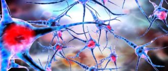
The main cause of atrial scotoma is a series of neurological disorders, the roots of which lie in a local decrease in the tone of the posterior cerebral artery. Against this background, ischemia (insufficient blood supply) develops in areas of the cerebral cortex involved in the analysis of visual signals, as well as in the retina itself. Alternatively, ocular migraine occurs due to compression of the 3rd pair of cranial nerves by the dilated cavernous sinuses or carotid arteries.
An attack of atrial scotoma can be triggered by:
- violation or prolonged non-compliance with the work and rest regime;
- sleep disorders;
- change of place of residence, especially with a change in time zones;
- suffered stress, emotional tension;
- hormonal imbalances, including cyclical changes in women;
- long-term stay in rooms with high gas pollution;
- exposure to flickering light sources, including older PC monitors.
The risk of experiencing an ocular migraine attack is higher in patients with abnormalities in the structure of blood vessels: aneurysms, malformations. In adolescents, an attack can be triggered by academic stress and a growth spurt.