A neuron, or structural and functional unit of the human nervous system, “silent” in itself does not mean anything. And even a set of neurons is also meaningless until they are busy with their most important task - generating and conducting a nerve impulse. The nerve impulse is the phenomenon thanks to which we exist. Any physiological act, from the secretion of gastric juice to voluntary movement, is regulated by the nervous system through the conduction of impulses. The higher nervous activity of the brain is also a collection of impulses from the cerebral cortex.
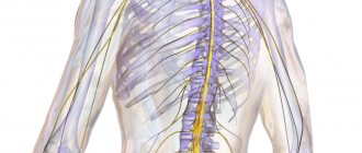
The impulse is carried along nerve fibers, which are nothing more than analogues of electrical wires, because a nerve impulse is a rapid change in the potential of the membrane of the nerve process, which must often be transmitted over a long distance. For example, the axons of the neurons of the anterior horns of the spinal cord, lying in the lower lumbar segments, form the lumbar plexus, from which its longest branch, the sciatic nerve, is formed. As part of this nerve axons go to the periphery and end with the branches of the peroneal nerve, on which, for example, extension of the big toe depends.
And nowhere are these axons interrupted; from the anterior horns of the spinal cord to the synapses in the muscles of the foot there is a dense bundle of neuron processes that form the longest nerve in our body. The pulse speed in it reaches 120 m/s. Thus, the length of a nerve cell, including its axon, in the human body can reach a length of more than a meter. How can you store and conduct an electrical impulse in the “wet environment” of the body without loss, and deliver it where it is needed? There is a special substance for this – myelin. The myelin sheath of nerve fibers is nothing more than the insulation of an electrical wire, without which the nerve impulse will “spark”, be distorted, or not be carried out at all . How are the myelin sheaths of nerves arranged in the human body, and what does their destruction lead to?
Principles of treatment of shell defects
Diseases associated with the destruction of the pulp membrane are very difficult to treat. Therapy is aimed mainly at relieving symptoms and stopping destruction processes. The earlier the disease is diagnosed, the greater the chances of stopping its progression.
Possibilities of myelin restoration
Thanks to timely treatment, the process of myelin restoration can be started. However, the new myelin sheath will not perform its functions as well. In addition, the disease may enter a chronic stage, and the symptoms will persist, only slightly smoothing out. But even minor remyelination can stop the course of the disease and partially restore lost functions.
Modern drugs aimed at myelin regeneration are more effective, but are very expensive.
- What are these areas of the brain and what is known about them?
- What does it consist of?
- Functions
- Biotin in myelin repair in MS
- Molecular organization of myelin
- Learning activates the brain
- Lecithin delivery
- Compound
- The role of microglia in the destruction of myelin structure
- Conclusion
- What did you find and what conclusions did you draw?
Therapy
The following drugs and procedures are used to treat diseases caused by the destruction of the myelin sheath:
- beta interferons (stop the course of the disease, reduce the risk of relapses and disability);
- immunomodulators (affect the activity of the immune system);
- muscle relaxants (help restore motor functions);
- nootropics (restore conductive activity);
- anti-inflammatory (relieve the inflammatory process that caused the destruction of myelin);
- neuroprotectors (prevent damage to brain neurons);
- painkillers and anticonvulsants;
- vitamins and antidepressants;
- cerebrospinal fluid filtration (a procedure aimed at cleansing cerebrospinal fluid).
Neural insulation defects
Fetal brain development is a complex process in which rapid changes in the morphology and microstructure of nervous tissue occur. In some areas of the brain, the process of myelin formation begins as early as 18-20 weeks of pregnancy, and continues until approximately ten years of age.
Not everyone knows that myelin is many layers of cell membrane, “wound” many times around an axon. Myelin is formed by flat outgrowths of “service” glial cells, in which there is practically no cytoplasm. The myelin sheath is not continuous, but discrete, with gaps (nodes of Ranvier). Therefore, the axon has faster saltatory conduction: the speed of signal transmission along fibers with and without myelin can differ hundreds of times. As for the molecular composition of the “insulator”, it, like all cell membranes, consists mainly of lipids and proteins
It is myelination disorders that often underlie delays in the physical and mental development of a child, and also cause the formation of a number of neurological and psychiatric pathologies. In addition to diseases such as stroke, delays in fetal brain development with impaired myelination are sometimes observed in multiple pregnancies. At the same time, desynchronization in the development of the brain of twins is quite difficult to assess “by eye”.
But how to identify myelin defects during fetal development? Currently, obstetricians and gynecologists use only biometric indicators (for example, brain size), but these are highly variable and do not provide a complete picture. In pediatrics, even in the presence of obvious functional abnormalities in the child’s brain activity, traditional MRI or neurosonography
(ultrasound examinations of the brain of newborns) often do not show structural abnormalities.
Therefore, the search for accurate quantitative criteria for assessing myelin formation during pregnancy is an urgent task, which also needs to be solved using non-invasive diagnostic methods that have already been tested in obstetrics. Specialists from the Novosibirsk International Tomography Center SB RAS proposed using for these purposes a new method of quantitative neuroimaging, already adapted for prenatal ( prenatal)
) research.
What are these areas of the brain and what is known about them?
The nucleus accumbens is part of the interconnected ventral area-nucleus accumbens (VTA-NAc) system. This system is critical for obtaining “rewards” through the release of dopamine. VTA dopaminergic neurons also innervate several areas of the prefrontal cortex (PFC), central amygdala, basolateral amygdala (BLA), and hippocampus, as well as other areas (Fig. 2). All of these so-called reward regions of the brain are interconnected in complex ways: for example, the NAc receives dense glutamatergic innervation from the PFC, amygdala, and hippocampus; The PFC, amygdala, and hippocampus form reciprocal glutamatergic connections with each other. The functional output of each of these areas is modulated by several types of GABAergic interneurons.
Figure 2. Diagram of the ventral area-nucleus accumbens (VTA-NAc) reward system. Simplified diagram of the major dopaminergic, glutamatergic, and GABAergic connections around the ventral area (VTA) and nucleus accumbens (NAc) in the rodent brain. The primary circuitry of the reward system involves dopaminergic connections from the VTA to the NAc, which release dopamine in response to reward-associated stimuli (and in some cases aversive-associated stimuli). There are also GABAergic projections from the NAc to the VTA; The NAc also receives dense innervation from glutamatergic monosynaptic circuits of the medial prefrontal cortex (mPFC), hippocampus (Hipp), and amygdala (Amy), as well as other regions. The VTA receives glutamatergic stimuli from the lateral dorsal segment (LDTg), the lateral leash of the epithalamus (LHb), and the lateral hypothalamus (LH). These various glutamatergic inputs control reward-related aspects of perception and memory. Dashed lines show internal inhibitory projections. Red lines—glutamatergic connections; green - dopaminergic; blue - GABAergic.
The release of dopamine into the nucleus accumbens is the way the brain receives pleasure signals
Feelings of pleasure motivate us to repeat behaviors that are vital for survival, and disruptions in the acquisition of dopamine rewards are associated with symptoms such as anhedonia and impaired reward-related perception and memory
The medial prefrontal cortex is the part of the prefrontal cortex that is also associated with the reward system, as shown above. Research has shown that patients with depression have less cortical volume, including less white matter in this area, which leads to impaired perception of events that previously brought joy.
What does it consist of?
Such an amazing biological insulating function of myelin turned out to be possible due to its structure. Don’t think that myelin is just an insulating layer wrapped around a neuron. Let us remember that in nature everything consists of cells, and the myelin of a peripheral nerve is simply an overgrown Schwann cell that has wrapped its cytoplasm around the axial cylinder of the neuron several times. It is myelin that gives the white color to nerve fibers, hence the concept of “white matter of the brain.” These are nothing more than bundles of nerve fibers that contain a lot of myelin. Their function is to be conductors of current. The pons, brain stem, midbrain are all areas consisting of an unimaginably large number of conductive bundles.
Therefore, myelin consists mostly of lipids, which repel water, and proteins. Myelin contains about 75% lipids, which is much higher than in most membranes. It's clear why this happens. After all, a membrane consisting of a bilipid layer should not only delimit the internal environment of the cell. This is a complex transport system that occurs with the help of carrier proteins. As for the myelin “wrappings” of the nerve, their task is very simple - to isolate the nerve fiber as much as possible. That’s why myelin is so “fat.” In the area of nodes of Ranvier, ions can enter the cytoplasm of the neuron, causing depolarization of the membrane, but not in the myelin areas. Thanks to this, the uninterrupted passage of impulses is ensured.
But there are situations in which myelin begins to break down. This process is called demyelination, and it manifests itself in a whole group of diseases of the same name. Why does this happen and how does it manifest itself?
Demyelination and its manifestations
Defects in the myelination of nerve fibers are called demyelination. This can occur due to genetic defects (called myelinopathy). Sometimes myelin is synthesized normally, but the physiological restoration of myelin occurs slowly or with damage. Demyelination is the process by which nervous tissue responds to pathological influences. Sometimes demyelination is secondary, that is, first the nerve is destroyed, and then its sheath. But still, most often myelin is lost first, and only then the process of the nerve cell is damaged.
Most often, immune inflammation is to blame for the primary destruction of myelin. Nerve insulation is destroyed by cytokines, enzymes and other active substances that are synthesized by plasma cells and macrophages. Antimyelin antibodies cause significant damage.
Important Glia
The most common causes of demyelination are the following processes:
intoxication (alcoholism, radiation, increased glucose levels in diabetes mellitus);
cerebrovascular diseases, strokes, atherosclerosis;
vasculitis and systemic collagenosis;
autoimmune post-vaccination and post-infectious reactions.
The most well-known disease from this group is multiple sclerosis, which can occur with very diverse clinical symptoms (paralysis, paresis, dysfunction of the pelvic organs, tremor, ophthalmoplegia, extinction of reflexes, impaired coordination of movements). In multiple sclerosis, symptoms depend on where the lesion is located and the severity of demyelination.
Demyelination also occurs from the action of physical factors. Very serious exacerbations of multiple sclerosis can occur accidentally if you do not follow the rules of behavior. It has long been established that myelin is destroyed by thermal procedures. Thus, patients are strictly prohibited from:
take a steam bath;
take hot baths and showers;
sunbathing and being in the sun with exposed parts of the body.
Also, serious exacerbations occur after ARVI, influenza, and other diseases that occur with fever syndrome. An increase in temperature in multiple sclerosis and similar diseases stimulates the breakdown of myelin.
How to repair/regenerate the myelin sheath
The myelin sheath helps nerves transmit signals. If it is damaged, memory problems arise, and a person often develops specific movements and functional impairments. Certain autoimmune diseases and external chemical factors, such as pesticides in food, can damage the myelin sheath. But there are a number of ways, including vitamins and foods, that will help regenerate this covering of nerves: you will need specific minerals and fats, preferably obtained through a nutritious diet. This is even more necessary if you suffer from a disease like multiple sclerosis: usually the body is able to repair damaged myelin sheath with some help from you, but if sclerosis has manifested itself, treatment can become very difficult. So, here are the remedies that will help support the restoration and regeneration of the myelin sheath, as well as prevent sclerosis.
You will need: – folic acid; – vitamin B12; – essential fatty acids; - vitamin C; – vitamin D; - green tea; – martinia; – white willow; – boswellia; - olive oil; - fish; – nuts; - cocoa; – avocado; – whole grains; – legumes; - spinach.
1. Provide yourself with dietary supplements in the form of folic acid and vitamin B12. The body requires these two substances to protect the nervous system and properly repair myelin sheaths. In a study published in the Russian medical journal Vrachebnoye Delo in the 1990s, scientists found that patients suffering from multiple sclerosis who were treated with folic acid showed significant improvement in symptoms and in myelin repair. Both folic acid and B12 can both help prevent the breakdown and regenerate damage to myelin.
2. Reduce inflammation in the body to protect myelin sheaths from damage. Anti-inflammatory therapy is currently the mainstay of treatment for multiple sclerosis, and in addition to taking prescribed medications, patients can also try dietary and herbal anti-inflammatory agents. Natural remedies include essential fatty acids, vitamin C, vitamin D, green tea, martinia, white willow and boswellia.
3. Consume essential fatty acids daily. The myelin sheath is primarily composed of an essential fatty acid: oleic acid, an omega-6 found in fish, olives, chicken, nuts and seeds. Plus, eating deep sea fish will provide you with a good amount of omega-3 acids: to improve mood, learning, memory and overall brain health. Omega-3 fatty acids reduce inflammation in the body and help protect myelin sheaths. Fatty acids can also be found in flaxseed, fish oil, salmon, avocados, walnuts and beans.
4. Support your immune system. Inflammation, which causes damage to myelin sheaths, is caused by immune cells and autoimmune diseases in the body. Nutrients that will help your immune system include: vitamin C, zinc, vitamin A, vitamin D, and vitamin B complex. In a 2006 study published in The Journal of the American Medical Association, vitamin D has been cited as significantly helping to reduce the risk of demyelination and manifestations of multiple sclerosis.
5. Eat foods high in choline (vitamin D) and inositol (inositol; B8). These amino acids are critical for the restoration of myelin sheaths. You can find choline in eggs, beef, beans and some nuts. It helps prevent fat deposits. Inositol supports nervous system health by helping to create serotonin. Nuts, vegetables and bananas contain inositol. The two amino acids combine to produce lecithin, which reduces “bad” fats in the bloodstream. Well, cholesterol and similar fats are known for their ability to interfere with the restoration of myelin sheaths.
6. Eat foods rich in B vitamins. Vitamin B-1, also called thiamine, and B-12 are physical components of the myelin sheath. We look for B-1 in rice, spinach, and pork. Vitamin B-5 can be found in yogurt and tuna. Whole grains and dairy products are rich in all B vitamins, and they can also be found in whole grain bread. These nutrients enhance metabolism, which burns fat in the body, and they also transport oxygen.
7. You also need food containing copper. Lipids can only be created using copper-dependent enzymes. Without this help, other nutrients won't be able to do their job. Copper is found in lentils, almonds, pumpkin seeds, sesame seeds and semi-sweet chocolate. Liver and seafood may also contain copper in lower doses. Dried herbs like oregano and thyme are an easy way to add this mineral to your diet.
Additions and warnings:
– Milk, eggs and antacids can interfere with the absorption of copper;
– In culinary recipes, replace liquid olive oil with solid oil (this also happens!);
– If you drink too many B vitamins, they will simply leave the body without causing harm;
– Copper overdose can cause serious problems in the mind and body. So natural consumption of this mineral is the best option;
– Even natural methods, such as food selection and other things, should be supervised by a medical representative.
Functions
The main functions of the myelin sheath are:
- axon isolation;
- acceleration of impulse conduction;
- energy savings by maintaining ion flows;
- nerve fiber support;
- axon nutrition.
How pulses work
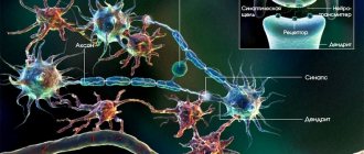
Nerve cells are isolated due to their membrane, but are still interconnected. The areas where cells touch are called synapses. It is where the axon of one cell and the soma or dendrite of another meet.
An electrical impulse can be transmitted within a single cell or from neuron to neuron. This is a complex electrochemical process that is based on the movement of ions through the membrane of the nerve cell.
In a calm state, only potassium ions enter the neuron, and sodium ions remain outside. At the moment of excitement they begin to change places. The axon is positively charged from within. Then sodium stops flowing through the membrane, but the outflow of potassium does not stop.
The change in voltage due to the movement of potassium and sodium ions is called the "action potential". It spreads slowly, but the myelin sheath that envelops the axon speeds up this process by preventing the outflow and influx of potassium and sodium ions from the axon body.
Inflammation of the oral cavity - stages of treatment of the mucous membrane and methods of eliminating inflammation
Passing through the node of Ranvier, the impulse jumps from one part of the axon to another, which allows it to move faster.
Once the action potential crosses the break in the myelin, the impulse stops and the resting state returns.
This method of energy transfer is characteristic of the central nervous system. As for the autonomic nervous system, it often contains axons covered with little or no myelin. There are no jumps between Schwann cells, and the impulse travels much more slowly.
Definition of the myelin sheath
The myelin sheath is a fatty insulation that surrounds the nerve cells of jawed vertebrates, or gnathostomes. All extant members of the Gnathostomata, from fish to humans, have a myelin sheath on the axon of their nerve cells. The oldest known members of the jawed fishes, the extinct placoderms, were armored fish that appear to have more primitive nerves that are not covered with myelin sheaths. Soft placoderm tissues have been discovered in an organism that lived more than 400 million years ago during the late Devonian period in Australia. Current evidence suggests that the myelin sheath evolved some time after this, as the current vertebrate lineage diverged from the armored placoderms. Some invertebrates have developed myelin sheath-like substances that perform a similar function.
The myelin sheath is produced through a process called myelination, which can be seen in the image above. The myelin sheath of nerve cells usually forms during the early stages of development. Special cells called oligodendrocytes or Schwann cells create and store large amounts of myelin. The oligodendrocytes then wrap around the axon of the nerve cell. Many oligodendrocytes are required to cover the long axons in mammals, which can be up to a meter in length. Myelin itself is a fatty substance that appears white. In the part of the brain and nervous system called white matter, there is an excess of myelin sheath. In the gray matter, more cell bodies are present. Myelin is chemically composed of various lipids and proteins that also absorb water.
Some degenerative diseases, such as multiple sclerosis, lead to degradation of the myelin sheath. This can lead to a large number of side effects, and ultimately to complete paralysis and death. The myelin sheath is necessary to insulate nerves from each other and to speed up the passage of signals along long nerves. Without these functions, signals become mixed and normal movements become impossible. Blindness and other neurological conditions associated with nerve damage occur when the myelin sheath is removed.
Biotin in myelin repair in MS
Biotin is a vitamin involved in both energy metabolism and the creation of fatty acids in the body. It is found in dietary supplements for hair, skin and nail growth, as well as in multivitamins and prenatal vitamins.
Because the myelin sheath is a fatty sheath, researchers hypothesize that by giving people large doses of biotin (for example, 300 mg per day), the myelin sheath can be repaired.
In addition to restoring myelin (fatty covering), some scientists believe that biotin may reduce axonal degeneration by increasing energy production. Axonal degeneration occurs predominantly in progressive types of MS and refers to the loss of nerve fibers and ultimately the death of nerve cells.
The big picture is that experts suggest biotin may provide therapeutic benefits in two ways—double bonus. However, to date, scientific evidence supporting the role of biotin in the treatment of MS is sparse and inconclusive.
Let's take a closer look at a few studies showing these mixed results.
Thumbs up for biotin
In a small study called Multiple Sclerosis and Related Disorders, 23 people with primary or secondary progressive MS were given high doses of biotin and improvements were noted in a range of symptoms, particularly visual acuity, spinal cord problems and fatigue.
Mixed Results for Biotin
A large study in Multiple Sclerosis found that high doses of biotin improved MS-related disability in about 12 percent of participants with progressive MS. However, the fact that only 12 percent showed improvement suggests that potentially only a proportion of people with MS may benefit from biotin.
Also of concern is the fact that the researchers in this study noted that those taking biotin had more new or enlarged brain lesions on their MRI compared to those in the placebo group. The researchers questioned whether biotin causes inflammation answer (that wouldn't be good).
Thumbs down for biotin
The third study discusses the issue of biotin. In this study, there was no improvement in MS-related disability in people with progressive MS. In fact, about one-third of participants experienced worsening of their disease, particularly with greater leg weakness, poorer balance, and more falls.
Of course, the worsening of their disease may not be due to biotin and due to the natural progression of multiple sclerosis. However, the study researchers wondered whether high-dose biotin had anything to do with it. It is possible that biotin shifted the body's metabolic needs away from protecting the brain and spinal cord, allowing the immune system to wreak havoc.
Important Histrionic personality disorder (hysterical psychopathy)
Myelin is the sheath of neural “wires”
Nerve cells have two types of processes - small and extremely branched dendrites, with the help of which the neuron collects impulses from other nerve cells, and very long axons, which send impulses further. Almost all axons in the central nervous system (that is, in the brain and spinal cord) are covered with myelin, a light-colored substance consisting primarily of lipids. Myelinated nerve fibers are also abundant in the peripheral nervous system, that is, in the nerves that leave the brain and spinal cord and go to other organs.
Oligodendrocyte and myelin sheath. One oligodendrocyte forms a myelin sheath on several axons at once, but on each of them it creates only one segment of the sheath (from one node of Ranvier to another). Illustration: Holly Fischer/Wikimedia Commons/CC BY 3.0.
Destruction of the myelin sheath in multiple sclerosis. Photo: edesignua/ru.depositphotos.com.
‹
›
Myelin simultaneously accelerates the electrochemical impulses traveling along the axons and insulates them from each other, preventing a “short circuit” between the neural “wires”. To understand how myelin accelerates impulses, we need to remember that any impulse in a neuron is a rearrangement of ions between the outer and inner sides of the cell membrane. When ion channels open on a certain section of the membrane, the same ion flows immediately open on a neighboring section of the membrane, then on a section a little further away, etc. The electrical properties of the membrane change sequentially along the neuronal process - this is a running impulse. Myelin does not envelop the axons completely from beginning to end. There are gaps in the myelin coil where the membrane is not covered by myelin - nodes of Ranvier (named after the French physiologist Louis Antoine Ranvier who discovered them). And when the impulse propagates along the axon, the rearrangement of ions occurs precisely at the nodes of Ranvier. That is, the impulse does not creep slowly between areas that are close to each other, but jumps from one interception to another. And if in an axon without myelin an impulse travels at a speed of 0.5-10 m/s, then in the same axon, but with myelin, the impulse speed reaches 150 m/s.
Clusters of axons wrapped in myelin appear lighter, so areas in the brain where axonal “wires” predominate are called white matter. (Clusters of dendrites that do without myelin form gray matter. Since dendrites are much shorter than axons, they do not transmit impulses over long distances and speed is not so important for them.) Neurons do not produce myelin themselves; there are special cells for this purpose - oligodendrocytes in the central nervous system and Schwann cells in peripheral nerves. Both of them belong to glia, or neuroglia - this is the name given to the collection of various cells of the nervous system that serve neurons, creating conditions for them to work. Recently, more and more evidence has emerged that glial cells not only serve neurons, but directly interfere with their work (see the article “Immune “electricians” of the brain,” “Science and Life” No. 8, 2021). The job of oligodendrocytes and Schwann cells is to provide the myelin sheath for neurons. An oligodendrocyte or Schwann cell protrudes its own membrane and wraps around the axon, the membrane grows - and as a result, a layered lipid roll is formed around the axon. The glial cell remains alive and maintains the integrity of the myelin coil in the section of the axon for which it is responsible.
Destruction of the myelin sheath leads to neurological symptoms of various types and varying degrees of severity. There are many diseases associated with the loss of myelin on axons, and multiple sclerosis is the most famous among them. This is one of the autoimmune diseases, when the immune system for some reason attacks the body's own cells and molecules. In multiple sclerosis, different immune mechanisms are triggered, in which both immune cells of the brain and immune cells that enter the brain from the blood are involved. But, one way or another, it all ends with the myelin sheath around the axons being destroyed, and sometimes the axons themselves are destroyed. Astrocytes come to the site of the disease - this is the name of another type of glial cells. Their job is to support and nourish neurons and heal damage; This is exactly what they do, trying to heal the diseased area and forming a characteristic plaque. It is worth adding that multiple sclerosis usually affects the central nervous system; peripheral nerves are rarely affected.
Molecular organization of myelin
A unique feature of myelin is its formation as a result of the spiral entanglement of glial cell processes around axons, so dense that there is practically no cytoplasm left between the two layers of the membrane. Myelin is this double membrane, meaning it consists of a lipid bilayer and proteins associated with it.
Myelin proteins include so-called intrinsic and extrinsic proteins. The internal ones are integrated into the membrane, the external ones are located superficially and therefore are weaker connected to it. Myelin also contains glycoproteins and glycolipids.
Proteins make up 25-30% of the dry matter mass of the myelin sheath of neurons in the central nervous system of mammals. Lipids account for approximately 70-75% of the dry mass of the brain. Spinal cord myelin has a higher percentage of lipids than brain myelin. Most of the lipids are phospholipids (43%), the rest are cholesterol and galactolipids in approximately equal proportions.
Learning activates the brain
When we learn something new, whether programming in Ruby on Rails, consulting on the phone, playing chess, or making a cart wheel, our brains begin to function at a higher level.
Any new experience, be it playing the guitar or even computer games, promotes the production of myelin in the brain, which leads to strengthening mental abilities
Science has long proven that our brain is very mobile, that is, it does not stop developing even at 25. Of course, many things, especially languages, are easier for children than for adults. But there are many examples in the world of how older people learn something new.
But how does this happen? To perform any task, we must activate different parts of the brain. For example, to explain something, our brain coordinates a series of actions that include motor function, visual and auditory processing, speech, and more.
At first our explanation will be terse and chaotic.
We may forget to say something important. But with practice, our speech becomes smoother, more natural and softer.
Practice helps the brain optimize and coordinate all its actions through a process called myelination.
Lecithin delivery
Lecithin, also known as phophatidylcholine, is a fatty substance made up of choline, fatty acids and other lipid molecules. Lecithin is important for nerve transmission and can serve as a source of choline for myelin production. Dietary sources of lecithin include egg yolks, soybeans, wheat germ and liver. Brussels sprouts, shrimp, peanut butter and chocolate also contain significant levels of lecithin. If you follow a vegetarian diet that excludes dairy or eggs, you risk becoming deficient in lecithin and choline. Taking a vegetarian lecithin supplement can help ensure you're getting enough of these important nutrients.
Compound
The myelin layer consists of two layers of lipids and three layers of protein. There are much more lipids in it (70-75%):
- phospholipids (up to 50%);
- cholesterol (25%);
- glactocerebroside (20%), etc.
The high fat content causes the myelin sheath to be white, which is why the neurons covered by it are called “white matter.”
Protein layers are thinner than lipid layers. The protein content in myelin is 25-30%:
- proteolipid (35-50%);
- myelin basic protein (30%);
- Wolfgram proteins (20%).
There are simple and complex proteins of nervous tissue.
The role of lipids in the structure of the shell
Lipids play a key role in the structure of the pulp membrane. They are the structural material of nervous tissue and protect the axon from energy loss and ion flows. Lipid molecules have the ability to restore brain tissue after damage. Myelin lipids are responsible for adaptation of the mature nervous system. They act as hormone receptors and communicate between cells.
The role of proteins
Protein molecules are of no small importance in the structure of the myelin layer. They, along with lipids, act as building materials for nervous tissue.
Their main task is to transport nutrients into the axon. They also decipher signals entering the nerve cell and speed up reactions in it. Participation in metabolism is an important function of the myelin sheath protein molecules.
The role of microglia in the destruction of myelin structure
Microglia are macrophages capable of phagocytosis and able to recognize various pathogenic particles - antigens. Thanks to membrane receptors, these glial cells produce enzymes - proteases, as well as cytokines, for example, interleukin 1. It is a mediator of the inflammatory process and immunity.
The myelin sheath, whose functions are to insulate the axial cylinder and improve the conduction of nerve impulses, can be damaged by interleukin. As a result of this, the nerve is “exposed” and the speed of excitation is sharply reduced.
Moreover, cytokines, by activating receptors, provoke excessive transport of calcium ions into the neuron body. Proteases and phospholipases begin to break down the organelles and processes of nerve cells, which leads to apoptosis - the death of this structure.
It is destroyed, breaking up into particles, which are devoured by macrophages. This phenomenon is called excitotoxicity. It causes degeneration of neurons and their endings, leading to diseases such as Alzheimer's disease and Parkinson's disease.
Function of the myelin sheath
The myelin sheath has a number of functions in the nervous system. Primary functions include protecting nerves from other electrical impulses and speeding up the time it takes for a nerve to travel through an axon. Unmyelinated nerves should send a wave along the entire length of the nerve. In large organisms this is a problem because it takes a long time to reach the end of the nerve. Due to greeting conduction, individual myelin sheaths cause the signal to jump from one node of Ranvier to another. The myelin sheath prevents an action potential from forming where it spans the axon. Instead, the electrical wave created at each node propels the action potential to the end of the nerve, where it can be transmitted to the next neuron. This method of propagating the signal down the axon also saves energy, since the sodium and potassium pumps that regulate the action potential are not needed beneath the myelin sheath. Although the myelin sheath is only visible in vertebrates, greeting conduction is observed in many animals. This phenomenon can be seen in the graph below.
- Neuron – A nerve cell that transmits a signal to other nerve cells in the nervous system.
- axon – Long extensions of neurons that carry nerve impulses to the next neuron.
- dendrite – Small structured neurons used to communicate with each other.
- Node of Ranvier - Small spaces between individual cells of the myelin sheath.


