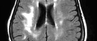
The role of venous circulation disorders in the origin and course of cerebrovascular diseases has been underestimated for a long time. This is explained by the previously existing difficulties of intravital assessment of cerebral venous hemodynamics when using traditional methods of recording venous blood flow in the vessels of the brain, as well as insufficient attention from researchers to this section of angiology.
The emergence of modern neuroimaging methods, in particular MRI diagnostics, has greatly facilitated the identification of such diseases.
Let's consider several examples of pathological processes detected by MRI diagnostics using MR venography.
THROMBOSIS OF THE VEINS AND SINES OF THE BRAIN
The cause of the development of thrombosis of the veins and sinuses of the dura mater can be septic lesions, trauma, compression of the sinus by a tumor, and systemic lesions of the connective tissue.
In addition, sinus thrombosis can develop due to thrombophlebitis of the extremities or inflammatory foci in the body (in the postpartum period, after an abortion, with infectious diseases, as well as diseases of the ears and paranasal sinuses).
Taking into account the patient’s age, the degree of development of collateral circulation, as well as the localization of the pathological process, the clinical manifestations of venous thrombosis are quite variable and nonspecific.
It is very difficult to identify typical clinical manifestations of sinus thrombosis, but the most common initial symptoms are the following: 1. headache 2. papilledema (a sign of intracranial hypertension) 3. focal neurological deficit 4. disturbances of consciousness (appear in case of damage to the brain substance in the form of progressive edema, developing infarction or hemorrhage).
In case of damage to the sinuses, general cerebral symptoms depend on the size and rate of increase of thrombosis.
Focal symptoms develop when brain matter is involved in the process, i.e. with the development of cortical venous infarction. Cortical motor deficits, cortical symptoms, and seizures appear according to the location of the affected sinus.
When a clinical picture of thrombosis of the veins and sinuses of the brain occurs, MRI in most cases reveals signs of extensive areas of ischemia and hemorrhage. However, in some cases, standard neuroimaging methods fail to detect changes in the brain parenchyma.
The method of choice in such cases is magnetic resonance imaging (MRI) of the brain using MR venography.
Thrombosis of the right transverse sinus - hypointense areas on T2 (intracellular deoxyhemoglobin).
To confirm venous sinus thrombosis and determine the exact location and extent of the thrombus, MR venography is necessary.
MR venography – lack of visualization of blood flow in the right transverse sinus and jugular vein.
MRI of the brain: on the right (green arrow) the T2-weighted image shows the normal phenomenon of “empty flow” from the right sigmoid sinus and jugular vein. On the left (orange arrow) there is an abnormally high signal, most likely resulting from thrombosis. To confirm sinus thrombosis and definitively determine the location and extent of thrombosis, MR venography is necessary.
MR venography: thrombosis of the left transverse sinus. There is loss of MR signal from the left transverse sinus. The presence of sinus visualization on “raw” data or brain MRI confirms sinus thrombosis and excludes its hypoplasia.
MR venography: thrombosis of the right transverse sinus. There is loss of MR signal from the right transverse sinus. The presence of sinus visualization on “raw” data or MRI of the brain confirms sinus thrombosis and excludes its hypo- and aplasia.
Thrombosis of the right transverse sinus. Absence of the phenomenon of “empty flow” from the right transverse sinus on MRI of the brain. Absence of visualization of the right transverse sinus on MR venography.
As mentioned above, in cases of a clinical picture of cerebral venous thrombosis along the veins and sinuses, an MRI of the brain in some cases reveals an area of ischemia and hemorrhage.
MRI of the brain: a combination of vasogenic (orange arrow), cytotoxic edema and hemorrhage (green arrow) is noted. This MR picture, as well as the location of the pathological zone in the projection of the temporal lobe, makes us think about hemorrhagic venous cerebrovascular accident due to thrombosis of the vein of Labbe. Confirmation requires MR venography or contrast-enhanced MRI.
STENOSIS, AREAS OF PATHOLOGICAL EXTENSION AND HYPOPLASIA OF VENOUS STRUCTURES OF THE BRAIN
MRA picture of pronounced asymmetry of the venous network with a predominance and mild dilatation of the veins of the right hemisphere (transverse, sigmoid sinuses and jugular vein on the right); hypoplasia of the left transverse and sigmoid sinus. Single areas (2) of local dilatation of veins in the parasagittal parts of the left hemisphere, the great vein of the brain. Asymmetrical, dilated and clearly convoluted venous structure of the extracranial sections on the right.
MRA signs of slight dilatation of the superior sagittal sinus, local decrease in blood flow and narrowing of the lumen of the distal straight sinus; asymmetry of the lumen of the transverse, sigmoid sinuses and internal jugular veins.
VASCULAR MALFORMATIONS
What is hypoplasia? Causes, signs, treatment of hypoplasia
Hypoplasia is the underdevelopment of any organ or tissue - this is a common and main feature of pathology, be it hypoplasia of the kidneys, brain, or hypoplasia of a segment of the right or left vertebral artery.
In humans, underdevelopment of individual organs most often occurs. So, this pathology can affect the brain, but not the entire central nervous system, or one lung without involving the second and upper respiratory tract.
Pathology occurs due to a malfunction during intrauterine development. The most common causes of hypoplasia development:
- a failure that occurs during the initial formation of germ cells;
- incorrect intrauterine position of the fetus;
- scanty amount of amniotic fluid;
- bad habits of the expectant mother - smoking, drinking alcohol and drugs (most often they cause brain hypoplasia);
- infectious diseases of women during pregnancy;
- abdominal injuries of a pregnant woman;
- exposure to physical factors - elevated temperature, radiation;
- exposure to toxic compounds and harmful metabolic products.
Hypoplasia is not always detected on time. The success of diagnosis depends on the degree of underdevelopment and which organ is affected. Thus, to identify signs of tooth enamel hypoplasia in a child, an examination by an experienced dentist is often sufficient. While diagnosing renal underdevelopment will require additional instrumental examination methods.
Underdevelopment of the entire organism is called dwarfism.
results
A 19-year-old patient came to the SSMU clinic on the referral of a neurologist from the district clinic for a triplex scan of the brachiocephalic arteries (TS BCA) with complaints of frequent severe dizziness, recurrent headaches, and lack of coordination.
Similar symptoms have been bothering him for 5 years; he denies any connection with the injury. According to the results of TS of the BCA, bilateral narrowing of the diameter of both VAs was revealed. On the left, the diameter in the V1 segment is no more than 1.8 mm, then at the level of the V2 segment it is in the range of 1.5-1.7 mm, in the V3 segment it is 1.5 mm.
On the right, the diameter of the V1 segment does not exceed 1.5 mm, the diameter of the V2 segment is in the range of 1.3-1.6 mm, in the V3 segment the diameter reaches 1.4 mm. During a transcranial study at the level of V4 segments, the VA is located only on the left side.
There is a pronounced decrease in the linear velocity of blood flow (LBV) on both sides - at the level of the 2nd segment of the LSV, no more than 15-18 cm per second. At the level of the V4 segment on the left, the BSC is reduced to 10 cm per second. The basilar artery is not visualized during TS BCA.
When performing a rotation test to the left side, the LSC along the PA decreases to 4-6 cm per second. Both VAs originated from the anterior wall of the subclavian arteries in segment 1. No ultrasound signs of pathological tortuosity were detected on either side.
On both sides, the PA entered the bone canal at the level of the C6 vertebrae. The above-described ultrasound changes were regarded as signs of hypoplasia of both VAs with severe hypoperfusion in the vertebrobasilar region.
For the purpose of additional verification and detailed examination of the intracranial bed, computed tomography (CT) with contrast was performed, which revealed a sharp narrowing of both VAs at the level of the V4 segment with signs of hypoperfusion in the basilar artery. The patient was sent for planned hospitalization to the neurological department of the SSMU clinic.
Underdevelopment of the central nervous system. Brain hypoplasia
Intrauterine underdevelopment of the structures of the central nervous system is one of the most serious defects affecting the functioning of the entire organism.
You can guess about the underdevelopment of the central nervous system structures from the symptoms, which are detected almost from the moment of birth. Thus, with hypoplasia of the cerebellum, which is responsible for the coordination of movements, the child experiences incoordination and deterioration in muscle tone. And when he begins to walk, a balance disorder is revealed. Hypoplasia of the corpus callosum, which is responsible for the coordinated work of both hemispheres, can lead to a variety of consequences. First of all:
- muscle spasms;
- convulsions;
- impaired skin sensitivity and body temperature regulation;
- deterioration of auditory and visual memory.
Brain hypoplasia (or microcephaly) is a proportional underdevelopment of all its structures. In early childhood, such a disorder manifests itself as delayed growth and development, and in later childhood – serious intellectual impairment.
Consequences and complications
In general, VA hypoplasia significantly impairs quality of life. The most serious complications of VA hypoplasia primarily include a high risk of cerebrovascular accident/ stroke , which is caused by a decrease in blood supply to brain structures. In addition, frequent complications of the pathology of the vertebral arteries include neurological symptoms: headaches ( migraine ), irritability, depression, severe fatigue, disorders of the autonomic nervous system, impaired auditory/visual function, weakened cognitive abilities, dementia, decreased ability to work, arterial thrombosis
Underdevelopment of the endocrine system organs
Hypoplasia of any organ of the endocrine system, as well as hypoplasia of the central nervous system structures, is of systemic importance, since many body functions depend on the hormones they produce. Thus, underdevelopment of the adrenal glands provokes disruptions in the production of steroid hormones, without which normal metabolism and immunity are impossible. Hypoplasia of the thyroid gland in women and men is fraught with metabolic disorders. And underdevelopment of the pancreas, which produces insulin, can cause diabetes.
Self-massage in the collarbone area
Working out certain points improves blood flow through the jugular veins. This will help immediately reduce or completely eliminate headaches caused by blood stagnation. If you do this self-massage regularly, the veins will become more elastic, resilient and begin to work like pumps, draining blood from the head in time. Accordingly, headaches will stop without taking medications. This is important because any drugs have side effects, including those delayed over time. It is imperative to use the body’s natural ability to heal itself.
Self-massage technique step by step:
- Place two fingers in the middle of the collarbone - just above the jugular fossa.
- Lightly press on the point - the pressure should not be strong, but should feel pleasant.
- Start working the point with circular rotations clockwise or counterclockwise - whichever is more comfortable for you.
- Do 20-30 rotations.
- Move to the other side of the neck and repeat steps 1-4.
Important! Please note again that you cannot put pressure on the point. Throughout the self-massage there should be pleasant sensations, and after its completion - a feeling of vigor or pleasant relaxation (depending on the time of day). This is a marker that you are doing everything right.
How often to perform
Everything is individual here. For some, it will help the first time and the headache will subside for a long time, for others, several sessions in a row are needed, and then maintain the condition of the veins with preventive self-massage.
If you have a diagnosed migraine, you need to start working on the point as soon as the first signs of pain appear. As soon as there is the slightest hint of imminent pain, we immediately do it. This will certainly reduce the intensity of pain and will most likely stop its further development. That is, a terrible migraine attack, for which even pills don’t help, simply won’t happen, your head will hurt a little, but it will go away after working on the clavicular point.
To prevent migraines, if they happen several times a month or a year, it is useful to do self-massage every week. This will help maintain the elasticity of the veins and blood vessels, so the blood will circulate correctly, without stagnation. The risk of not only headaches, but also increased intracranial pressure will be reduced.
Underdevelopment of the genitourinary system
Underdevelopment of the genitourinary system manifests itself in the form of:
- kidney hypoplasia (possible underdevelopment of one kidney and absence of the second);
- underdevelopment of the testicles in men and the uterus in women, manifested by reproductive dysfunction.
During intrauterine development, a combination of both disorders is possible - from the urinary tract and the reproductive system.
In men, testicular hypoplasia can be accompanied by a reduction in the penis and scrotum. And in women with uterine hypoplasia, the ovaries, fallopian tubes and external genitalia (clitoris, labia) may not undergo such changes.
A woman’s reproductive function depends on the degree of endometrial hypoplasia of the uterus - if the inner layer of the uterus is underdeveloped, a fertilized egg will not be able to attach to the inner surface of this organ.
conclusions
The results of the study showed the important role in the pathogenesis and pathoplasty of the schizophrenic process of the features of the functional and anatomical pathology of the cerebral circulatory system, which is related to both the current neurodegenerative process and congenital disorders of brain development. The MRI signs of pathology of the vascular system of the brain, which were first identified, confirm the data on the disruption of the blood-brain barrier in this disease [12, 13] and may be associated with inflammatory changes in the vascular wall. The data obtained indicate the need for further research into the role of infectious agents, especially viral factors causing serous inflammation, in the pathogenesis of schizophrenia. In addition, the results of the study, even at this stage, clearly indicate the need to introduce means of normalizing cerebral hemodynamics into the treatment algorithm for patients with schizophrenia.
Treatment of hypoplasia: general principles
There is no pathognomonic treatment (directed against provoking factors and slowing down the development of the disease) - medicine is taking the first steps in curing fetal diseases during intrauterine development. Rather, it is necessary to take preventive measures (in particular during pregnancy), which will help prevent failure in the formation of organs and systems, leading to disruption of tissue formation.
In the treatment of hypoplasia after the birth of a child and in adulthood, replacement therapy is practiced. So, with hypoplasia of the thyroid gland, accompanied by its hypofunction, the patient is injected with gland hormones.
You can read more about the tactics and methods of treating hypoplasia on our clinic’s website https://dobrobut.com.
Related services: Ultrasound examination Consultation with an obstetrician-gynecologist during pregnancy
Pathogenesis
Currently, hypoplasia of the vertebral arteries is considered as one of the manifestations (during the formation of the fetal vascular system) of undifferentiated connective tissue dysplasia. Also in the pathogenesis of clinical manifestations, the development of vasospastic reflex reactions caused by irritation of the sympathetic plexus of the vertebral arteries is of significant importance. The resulting flow of afferent impulses irritates the overlying centers of vascular-motor regulation, which is manifested by local/diffuse reactions that predominantly affect the vessels of the vertebrobasilar system.
Forms of hypoplasia
Hypoplasia can be local or systemic. In the first case, we are talking about damage to several incisors, and in the second, about larger-scale violations. Both milk and molar teeth can suffer. In the first case, problems with enamel arise due to abnormalities in the mother's body. Hypoplasia of permanent teeth is more common. It occurs when pathology occurs in the child’s body. The first manifestations of the disease, which is systemic in nature, can be detected when the baby is 5–6 months old.
The localization and level of damage to the enamel coating greatly depends on the age at which the disease, which is a pathogenetic link, occurred:
- Diseases at the age of 5–6 months lead to damage to the cutting edges of the central teeth.
- At 8–9 months, the pathology also affects the erupted lateral incisors and the cutting edges of the canines.
- By the age of one year, all teeth suffer, but most of all those that erupted earlier.
If the pathology suffered has led to serious disturbances in metabolic processes, hypoplasia can spread over the entire surface of the teeth. The characteristics of the disease can be determined by the enamel. The more severe the form of hypoplasia, the more pronounced the uneven structure.
Causes of pathology
The problem may begin to form in the womb. When planning pregnancy, it is recommended to study all the factors that influence the occurrence of hypoxia. Many factors can provoke hypoplasia of the right transverse sinus. Main reasons:
- bruises and injuries;
- ionization and radiation waves;
- genetic predisposition.
Cases have been recorded when changes appeared without clearly defined reasons. Experts could not explain this phenomenon. In adults, pathology can occur as a result of:
- osteochondrosis in acute or chronic form;
- dislocation of the vertebrae;
- overgrowth of the nape membrane with bone tissue;
- thrombosis;
- a strong blow to the head;
- spinal bruise;
- atherosclerosis.
Age-related changes provoke progression of the disease. Doctors often make incorrect diagnoses, since the disorder is influenced by many factors.
Patchy hypoplasia
One of the most common forms of hypoplasia is spotted, when white or yellow spots with clear boundaries form on the front and side groups of teeth. As a rule, the lesions are symmetrical. The spots may be shiny or dull. In the first case, we are talking about minor lesions characterized by demineralization of the subsurface layer. As for dull spots, they occur during the final stages of enamel formation, so only the outer layer is affected.
Peculiarities:
- In the area of stain formation, the thickness of the enamel, as a rule, does not change.
- There is no pain or irritation when drinking hot drinks.
- X-ray examination does not reveal the disease.
- The disease has similar features to the spotted form of fluorosis and initial caries, and therefore requires differentiated diagnosis.
How it develops
The disease appears as a result of a disorder in the structure of the aorta. Defects develop during embryonic maturation. Pontocerebellar hypoplasia occurs due to a lack of CDG (carbohydrate glycoprotein). This deviation is clearly visualized.
The pathology refers to an autosomal recessive disease. There is a delay in the child's development and obvious microcephaly.
The cervical vessels are responsible for supplying blood to the brain; they form a dense circle. If the lumen narrows, life-threatening diseases appear.
Even with minor damage, the cerebellum ceases to receive normal nutrition. The child’s fine motor skills suffer and handwriting cannot be developed. If the brain stem is damaged, facial expressions of the entire face are disrupted, and the swallowing process fails.
With the development of microcephaly, the patient loses weight and volume of the head. The development of internal elements changes and defects appear.
The convolutions lag behind in development and are not deep. The visual tubercle does not protrude and looks smaller. The trunk and pyramid of the oblong part are also much smaller, which affects the size of the skull.
During the process of growth, the facial part of the child grows quickly and the brain part of the skull grows more slowly. As a result, the deformation of the head can be seen with the naked eye.
Right artery lesion
Hypoplasia of the right vertebral vessel is somewhat more common. Symptoms of hypoplasia of the right vertebral artery are similar to fatigue or malaise, the emotional background is unstable.
The right vessel in the spine is 2 times smaller than the left one, so if the lumen is narrowed, then the anomaly is not obvious.
The nutrition of the occipital region of the head is disrupted, so the emotional background will malfunction, and visual impairment is observed.
In rare cases, it affects the general condition of the patient, constant fainting, impaired coordination of movement, and dizziness after getting out of bed.
Hypoplasia of this side is dangerous because the resulting blood clot can block the lumen, which will lead to a stroke.


