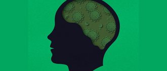
Article for the “bio/mol/text” competition: Prion diseases are a phenomenon discovered in the twentieth century, and in it they began to play a big role: an increase in life expectancy in developed countries has led to the fact that more and more people began to live to see “their Alzheimer’s” "or "your Parkinson's." The nature of neurodegenerative diseases continues to remain vague, and scientists are still studying only certain aspects of them - for example, the reason for their development in old age or the ability to be transmitted from one species of living beings to another.
“Bio/mol/text”-2012
This article was submitted to the competition of popular science works “bio/mol/text”-2012 in the category “Best News Message”.
The competition is sponsored by the visionary Thermo Fisher Scientific.
It all started when in the 20th century scientists became interested in the nature of unusual diseases in humans and animals: kuru, Creutzfeldt-Jakob, scrapie. The noticeable similarity in the pathology of these diseases gave rise to the hypothesis of their infectivity, which was subsequently confirmed experimentally. Then the question arose about the causative agent of these diseases. Before the answer was found, the extraordinary properties of the pathogens were revealed: they do not reproduce on artificial nutrient media, are resistant to high temperature, formaldehyde, various types of radiation, and the action of nucleases. Purification of the infectious material and its study made it possible to proclaim that “everything is to blame” for the protein, which 30 years ago received the name prion (from the English prion - protein infection).
Thus, famous American scientists - virologist and doctor D.K. Gaidushek, who discovered the infectious nature of prion diseases, in 1976, and biochemist S.B. Prusiner, who identified prions and developed the prion theory in 1997, were awarded Nobel Prizes. Their work became the impetus for subsequent research, thanks to which new types of prion infections were studied. But, even despite the undying interest in the “prion topic,” the formation of prions remains a mystery to this day.
4.Treatment
To date, there is no effective treatment for fatal familial insomnia. Therapy is forced-palliative, symptomatic in nature and is aimed at maintaining fading vital functions and correcting mental disorders. None of the known groups of sleeping pills are effective.
The most promising research in the field of treatment of prion infections, incl. hereditary familial insomnia are associated with the search for and attempts to activate prion-specific antibodies, as well as with progressive developments in the field of genetic engineering and therapy. However, there are no effective remedies and real results yet, and fatal insomnia remains, unfortunately, an incurable disease.
Biological essence of prions
Figure 1. The metaphor for neurodegenerative brain damage is that neural tissue becomes a sponge as a result of massive neuronal death.
The prion molecule is not something exotic: in a “normal” form it is present on the surface of the nerves of each of us. At the same time, we feel great, and our nerve cells are alive and healthy. However, this is all until our normal protein “degenerates” into an abnormal form. And if this happens, it will lead to terrifying consequences: the infectious form of prions has the ability to “stick together” with other molecules and, moreover, “convert” them into the same form, causing a “molecular epidemic”. As a result of this polymerization, toxic protein plaques appear on the nerve cell, and it dies [1]. In place of the dead cell, a void is formed - a vacuole filled with liquid. Over time, one neuron after another will disappear, and more and more “holes” will form in the brain, until finally the brain turns into a sponge (Fig. 1), which will inevitably be followed by death.
There is a simplified idea that polymerized prion fibrils “puncture” the neuron, causing its death. In fact, this is not entirely true: spherical prion aggregates preceding the fibrillar stage are also toxic (at least for Alzheimer's disease): “Alzheimer's neurotoxin: not only fibrils are poisonous.” - Ed.
But how can a normal natural protein (denoted PrPC) suddenly become pathological (PrPSc; Sc - from the word “scrapie”)? What is going to happen? As in the case of a “normal” infection, such transformation requires an encounter with an infectious prion molecule. There are two ways of transmitting this molecule: hereditary (due to mutations in the gene encoding the protein) and infectious. That is, the introduction of a prion can occur unexpectedly - for example, when eating insufficiently well-fried or cooked meat (which should contain the PrPSc form), during a blood transfusion, during organ and tissue transplantation, or when administering pituitary hormones of animal origin.
And then an amazing event occurs: normal protein molecules, in contact with pathological ones, themselves transform into them, changing their spatial structure (the mechanism of transformation remains a mystery to this day) [1]. Thus, the prion, as a real infectious agent, infects normal molecules, triggering a chain reaction that is destructive to the cell.
Diagnostics
Diagnosis of prion diseases is based on the detection of cognitive disorders, their progressive development, and already at an intermediate stage of development - non-specific EEG disturbances, gradually turning into specific patterns (burst suppression pattern) with myoclonus in the clinical and EMG picture, and epileptic seizures.
Only 14-3-3 protein is present in the cerebrospinal fluid.
In sporadic Creutzfeldt-Jacobs disease, T2-weighted MRI demonstrates hyperintensity in the nucleus basalis and caudalis. In the new variant of the disease, it is present in the pulvinar thalamus and has the shape of a club.
DNA testing can help identify genetic and familial forms of the disorder, which means identifying the E200K mutation.
The clinical diagnostic conclusion is confirmed by immunohistochemical and neurohistological examination of brain tissue.
Shown:
- typical neurocytic vacuolization;
- spongiomatous degeneration of the neuropil;
- loss of nerve cells;
- astrogliosis;
- presence of scrapie-associated fibrils in the affected brain.
Some information about prions
The researchers note:
- The prion protein includes 254 amino acid residues and weighs 33–35 kilodaltons [2];
- the gene encoding the PrP protein is found in humans, mammals, and birds [1];
- To completely destroy the prion protein, a temperature of at least 1000 degrees is required [1]!
- it is possible that prions take part in intercellular recognition and cellular activation [3];
- it is possible that the function of prions is to suppress age-related processes [3];
- with the development of clinical manifestations of prion diseases there are no signs of inflammation or changes in the blood;
- it is assumed that prions are involved in the development of schizophrenia and myopathy;
- The mechanism of action of prions and their transformation from a normal form to a pathological one remains unclear.
Do you remember how it all began
In the twenties of the last century, doctors were faced with a new and hitherto unknown disease. The German neurologist Hans Gerhard Creutzfeldt observed one patient in his clinic - a 20-year-old girl. At the initial stage of the disease, she had impaired sensitivity in her arms and legs, disorders of memory and nervous activity rapidly progressed, and the patient increasingly fell into an unconscious state. A few months later, the girl died from respiratory and cardiac disorders. A neurologist, who would later become a prominent Nazi doctor and take part in the Euthanasia program, documented the course of the disease.
A few months later, Dr. Alfons Maria Jacob from Hamburg encountered three similar patients. The young people suffered from disorders of nervous activity, swallowing, were practically unaware of what was happening around them, and soon died. During the autopsy, Jacob saw an interesting phenomenon that doctors had never seen before - only the brain was affected in the patients. Massive death of cells in the gray matter of the brain was recorded, and the remaining neurons were characterized by unusual swelling. No pathological changes were recorded in any other organ. In memory of the two discoverers, the disease was named Creutzfeldt-Jakob disease.
Related links
- Cardiac arrest and cerebral coma: clinical death from a medical point of view
- The mystery of “blue blood”: The tragic fate of the creator of perftoran
In those early years, virology as a science was still in its infancy. Therefore, the disease was destined to remain forgotten for a long time. This was facilitated by the Great Depression and World War II. It was only in the fifties of the last century that scientists began to actively take an interest in what actually happened to people who were unlucky enough to catch Creutzfeldt-Jakob disease.
At the same time, scientists are discovering two more diseases, which in their symptoms and course are very, very reminiscent of the terrible illness described above - kuru and scrapie. The first disease was common among the Fore people on the island of Papua New Guinea, and the second affected sheep around the world. But something else turned out to be important: the symptoms of the diseases were somewhat different from Creutzfeldt-Jakob disease, but the nature of the lesions was almost identical - the formation of voids in the brain tissue and the massive death of nerve cells.
It would seem that everything is clear. There is a disease, it is caused by some kind of virus or bacteria, let's figure out who is the causative agent and eliminate the cause. But it was not there! Everything turned out to be not so simple...
"Educational films": Vaccines
Conditions for the occurrence of diseases
The conditions for the occurrence of prion diseases are unique. They can form according to three scenarios: as infectious, sporadic and hereditary lesions. In the latter option, genetic predisposition plays a major role [2].
Renowned prion researcher Stanley Prusiner identifies two striking features of neurodegenerative diseases such as Creutzfeldt-Jakob disease, Alzheimer's disease, and Parkinson's disease. The first is that more than 80% of cases of the disease are sporadic (that is, random, occurring “by themselves”). Second: despite the fact that a large number of mutant proteins specific to a particular disease are expressed during embryonic development, the patterns of inheritance of these neurodegenerative diseases appear later. This suggests that certain processes occur during aging that release disease-causing proteins [5]. More than 20 years ago, the author argued that this process involves the random refolding of a protein into an incorrectly folded one, which corresponds to the transition to an infectious state - a prion.
Interesting facts about Alzheimer's disease: Chronic sleep deprivation may increase the risk of Alzheimer's disease ("New step in understanding Alzheimer's disease: sleep deprivation may be a risk factor"), and Alzheimer's neuropeptide itself (amyloid-β Aβ) may be part of the innate immune system ( "Perhaps Alzheimer's β-amyloid is part of the innate immune system." - Ed.
In the last decade, interest in this topic has renewed due to the possibility of developing diagnostics and effective therapy [5]. Many different explanations have emerged for age-related neurodegenerative diseases, such as oxidative modification of DNA, lipids and/or proteins; somatic mutations; altered innate immunity; exogenous toxins; DNA-RNA mismatches; disruption of chaperone function; absence of one of the gene alleles [5]. An alternative complex explanation is that different groups of proteins can form prions. Although small amounts of prions can be eliminated through protein degradation pathways, excessive accumulation over time allows prions to self-propagate throughout the body (Figure 2), resulting in central nervous system dysfunction [5].
Figure 2. Processes of neurodegeneration caused by prions. Top: The accumulation of a “normal” prion protein increases its likelihood of transitioning to a toxic conformation, which is described by a greater content of β-structure. Prions are most pathogenic in the form of oligomers; After fibril formation, toxicity decreases. Depending on which specific prion protein we are talking about, in a pathological state it can form plaques, tangles or inclusion bodies. Possible routes of drug intervention: (I) reduction of con precursor protein; (II) inhibition of the formation of the prion form; (III) destruction of toxic aggregates. Bottom: Hereditary senile neurodegeneration is explained by two events: the presence of a mutant form of the precursor and the formation of a prion from it, ready for oligo- and polymerization to form toxic forms.
[5]
Prions. History of slow infections.
Prions (English prion from protein - “protein” and infection - “infection”). The term was proposed by the person who laid out the foundations of modern knowledge about these proteins - Stanley Prusiner in 1982. Now we know that these are pathological proteins that cause a number of encephalopathies in humans (Creutzfeldt disease - Jacobus syndrome Gerstmann - Straussler - Schenker, fatal familial insomnia, Kuru, etc.), livestock (mad cow disease, scrapie in sheep) and birds. Type of transmission, pathogenesis, etc. in these diseases it is not similar to viruses or bacteria. But first things first.
Story
The first disease from the list of prion diseases described by humans is scrapie - sheep scabies. In 1700, in England (the country with the largest population of domestic sheep at that time), the following symptoms were described - severe itching, pain in the limbs when moving, and convulsive seizures. The disease progressed within a week. Outbreaks occurred in different counties. Veterinarians and doctors shrugged their shoulders, not knowing the source of the disease. All symptoms pointed to brain damage. By the 20th century, no new data had been added about what kind of disease affected the poor sheep. And so, in the 1920s, Hans Gerhard Creutzfeldt and Alfons Maria Jacob separately (both in 1920, but Creutzfeldt earlier) described an incurable lesion of the human nervous system, which was later named after them. The pathological picture was described (focal lesions of brain tissue). A first attempt was made to classify symptoms. The definition of the disease was “spastic pseudosclerosis or encephalopathy with scattered foci in the anterior horns of the spinal cord, extrapyramidal and pyramidal system.”
Histological specimen of the brain showing microcavities
Hans Gerhard Creutzfeldt, as a German neurologist, had his ties to Nazism. Not being a party member, he acted as a medical expert on issues of heredity, that is, he resolved issues of forced sterilization and euthanasia. There are different versions about his activities in this area. Some write that Creutzfeldt saved people from these procedures by concealing pathologies and correcting medical histories, others that the doctor was still involved in sending people to death camps and to the Kiel clinic (where “euthanasia” was carried out). In any case, the British occupation authorities found no traces of crime in his case and the 60-year-old doctor continued his activities in Munich. The history of prion diseases developed further. In 1957, Carlton Gajdushek and Vincent Zigas discovered a disease similar in clinical picture to Creutzfeldt-Jakob disease (the disease now bore the names of these two doctors). If the disease discovered in the 20s affected residents of Western Europe, then the new one affected representatives of a tribe in New Guinea. It was suggested that this pathology is caused by a virus. The clinical picture, characterized by tremor, convulsions, and headache, was studied. Based on the fact that neighboring tribes did not practice cannibalism and brain eating and did not suffer from similar pathologies, theories began to appear that the virus was located in brain tissue and could be transmitted through nutrition. In 1967, the first successful experiment was carried out with the infection of experimental mice with the biological fluids of sheep infected with scrapie. The result was positive. The mice developed the same symptoms as the “donors.” The evidence for the contagiousness of the disease has increased. Interestingly, in 1976, Gaidushek was awarded the Nobel Prize for discoveries concerning new mechanisms of the origin and spread of infectious diseases associated with the study of the Fore tribe disease. Until the end of his life, he was sure that it was caused by viruses.
Fore tribe
Stanley Prusiner
As mentioned above, the foundations of knowledge about prions were laid by Stanley Prusiner. A little from his biography. Born in the USA in 1942. His ancestors are emigrants from the Russian Empire, of Jewish origin, forced to leave the country due to Jewish pogroms. Stanley Prusiner himself graduated from the University of Pennsylvania in 1968 and worked as a neurology resident at the University of California School of Medicine (San Francisco). In 1970, he first encountered Creutzfeldt-Jakob disease. The patient who was being treated by Prusiner had no pathogen detected. Having become closely involved in this research, the neurologist turned to the works of another doctor, Siggurdson, who identified certain patterns in diseases that were incomprehensible at that time.
These patterns became:
- unusually long (months or years) incubation period;
- slowly progressive nature of the course;
- unusualness of damage to organs and tissues;
- the inevitability of death.
The diseases known at that time that met these criteria were Creutzfeldt-Jakob disease, Kuru in humans, and scrapie, which began to affect not only sheep, but also goats. It was from biological fluids (cerebrospinal fluid, urine, seminal fluid, saliva) of sheep suffering from frequent diseases that preparations for infection and further research were prepared. Experiments were carried out on mice. It was found that the incubation period lasts 100-200 days. The disease develops in all experimental mice. Progress was made after the appearance of hamsters in the laboratory. Their incubation period was much shorter, but the clinical manifestations were still the same. So, after 10 (!) years of painstaking work on infection, slaughter of animals, cleaning and examination of the material, a pathogenic object was identified. Experiments strongly suggested that it consisted of a single protein, which Prusiner called a prion. Despite the enormous evidence base collected over years of research, the theory has not received universal acceptance. Most virologists of that time (and it was already 1982) treated the statement with distrust. The main reason for this was the pathogen’s lack of its own genotype. There were only amino acids, no nucleic acids. Without losing his inspiration, Siggurdson continued to study the strange agent. Its amino acid sequence was revealed. Further, the production of antibodies to the prion protein made it possible to determine its localization in the cell membrane. The scientist's career was going well. In 1980 he became a professor of neurology and in 1988 a professor of biochemistry. In 1982, he published a scientific paper on a completely new type of pathogen. The doctor and scientist received universal recognition in the 90s. In 1997, he received the Nobel Prize for his discovery of prions, a new biological principle of infection. Another reason for the increasing interest in this pathology is the epidemic of mad cow disease, or bovine spongiform encephalopathy, that swept the UK (there were 179 thousand heads of cattle with symptoms of the disease).
What are prions and what is their mechanism of action on the body (modern ideas)?
In fact, humans and many other living things contain PrPC proteins. In Russian - the normal form of prion proteins (they were discovered after Siggurdson’s research, which is why the name is so strange). Its length, amino acid sequence, and secondary structure are known. It is important to know that the final structure consists of three α-helices and a double-stranded antiparallel β-sheet. They have an interesting property, namely they are precipitated by high-speed centrifugation, which is a standard test for the presence of prions. There is evidence that PrP plays an important role in cell attachment and the transmission of intracellular signals, and therefore may be involved in the communication of brain cells. However, the functions of PrP are not well understood.
(a) normal (b) pathology
Experiments in mice lacking these proteins show that the absence of PrP leads to nerve demyelination. It is possible that prion proteins normally support long-term memory. But this is normal. Sometimes “problems” happen and proteins called PrPSc appear - infectious prions. They differ in that instead of α-helices, β-sheets predominate in them. This leads to changes in the interaction of other proteins with the new protein. It wouldn’t be so bad if only one protein was produced per body. The trouble is that, once formed, the protein (!) itself begins to change the structure of others. Let's consider the main mechanisms of PrPSc reproduction
It is believed that prion disease can be acquired in 3 ways: through direct infection, hereditary or sporadic (spontaneous) or combinations thereof. Sporadic (that is, spontaneous) prion disease occurs in a population in a random individual. This is, for example, the classic version of Creutzfeldt-Jakob disease. There are two main hypotheses regarding the spontaneous occurrence of prion diseases. According to the first of them, a spontaneous change occurs in the hitherto normal protein in the brain, that is, a post-translational modification takes place. An alternative hypothesis is that one or more cells in the body at some point undergo a somatic mutation (that is, not inherited) and begin to produce the defective PrPSc protein. Be that as it may, the specific mechanism for the spontaneous occurrence of prion diseases is unknown. The second is infection. According to modern research, the main way to acquire prion diseases is by eating contaminated food. It is believed that prions can remain in the environment in the remains of dead animals, and are also present in urine, saliva and other body fluids and tissues (blood, cerebrospinal fluid). Because of this, infection with prions can also occur during the use of unsterile surgical instruments. This makes it difficult to sterilize surgical instruments or devices in the slaughterhouse. Prions are generally resistant to proteases, heat, radiation, and storage in formalin, although these measures reduce their ability to become infected. Effective disinfection against prions must involve hydrolysis or damage/destruction of their tertiary structure. This can be achieved by treating with bleach, sodium hydroxide and strongly acidic detergents. Spending 18 minutes at 134°C in a sealed steam autoclave will not deactivate prions. Ozone sterilization is currently being studied as the main modern method for deactivating and denaturing prions. Renaturation of a completely denatured prion to an infectious state has not been recorded, but for partially denatured prions under some artificial conditions this is possible. It is also worth remembering that these proteins can remain in the soil for a long time due to binding to clay and other soil minerals. Don't get paranoid, but theoretically they could be everywhere. In 2011, the discovery of prions transmitted through the air in aerosol particles (i.e., airborne droplets) was reported. Also in 2011, preliminary evidence was published that prions can be transmitted by urine-derived human menopausal gonadotropin, used to treat infertility. Theoretically, with just one sick animal with prion disease, it is possible to destroy entire nations and countries by simply adding its bone meal to feed additives and selling them to the desired state. A similar situation occurred in the late 80s in Britain (mad cow disease epidemic). Then, most likely out of ignorance (and not out of malicious intent), the above process occurred, which claimed the lives of about 200 people (as of 2009) and 179 thousand heads of cattle.
Prion propagation
The third mechanism is genetic. It was opened recently and does not fit into the overall picture at all. The gene encoding the normal PrP protein, PRNP, localized on chromosome 20, was identified. In all hereditary prion diseases, a mutation of this gene occurs. A “distorted” prion protein that has entered in one way or another begins to change the structures of proteins close to it in structure, turning them into the same pathogenic agents. The main hypothesis that most closely reflects this process is very simple. One molecule of PrPSc attaches to one molecule of PrPC and catalyzes its transition to the prion form. The two PrPSc molecules then separate and continue to convert other PrPCs into PrPSc. But the scheme raises more questions than answers.
Clinic
Let's talk about diseases and clinical manifestations. Theoretically, it can occur in all living creatures that have PrPc. Here are some examples. In sheep and goats, as mentioned above, the main manifestation is scrapie. Cows are characterized by mad cow disease (bovine spongiform encephalopathy). In minks, transmissible encephalopathy of minks. And so on. Manifestations of diseases have been recorded in cats, wild artiodactyls, and ostriches. But we are interested in human diseases.
Creutzfeldt-Jakob disease. ICD-10 code A81.0; F02.1. Code A corresponds to infectious diseases (A81 - infectious diseases of the nervous system). Code F – mental disorders, F02 – dementia.
Dark green spread K-Y
Light green - mad cow disease
Basic clinical criteria for diagnosis:
- rapidly progressing - over 2 years - (“devastating”) dementia with disintegration of all higher cortical functions; pyramidal disorders (spastic paresis);
- extrapyramidal disorders (choreoathetosis);
- myoclonus;
- ataxia, akinetic mutism;
- dysarthria;
- epileptic seizures;
- visual disturbances (diplopia)
Stages of the disease:
- Prodromal period - symptoms are nonspecific and occur in approximately 30% of patients. They appear weeks and months before the onset of the first signs of dementia and include asthenia, disturbances in sleep and appetite, attention, memory and thinking, weight loss, loss of libido, and changes in behavior.
- Initial period - the first signs of the disease are usually characterized by visual disturbances, headaches, dizziness, unsteadiness and paresthesia. In the majority of patients, it gradually develops, less often - an acute or subacute onset. In some cases, as in the so-called amyotrophic forms, neurological signs may precede the onset of dementia.
- Advanced period - usually there is progressive spastic paralysis of the limbs with accompanying extrapyramidal signs, tremor, rigidity and characteristic movements. In other cases, there may be ataxia, decreased vision, or muscle fibrillation and upper motor neuron atrophy.
There are several clinical forms:
Spontaneous - classical (sCJD) According to modern concepts (prion theory), prions in this form of the disease arise spontaneously in the brain, without any visible external cause. The disease usually affects people over the age of 50 and occurs with a probability of 1-2 cases per million inhabitants. Initially, it manifests itself in the form of brief memory loss, mood changes, and loss of interest in what is happening around. Further, the symptoms of dementia progress with all the ensuing consequences.
Hereditary (fCJD) The disease occurs in families where damage to the gene for the prion protein is inherited. A defective prion protein is much more susceptible to spontaneous conversion into a prion. The signs and course of the disease are similar to the classical form.
Iatrogenic (1CJD) The disease is caused by the unintentional introduction of prions into the patient's body during medical intervention. The source of prions used to be certain drugs, instruments or meninges that were taken from dead people and used to close the wound during brain surgery. The signs and course of the disease are similar to the classical form. New variant (nvCJD) The disease first appeared in 1995 in the UK and since then no more than 100 people have died from it. Most likely, they became infected with meat products containing bovine prions.
- mental disorders and sensory impairments,
- Global cognitive impairment and ataxia are characteristic.
- Several cases of the disease that began with cortical blindness (Heidenhain variant) have been described.
- The episyndrome is also represented by myoclonic seizures.
- cerebellar symptoms are detected in 100%.
The main diagnostic method is intravital brain biopsy.
MRI and PET methods are also used. There are pathognomonic symptoms of electroencephalography. Gerstmann-Straussler-Scheinker syndrome is a rare, usually familial, fatal neurodegenerative disease that affects patients between 20 and 60 years of age. Code A81.9. Nine here means “Slow viral infections of the central nervous system, unspecified.” The syndrome occurs in people aged 40-50 years and is characterized mainly by cerebellar ataxia, swallowing and phonation disorders, progressive dementia over 6 to 10 years (the average duration of the disease is 59.5 months), after which death occurs. The incubation period lasts from 5 to 30 years. Little studied. Research is being conducted on laboratory mice and hamsters. Fatal familial insomnia is a rare, incurable hereditary (dominantly inherited prion) disease in which the patient dies from insomnia. Only 40 families are known to be affected by this disease. The ICD code is the same as the previous one. The disease begins between the ages of 30 and 60, with an average of 50. The disease lasts from 7 to 36 months, after which the patient dies. There are 4 stages of disease development.
- The patient suffers from increasingly severe insomnia, panic attacks and phobias. This stage lasts on average 4 months.
- Panic attacks become a serious problem, and hallucinations join them. This stage lasts on average 5 months.
- Complete inability to sleep, accompanied by rapid weight loss. This stage lasts on average 3 months.
- The patient stops speaking and does not react to his surroundings. This is the last stage of the disease, lasting on average 6 months, after which the patient dies.
Sleeping pills don't help.
At all. Kuru is almost never found nowadays, due to the eradication of cannibalism. Interestingly, in 2009, American scientists made an unexpected discovery: some members of the Fore tribe, thanks to a new polymorphism of the PRNP gene that appeared in them relatively recently, have innate immunity to kuru.
Currently, there is not a single means of stopping or inhibiting the development of prion diseases.
There is a lot of research going on. Main directions:
- Medicine – a drug that can cure or stop/slow down the progression of a disease
- Vaccine is a means to prevent disease
- Genetic engineering methods are also used to produce animals that are immune to prion diseases.
How changes in the genotype and protein composition will affect their life is still a mystery.
Laboratory diagnosis and treatment
Diagnosis is based on intracerebral infection of mice or hamsters, which slowly (up to 150 days) develop the corresponding disease if the patient was sick [2]. Histological examination of the brains of dead animals is often carried out [2].
Unfortunately, to date, effective methods for treating prion diseases have not yet been developed, although attempts are being made to prevent the conformational transition of a normal protein into an abnormal one. Therefore, the most reliable way to prevent the development of infectious forms is prevention [2].
The solution to the “prion issue” is becoming especially relevant in connection with the growing threat of an epidemic through invasive medical operations and even when taking medications.
Animal diseases
The risk of animal diseases for humans is very speculative (the problem is best characterized by the connection between BSE and a new variant of Creutzfeldt-Jacobs disorder). In animals, disorders are manifested by aggressiveness and impaired motor abilities:
- bovine spongiform encephalopathy (BSE);
- scrapie;
- chronic wasting disease (CWD);
- felt spongiform encephalopathy;
- transmissible encephalopathy.
Prospects
Apparently, interest in prions will not fade until assumptions about them are fully confirmed and effective methods for treating prion diseases are found. The article [6] talks about the need for modern research, which requires careful consideration of foreign prions in extraneuronal tissues.
The authors used mice as model objects: two lines that transgenically expressed sheep prion protein, and one line that expressed human prion protein (Fig. 3). The goal was to compare the effectiveness of interspecies transmission of infection through brain and spleen tissue. Intracerebral infection with a foreign prion protein resulted in the absence or small amount of the infectious agent in the brains of these mice. However, infectious foreign prions were detected in the spleen at earlier stages of infection compared to when neurotropic prions were used, suggesting that lymphatic tissue may be more permissive to the spread of foreign prions compared with the brain.
Figure 3. The ability of the Sc237 hamster prion to infect and transmit when injected into the brain or spleen of transgenic mice having the sheep (tg338; white mice) or human (tg7; gray mice) PrP prion protein. The number of diseased/injected mice is shown in parentheses; Below is the average lifetime (in days).
[6]
What causes this preferential replication of prions in lymphatic tissues is still unknown. However, the findings indicate that humans may be more sensitive to foreign prions than previously thought based on the presence of prions in the brain, and for this reason, asymptomatic carriers of prion disease may not be recognized. This once again confirms that such a powerful biomolecule as the prion is fraught with many mysteries, the disclosure of which may help in understanding a number of insoluble problems of humanity...
How to slow down the progression of prion disease
Until recently, it was difficult to slow down prion disease, let alone cure it.
Modern neurosurgery can alleviate degenerative complications by transplanting neurons for prion disease.
A revolutionary attempt to stop pathogenic proteins was pioneered by a team of doctors from the University of Pittsburgh. Dr. Kondziolka's team transplanted neurons in vitro (2-6 million LBS neurons) into 12 patients as part of the study. 12 months after surgery, none of the recipients had any adverse effects from the transplant, and all received new cells. In 6 patients there was a significant improvement in condition.
Several years ago, the journal Nature reported on the development of antibodies against prions in England by a team of scientists led by Simone Hawke (Imperial College). In tests on infected mice, rodents that received the antibodies were cured within a month of infection.
Doctors intend to use this opportunity to clear neurons of pathogenic proteins to treat a new variant of Creutzfeldt-Jacobs disease in humans. Further research should help improve drugs that can clear prions from the brain and its microglia.
Recently, M. Pfeiffer and several other German scientists discovered and confirmed a method to clear the brain of prions. They proved that RNA interference can successfully treat diseases by removing from the RNA cell the part responsible for the production of pathogenic proteins. This prevented them from mutating into dangerous proteins.
A similar method is the effect of artificially synthesized primers (oligonucleotides) against prions. Like RNA interference, primer design is under investigation.
Theoretically, the PrP13 peptide, similar to the structural product found in these diseases, may be promising in the treatment of diseases. The substance targets the PRNP gene, which should affect the conversion of PrPC to PrPres. The last pathogenic molecule named changes to a PrPC-like structure. The substance goes by the acronym PrP13, and animal studies have been able to prolong survival by 50-300%.
Literature
- Abramova Z.I. Study of proteins and nucleic acids. Kazan: Kazan State University, 2006. - 157 pp.;
- Novikov D.K., Generalov I.I., Danyushchenkova N.M. Medical microbiology. Vitebsk: VSU, 2010. - 597 pp.;
- Prudnikova S.V. Microbiology with basics of virology. Krasnoyarsk: IPK SFU, 2008;
- Pozdeev O.K., Pokrovsky V.I. Medical microbiology. M.: Geotar-med, 2001. - 765 pp.;
- S. B. Prusiner. (2012). A Unifying Role for Prions in Neurodegenerative Diseases. Science
.
336 , 1511-1513; - V. Beringue, L. Herzog, E. Jaumein, F. Reine, P. Sibille, et. al.. (2012). Facilitated Cross-Species Transmission of Prions in Extraneural Tissue. Science
.
335 , 472-475; - Carolina Pola. (2012). Prion escape to spleen. Nat Med
.
18 , 360-360; - Elements: “10 facts about prions and amyloids”;
- Elements: “Geometry of protein bodies”;
- Charles Weissmann. (2012). Mutation and Selection of Prions. PLoS Pathog
.
8 , e1002582.
