Carotid angiography is an invasive X-ray imaging procedure of the carotid arteries that is performed through a catheter inserted into a blood vessel in the arm or leg. A contrast agent is injected through the catheter, which is opaque to X-rays and therefore shows the internal lumen of the carotid arteries. This procedure is the gold standard for visualization of the carotid and cerebral vessels.
Preparing for the study
- Preparation for the study involves shaving the access site (usually the groin area).
- You should tell your doctor about any allergic reactions you have.
- If you are diabetic, tell your doctor so your medications can be adjusted on the day of the test.
- Tell your doctor if you are allergic to anything, especially iodine, shellfish, X-ray contrast, latex, or rubber products (such as rubber gloves).
- It is advisable to drink enough fluids before the examination to reduce the possible effects of contrast on the kidneys.
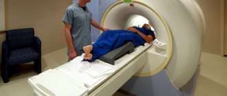
How is the procedure done?
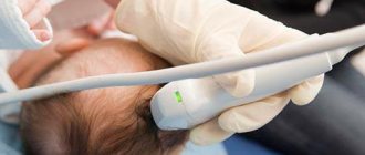
To perform MRA, a conventional magnetic resonance scanner is used. The procedure, depending on the type of study, takes from 10 minutes to an hour. The main condition for a successful study is immobility during scanning. If the patient finds it difficult to remain in one position, he is prescribed a sedative to put him into a short-term sleep.
During the study, the patient may hear noises made by operating equipment. To reduce discomfort from constant loud sounds, the patient is asked to wear headphones, through which the doctor will tell the person being examined how the procedure is going.
After research
Tell your doctor or nurses if you feel:
- Itching, dry throat
- Nausea
- Chest discomfort
- Vision problems
- Weakness in one or more limbs
At the end of the study, you will be transferred to a ward. It is necessary to remain in bed for several hours. The doctor will periodically examine the access site and assess the general condition. If there are no problems, then discharge can be made the next day.
MR angiography with contrast
Magnetic resonance imaging of the circulatory system involves various scanning modes:
- time-of-flight;
- phase contrast;
- MRI angiography of vessels with an intensifier.
Time-of-flight tomography provides T1-weighted images. The liquid medium under study is brightly colored against the background of the surrounding soft tissue.
Phase contrast MR angiography involves receiving a signal from blood moving through the vessels. The method allows you to determine the characteristics of fast arterial and slow venous flow with equal accuracy. The simultaneous use of time-of-flight and phase-contrast MR angiography helps to differentiate between hematomas and other static areas.
Magnetic resonance imaging with enhancement visualizes the bloodstream in the area of interest. A solution of gadolinium salts is used as a contrast agent. The drug has paramagnetic properties and provides a hyperintense signal.
Contrast for vascular MRI angiography is administered intravenously. The injection is given using a catheter connected to an automatic device. This system allows the “coloring” solution to be supplied at a constant speed.
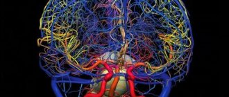
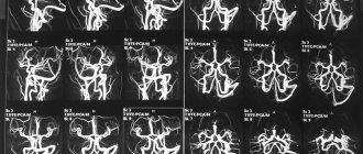
Layer-by-layer images during MRI angiography of cerebral vessels
Data recording begins after the lumen of the bloodstream is filled with contrast. 1-2 ml of solution is pre-injected, which helps to identify the initial moments of the arterial and venous phases. In this way, the signal registration time during vascular MRI angiography is determined. For further measurements, the peak arterial concentration of the gadolinium solution is used.
Angiograms make it possible to study the nature of the blood supply to a certain area. A broken line with a hyperintense signal indicates stenosis and occlusion of the vascular bed. The consequence of the pathology is ischemia and necrosis of the underlying tissues.
MRI angiography of vessels with contrast does not show calcifications, blood clots, or cholesterol plaques. In case of atherosclerosis, embolism, the images show changes in the lumen, fullness, blood channel, and parietal foci with a hypointense signal.
Contrast MR angiography for MRI diagnostics of neoplasms visualizes arteries and veins in the area of interest, shows the condition of surrounding tissues, and helps determine the nature of the disease.
The hemodynamics of the tumor circulatory network depends on the malignancy of the process. The vessels of a malignant tumor have a large number of anastomoses and bends, so the tumor tissue accumulates and releases the contrast agent more slowly.
Subarachnoid hemorrhage on MRI with MR angiography
The final diagnosis is made by the attending physician. The results of MRI angiography of veins and arteries for vascular neoplasms are supplemented with data from clinical tests, biopsy and other research methods.
Possible complications of carotid angiography
Complications are rare - the risk is 0.5%. The most dangerous are complications from the brain. They can develop as a result of the effect of contrast or when pieces of plaque are torn off during catheter insertion. Complications include:
- Transient ischemic attack - short-term disturbance of cerebral circulation
- Ischemic stroke is a persistent disorder of cerebral circulation
- Bleeding from the arterial access site
- Carotid artery dissection with thrombosis
- Allergic reaction to contrast
Timely diagnosis of complications allows you to minimize their consequences.
Why is MR angiography needed?
The study of the circulatory system is used in surgery, oncology, neurology and other areas of medicine. MRI of a given area with MR angiography allows you to assess the functionality, fullness, lumen, and condition of the walls of veins and arteries.
Based on the scan results, the nature of the blood supply to the area under consideration is determined, and foci of ischemia and necrosis are identified. In case of injuries, tomograms visualize hematomas, a violation of the integrity of the vascular wall.
Indications for prescribing MRI scanning in angiography mode are:
- dizziness, fainting;
- lack of coordination;
- severe headaches (cephalgia) that cannot be relieved with medication;
- decreased visual acuity, flickering of spots before the eyes;
- hearing loss, tinnitus;
- paresis and paralysis of the facial muscles;
- numbness, weakness of the upper and lower extremities;
- speech disorder;
- memory impairment;
- “shaky” gait;
- convulsions;
- leg pain;
- swelling of the lower extremities;
- dryness, peeling, ulceration of the skin of the legs, etc.
Decreased tone, decreased lumen, and ruptures of vessel walls lead to the development of pathological changes in surrounding tissues and organs. Impaired patency of the coronary arteries causes destructive, dystrophic processes in the heart muscle, which can cause a stroke or heart attack.
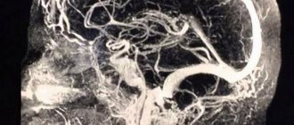
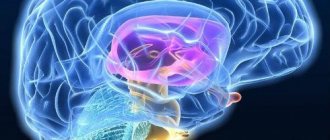
MRI angiography of cerebral vessels (sagittal section)
Pathologies of the aorta, superior and inferior vena cava are accompanied by dysfunction of the abdominal and pelvic organs and contribute to the development of diseases of the lower extremities.
In the diagnosis of cerebral vascular lesions, MRI using the MR angiography mode visualizes the slightest changes in the functioning of the circulatory system.
The procedure allows you to diagnose:
- stenosis, occlusion - narrowing and blockage of the vascular bed;
- acute disturbance of blood circulation in tissues;
- aneurysm - protrusion of the arterial wall;
- thrombosis - the formation of a clot in the lumen of a vein;
- vascular wall ruptures;
- atherosclerosis - cholesterol deposits in the lumen of the artery;
- vasculitis - inflammatory changes, thickening and stratification of the walls;
- embolism - blocking the lumen of the artery with foreign particles (gas, parasitic, etc.);
- extravasal compression syndrome - external compression of the vascular bed;
- arteriovenous malformations - pathological anastomoses (connections) of blood channels;
- congenital anomalies of the vascular system - kinks, bifurcations, etc.;
- dissection (dissection) of the aorta;
- thrombophlebitis - inflammation of the walls of the veins with the formation of bloody clots;
- vascular tumors.
The results of magnetic resonance angiography are analyzed by an MRI physician. The study protocol indicates:
- diameter, patency of the vascular bed;
- the condition of the walls, the fullness of the vessel;
- the presence of foreign particles (emboli) in the lumen;
- localization of the affected area;
- condition of surrounding tissues;
- functionality of veins and arteries;
- degree of circulatory disturbance in tissues;
- causes of hemorrhagic and ischemic phenomena.
MRI angiography examination is prescribed before surgery. Based on the scan results, the location and extent of the affected area are determined, the nature of the pathology and the scope of the upcoming operation are clarified.
During the rehabilitation period, recovery processes are monitored using MR angiography; MRI helps to timely diagnose the development of complications.
Scanning allows you to track the course of chronic diseases. In this case, vascular MRI angiography is prescribed at a frequency of 2 times a year. Early detection of relapses facilitates timely correction of treatment and helps achieve stable remission.
There are two types of MR angiography in MRI. The methods are identical, the difference lies in the purpose of the study.
MRI of the neck with MR angiography
Venography is aimed at studying the work of the vessels of the same name. Scanning allows you to diagnose inflammatory, degenerative processes, signs of reverse flow, and determine the causes of pathological phenomena.
Arteriography is indicated in case of symptoms of vascular insufficiency. If necessary, a comprehensive study of the vascular system is performed.
Angiography is a universal method for diagnosing vascular diseases
Many people all over the planet suffer from vascular diseases. A diagnostic method, angiography, helps in identifying pathologies associated with damage to the cardiovascular system. During this procedure, a contrast agent is injected into the vessels, which is illuminated with an X-ray. This is how possible or existing vascular defects can be identified.
Indications for angiography
The method allows you to identify narrowing, thinning, rupture and other problems with blood vessels. An examination may be prescribed if the following symptoms are present:
- shortness of breath, chest pain, difficulty breathing;
- surgery or injury to the sternum;
- examination of blood vessels during upcoming surgical treatment;
- diagnostics for suspected congenital heart defects or if they are present;
- if treatment with medications for diseases of the heart, veins, or blood vessels did not bring the expected effect, or the symptoms of the disease became brighter and more dangerous.
Angiography includes studies:
- cerebral blood flow (cerebral angiography);
- blood flow of the heart vessels (coronary angiography);
- examination of the veins of the extremities (phlebography);
- pulmonary vessels (angiopulmonography);
- examination of the aorta and its branches (renal aortography).
If angiography is done on time, many diseases can be prevented, as well as blood clots can be identified and eliminated in a timely manner.
8
24/7
Types of angiography
There are 2 methods of angiography: invasive and non-invasive.
In the invasive form, contrast (usually iodine) is injected into the vessels or arteries, and thanks to X-rays it becomes possible to study them. Despite the fact that the technique is considered outdated, a more accurate and informative alternative to the method has not yet been found.
Non-invasive angiography includes examination methods:
- Ultrasound;
- MRI;
- CT.
CT scan
It allows you to obtain images of all human organs and systems using an X-ray beam. The information that the computer received during the study is processed and a three-dimensional three-dimensional image of the examined organ is recreated.
In CT scanning, contrast is injected during the examination. Typically the insertion site is a vein in the forearm. This method of examination does not require anesthesia or hospitalization of the patient.
MR angiography
Magnetic resonance imaging uses electromagnetic waves and magnetic fields. The value of this method lies in diagnosing the vessels of the head and neck.
The study is performed both with and without contrast. MRI can be performed on people who are allergic to contrast agents. The disadvantages of this method are the time of the procedure (it is slightly longer than with CT), as well as the presence of contraindications:
- fear of confined spaces;
- psychological illnesses;
- metal prostheses implanted into the body.
Angiography of different parts of the body
Angiography is applicable for diagnosing different parts of the body. It has specific indications for each case.
- Angiography of cerebral vessels. After administration of the contrast agent, pictures are taken in different projections. For better diagnosis, contrast can be injected twice. It is prescribed if there are problems with the blood flow to the brain, headaches of unknown origin and the patient is bothered by other symptoms.
- Angiography of the extremities. It is usually prescribed for pain in the limbs, limited movement, swelling and pallor of the skin. The procedure allows you to study the surrounding tissues, lumens, and vessel walls.
- Angiography of the lower extremities. It is carried out in the presence of the following diseases:
- atherosclerosis of leg arteries, thrombosis;
- thrombophlebitis, phlebitis;
- traumatic damage to blood vessels of the lower extremities;
- arterial defects;
- impaired blood flow in the foot with diabetes;
- as a control after leg surgery.
- Angiography of the neck. Indications for its implementation are:
- injuries, neck wounds;
- headaches of unknown etiology;
- dizziness, fainting for unknown reasons.
There is also angiography of arteries, internal organs, coronary vessels.
8
24/7
Indications and contraindications
The main indicators for conducting an angiographic examination are the following factors:
- thromboembolism;
- vascular aneurysms;
- malformation;
- tumors and cysts;
- atherosclerosis;
- diseases of internal organs;
- heart disease;
- diagnosing retinal defects;
- control after operations.
In addition to the indications, angiography has contraindications:
- renal, liver, heart failure of the acute stage;
- venereal diseases;
- tuberculosis;
- thyroid dysfunction;
- psychological disorders;
- Carrying a child.
It is worth noting that angiography is performed only when the patient’s health condition can be regarded as satisfactory.
Preparation for the procedure
Before starting the procedure, it is necessary to exclude contraindications in the patient, including an allergic reaction to iodine and anesthetic drugs. You also need to do a fluorography and cardiogram, and take a routine and extended blood test.
You should avoid drinking alcohol for about 10-15 days. To prevent allergies, antihistamines are prescribed before angiography. To protect the kidneys from excess iodine in the body and facilitate its elimination, the body is saturated with fluid before the procedure. Do not eat 4-5 hours before the start of the study. Before the event itself, be sure to remove all jewelry and metal objects so that they do not interfere with the penetration of X-ray radiation.
The night before, take a shower and shave the hair in the area where the puncture is planned.
It is mandatory to have the patient's written consent for the procedure. Preparing for a study of patients with chronic diseases is somewhat different from the usual preparatory procedure.
- If blood pressure is high, the patient is first prescribed a course of medications to normalize it.
- For people with heart rhythm disturbances, medications containing potassium and glycosides are prescribed.
- For ischemic heart disease and heart pain, nitrates are used.
- If a person suffers from chronic bronchitis, tonsillitis and other inflammatory diseases, then he undergoes a preliminary course of antibiotic treatment for 2 weeks.
Performing angiography
The angiography procedure consists of several stages.
- The patient is placed on a special angiographic table and fixed in a stationary position.
- Next, they are connected to a heart monitor.
- Antiallergic drugs and a tranquilizer are administered.
- Clean the puncture site with alcohol or other antiseptic.
- For pain relief, lidocaine is injected subcutaneously.
- An incision is made in a small area of skin to gain access to the vessel.
- An introducer (short hollow tube) is inserted.
- To relieve vessel spasm and reduce irritation by contrast, Novocain is administered.
- A catheter is inserted inside the hollow tube and advanced to the beginning of the vessel, monitoring the process with X-rays.
- Contrast is administered, usually iodine preparations (Uragrafin, Cardiotrast, Triyotrast). Next, scanning is carried out in different protection. If necessary, this step is repeated several times.
- After completing the previous stage, the catheter and introducer are removed.
- If bleeding occurs, it is stopped.
- A pressure bandage is applied to the puncture site and left in place for 24 hours.
After angiography, it is recommended to drink more water, which helps to accelerate the removal of iodine from the body. It is also necessary to maintain bed rest for 6-12 hours after the procedure to avoid thrombosis.
The event is carried out by highly qualified specialists: anesthesiologist, cardiorheumatologist, radiologist.
Possible complications
Complications can be avoided if you carefully consider the patient’s medical history and avoid mistakes during the examination. However, sometimes unintended consequences happen. Possible complications:
- allergic reaction to contrast agent;
- hematoma, swelling at the site of the procedure;
- bleeding;
- injuries of blood vessels, arteries;
- If the procedure is carried out incorrectly, heart failure may develop.
Complications that arise during the study are treated immediately in a hospital setting.
Patient recovery, doctors' recommendations
The recovery process depends on the extent and success of the procedure. If the study was successful, the patient will be able to return to their normal lifestyle after 2-3 days, otherwise, they will have to spend some more time in the hospital until complete recovery.
The main recommendations include the following rules:
- diet and bed rest;
- emotional stability, absence of negative emotions;
- refusal to exercise for a while;
- use of antiallergic drugs;
- immediately seek help from a specialist if there is discomfort in the area being examined or a sudden deterioration in health.
Price
The pricing policy for angiographic examination varies somewhat depending on the region and the method of its implementation.
Angiography not only helps in diagnosis, but makes it possible to control the condition of blood vessels after surgery. A timely examination allows you to avoid complications and choose the right treatment tactics. If you have any symptoms of pathology, you should sign up for an angiography to avoid severe forms of the disease.
8
24/7
Interpretation of angiographic study results
Cerebral angiography requires coordinated action of an anesthesiologist, vascular surgeon and radiologist.
The results can be correctly interpreted only after carefully studying the images obtained and comparing them with the existing symptoms.
Table 3.
| Visual picture | Relevant pathology |
| The contrast agent passes from the arteries to the veins, excluding capillaries | Arteriovenous malformation Dural fistula |
| Abrupt break in the image of the vessel | Thrombosis, occlusion |
| Narrowing of the artery, slow spread of contrast agent | Atherosclerosis Vessel stenosis |
| Release of contrast into surrounding tissue | Aneurysm rupture (can usually be imaged intraoperatively during aneurysm embolization) |
| Uneven contours of the vessel, protrusion of the wall | Atherosclerosis Aneurysm |
| Asymmetry of blood flow in the venous sinuses, contrasting the cavernous sinus from the carotid artery | Carotid-cavernous anastomosis |