Neuromuscular response to strength training[edit | edit code]
Source: "Training Programs"
, scientific ed.
Author:
Prof. Dr. Tudor Bompa, 2021
Muscle structure[edit | edit code]
Muscle
is a complex structure responsible for movement.
Muscles are composed of sarcomeres, which contain a specific combination of fibrillar proteins - myosin (thick filaments) and actin (thin filaments), which play an important role in muscle contractions. Thus, a sarcomere
is a contractile element of muscle fiber, consisting of myosin and actin protein filaments.
In addition, the ability of a muscle to contract and exert force depends specifically on its type, cross-sectional area, and the length and number of fibers within the muscle. Fiber number is determined by genetics and cannot be influenced by training; however, training can change other variables. For example, the number and thickness of myosin filaments increases through rigorous training with maximum strength load. Increasing the thickness of muscle filaments increases muscle size and contraction strength.
The human body is made up of different types of muscle fibers, divided into groups, and each group belongs to one motor unit. Overall, our body contains thousands of motor units containing tens of thousands of muscle fibers. Each motor unit contains hundreds or thousands of muscle fibers that remain at rest until they need to act. The motor unit controls a set of fibers and directs their actions according to the “all or nothing” law. This law means that when a motor unit is stimulated, the impulse sent to its muscle fibers either spreads completely - thus irritating the entire set of fibers - or does not spread at all.
Different motor units respond to different training loads. For example, performing a bench press at 60% RM recruits a specific set of motor units, whereas larger motor units wait for a higher load. Since the sequential recruitment of motor units depends on the load, it is necessary to develop special programs to activate and adapt the main groups of motor units and muscle fibers that play a dominant role in the chosen sport. For example, training for short distance sprints and track and field events (such as shot put) should use heavy loads to help develop the strength needed to optimize speed and explosiveness.
Muscle fibers perform different biochemical (metabolic) functions; More specifically, some are better physiologically adapted to work under anaerobic conditions, while others work better under aerobic conditions. Fibers that use oxygen to produce energy are called aerobic, type I, red, or slow fiber. Fibers that do not require oxygen are called anaerobic, type II, white, or fast. Fast-twitch muscle fibers, in turn, are divided into subtypes IIA and IIX (sometimes called IIB, although type IIB is almost never found in humans [1]).
Slow and fast fibers exist in approximately equal proportions. However, depending on their function, some muscle groups (eg, hamstrings, biceps) contain more fast-twitch fibers, while others (eg, soleus) contain more slow-twitch fibers. In Table 2.1 we compare the characteristics of fast and slow fibers.
Comparison of fast and slow fibers
| SLOW FIBERS | FAST FIBER |
| Red, type I, aerobic | White, type II, anaerobic |
| • Slowly get tired • Nerve cell is smaller - innervates from 10 to 180 muscle fibers • Develop long, sustained contractions. • Used to develop endurance • Activated during low- and high-intensity activities | • Get tired quickly • Large nerve cell - innervates 300 to 500 (or more) muscle fibers • Develop short, strong contractions • Used to develop speed and strength • Activated only during high-intensity activities |
Training can influence these characteristics. Danish scientists Andersen and Aagaard [2][3][4][5][6] in their studies show that with volume loads or lactate-inducing training, IIX fibers acquire the characteristics of IIA fibers. That is, the myosin-rich chain of these fibers becomes slower and copes with lactate activity more effectively. These changes can be reversed by reducing the training load (tapering), causing IIX fibers to return to the original characteristics of the fastest fibers[3]. Strength training also increases fiber size, which produces more force.
The contraction of a fast motor unit is faster and more powerful than the contraction of a slow motor unit. As a result, the proportion of fast-twitch fibers tends to be higher in successful speed-strength athletes, but they also fatigue more quickly. Athletes with higher slow-twitch fibers, on the other hand, tend to excel in endurance sports because they can perform low-intensity activities for longer periods of time.
Activation of muscle fibers occurs according to the principle of magnitude, also known as Henneman's principle[7], according to which motor units and muscle fibers are activated from smaller to larger. Activation always begins with slow fibers. During low- to moderate-intensity exercise, the slow-twitch fibers are activated and do most of the work. Under heavy load, the slow fibers contract first, then the fast fibers are involved in the process. During repetitions to failure with a moderate load, fast-twitch fiber motor units are gradually activated to maintain force production while previously recruited motor units become fatigued (see Figure 1).
rice. 1. Sequential activation of motor units in a set of exercises until concentric failure
There may be differences in the distribution of muscle fiber types among athletes participating in different sports. This is illustrated in Fig. 2 and 2.3, representing the total percentage of fast and slow muscle fibers in athletes in selected sports. For example, the significant difference between sprinters and marathon runners makes it clear that success in some sports is at least partially determined by the genetic composition of the athlete's muscle fibers.
rice. 2. Distribution of fiber types in men in different sports. Note the predominance of slow fibers in athletes involved in aerobic sports and the predominance of fast fibers in athletes involved in speed-strength sports
Therefore, the peak power produced by athletes also relates to the distribution of fiber types—the higher the percentage of fast-twitch fibers, the more power the athlete produces. The percentage of fast-twitch fibers also relates to speed: the faster an athlete's speed, the higher the percentage of fast-twitch fibers they have. Such people make excellent sprinters and jumpers, and such natural talent should be channeled into speed-strength sports. Trying to train them for, say, distance running is a waste of talent; in such disciplines they will have only average success, while they can become excellent sprinters, baseball players or football players (the list of speed-strength sports does not end there).
rice. 3. Distribution of fiber types in women in different sports
Muscle contraction. The working principle of human muscles.
You already have an idea of how a muscle cell is structured and what a muscle is. But how does muscle contraction occur? What makes our muscles work?
In simple terms, muscle contraction occurs under the influence of nerve impulses that activate nerve cells of the spinal cord - motor neurons , the branches of which - axons - are connected to the muscle. If you take a closer look, inside the muscle the axon divides and forms a network of branches that, like electrical contacts, are “connected” to the muscle cell. Through such contacts, muscle contraction occurs.
It turns out that each motor neuron controls a group of muscle cells. Such groups are called neuromotor units, thanks to which a person can use part of the muscle in work. Therefore, we can consciously control the speed and force of muscle contraction.
So, we looked at the process of “launching” muscle contraction. Now let's take a closer look at what happens directly inside the muscle during contraction. This material is somewhat difficult to understand, but very important. You need to understand it, otherwise you will not be able to fully understand how our muscles grow.
Muscle contraction in rough approximation
First of all, it is necessary to understand that the myofibril consists of numerous strands of two proteins: myosin and actin, which are located along the myofibril. Moreover, myosin is thick filaments, and actin is thin filaments. This explains the light-dark striped structure of the myofibril (dark stripes - myosin, light stripes - actin).
In the literature, the dark areas of the myofibril are called the A-disc, and the light areas are called the I-disc. Actin filaments are attached to the so-called Z-line, which is located in the center of the I-disc. The myofibril segment between the Z-lines, including the myosin A-disc, is called a sarcomere, which can be considered a kind of contractile unit of the myofibril.
The sarcomere contracts as follows: with the help of lateral branches (bridges), thick myosin filaments draw thin actin filaments along themselves.
That is, the heads of the bridges engage with the actin filament and pull it between the myosin filaments. At the end of the movement, the heads disengage and engage again, continuing to retract. It turns out that muscle contraction is a combination of contractions of many sarcomeres.
If we consider the thin actin filament separately, it is a double helix of actin filaments, between which there is a double chain of tropomyosin.
Tropomyosin is also a protein that blocks the engagement of myosin bridges with actin in a relaxed muscle state. As soon as a nerve impulse is supplied to the muscle through a motor neuron, the charge polarity of the muscle cell membrane changes, as a result of which the sarcoplasm of the cell is saturated with calcium ions (Ca++), which are released from special stores located along each myofibril. The tropomyosin filament, in the presence of calcium ions, instantly deepens between the actin filaments, and myosin bridges are able to engage with actin - muscle contraction becomes possible.
However, after Ca++ enters the cell, it immediately returns to its storage and muscle relaxation occurs. Only with constant impulses emanating from the nervous system can we maintain a prolonged contraction - this condition is called tetanic muscle contraction.
Of course, contracting muscles requires energy. Where does it come from, how is the energy that supports the movement of the myosin bridge formed? You will learn about this in the next article Energy processes in a muscle cell. Energy of muscle contraction.
© Your Training
The materials in this article are protected by copyright law. Copying without providing a link to the source and notifying the author is PROHIBITED!
The mechanism of muscle contractions[edit | edit code]
As we described earlier, muscle contractions
occur as a result of a chain of events involving protein filaments - myosin and actin. Myosin filaments contain crossbridges—tiny bridges that extend laterally toward the actin filaments. The excitation leading to contraction stimulates the entire fiber, creating chemical changes that allow actin filaments to connect to myosin cross bridges. The binding of myosin to actin via cross-bridges releases energy, which causes the cross-bridges to rotate, thereby pulling up or performing a sliding motion linking the myosin filaments to the actin filaments. This sliding motion causes muscle contraction, which produces force.
To visualize this differently, imagine a rowing boat. The paddles are myosin filaments, and the waters are actin filaments. When the oars hit the water, the boat is pulled forward with force - and the more oars in the water, the greater the physical strength of the rower, the greater the force generated. Increasing the number and thickness of myosin filaments similarly increases force production.
The sliding filament theory described earlier provides insight into how muscles work to produce force. This theory includes mechanisms that promote efficient muscle contractions. For example, elastic energy release and reflex adaptation play a key role in optimizing athletic performance, but such adaptation only occurs when the correct stimulation occurs during training. For example, an athlete's ability to use stored energy to jump higher or throw a shot further is optimized through explosive movements like those used in plyometric training. However, muscle components—such as elastic components (including tendons, muscle fibers, and cross bridges)—cannot transport energy efficiently unless the athlete strengthens parallel elastic components (such as ligaments) and collagen structures (which provide stability and protect from injuries). If the body is to withstand the forces and impacts an athlete is exposed to in order to optimize the elastic qualities of the muscles, anatomical adaptation must precede strength training.
Reflex
is an involuntary muscle contraction caused by an external stimulus[8]. The two main components of reflex control are the muscle spindles and the neurotendon spindle. Muscle spindles respond to the magnitude and rate of muscle stretch[9], while the neurotendon spindle (which is located at the junction of muscle fibers with tendon bundles[8]) responds to muscle tension. When a muscle develops a high degree of tension or stretch, the muscle spindles and neurotendon spindle involuntarily relax the muscle to protect it from damage and injury.
When these inhibitory reactions are suppressed, athletic performance increases. The only way to achieve this is to adapt the body to a higher degree of tension, which increases the threshold for activation of reflexes. This adaptation can be achieved through strength training using progressively heavier loads (up to 90 percent of your rep max or even higher), thereby forcing the neuromuscular system to tolerate higher stress by continually recruiting more fast-twitch fibers. Fast-twitch fibers produce more protein, which helps increase strength.
All sports movements are performed according to a motor model called the stretch-shorten cycle and is characterized by three main types of contraction: eccentric (lengthening), isometric (static position) and concentric (shortening). For example, a volleyball player who quickly squats and immediately jumps up to block an attacking blow has completed the entire stretch-contraction cycle. The same applies to the athlete who lowers the barbell to his chest and quickly performs an explosive movement by extending his arms. To fully take advantage of the physiological qualities of the stretch-shorten cycle, the muscle must quickly move from lengthening to shortening [10] (Schmidtble-icher, 1992).
Muscle potential is optimized when all the complex factors that influence the stretch-shorten cycle are activated. Their influence can only be used to improve athletic performance when the neuromuscular system is strategically stimulated in the correct sequence. It is to achieve this goal that periodization of strength training bases the planning of stages on the physiological basis of the chosen sport. After compiling an ergogenic profile (assessing the contribution of energy systems) of the chosen sport, it is necessary to plan the stages of training step by step in order to transfer positive neuromuscular adaptation to practical indicators of human activity. Thus, understanding applied human physiology and establishing a goal at the end of each phase helps coaches and athletes integrate physiological principles into specific sport training.
Let's repeat:
The musculoskeletal system of the body is a combination of bones attached to each other by ligaments at the joints. The muscles that cross these joints provide the force to move the body. However, skeletal muscles do not contract independently of each other. The movements performed around a joint are produced by several muscles, each of which has a specific role, as mentioned above.
Agonists—or synergists—are muscles that interact with each other to perform a movement. In most cases, especially if we are talking about a skilled and experienced athlete, the antagonist muscles relax, making movement easier. Because the interaction of the agonist and antagonist muscle groups directly influences athletic movements, improper interaction between these groups can result in jerky or stiff movement. Therefore, the smoothness of muscle contraction can be improved by focusing on relaxing the antagonists.
For this reason, simultaneous contraction (simultaneous activation of agonist and antagonist muscles to stabilize the joint) is recommended only in the early stages of rehabilitation after injury. A healthy athlete, especially if he is involved in strength sports, does not need to perform exercises (for example, on an unstable surface) that cause simultaneous contractions. For example, one of the main characteristics of elite sprinters is very low myoelectric activity of antagonist muscles in each phase of the stride cycle[11].
Primary muscles are primarily responsible for joint action, which is part of volumetric force movement or technical ability. For example, during elbow flexion (biceps curl), the primary muscle is the biceps muscle, while the triceps muscle (triceps) acts as an antagonist and must be relaxed to allow unimpeded action. In addition to this, stabilizers, or anchors (usually smaller muscles), contract isometrically to anchor the bone so that the primary muscles have a strong base from which to begin pulling. Muscles of other limbs can also take part in this, acting as stabilizers, allowing the primary muscles to perform the necessary movements. For example, when a judoka pulls an opponent toward him by holding his judogi, the muscles of his back, legs, and abdomen contract isometrically to provide a stable base for the action of the elbow flexors (biceps), shoulder extensors (rear deltoids), and scapular adductors and depressors (trapezius). and latissimus dorsi).
Mechanics of muscle contractions[edit | edit code]
Source: “Sports Diagnostics”
Author:
Professor V.P. Guba, 2021
If a muscle is stimulated with a short electrical impulse, after a short latent period it contracts. This contraction is called a “single muscle contraction.” A single muscle contraction lasts about 10-50 ms, and it reaches maximum strength after 5-30 ms.
Each individual muscle fiber obeys the “all or nothing” law, i.e., when the force of stimulation is above a threshold level, a complete contraction occurs with the maximum force for a given fiber, and a stepwise increase in the force of contraction as the force of stimulation increases is impossible. Since the mixed muscle consists of many fibers with different levels of sensitivity to excitation, the contraction of the entire muscle can be stepwise depending on the strength of irritation, with strong irritations activating deeper muscle fibers.
Filament sliding mechanism[edit | edit code]
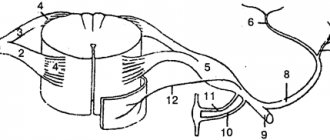
rice.
1. Scheme of the formation of cross-links - the molecular basis of sarcomere contraction Muscle shortening occurs due to the shortening of the sarcomeres that form it, which, in turn, are shortened due to the sliding of actin and myosin filaments relative to each other (and not the shortening of the proteins themselves). The theory of filament sliding was proposed by scientists Huxley and Hanson (Huxley, 1974; Fig. 1). (In 1954, two groups of researchers - H. Huxley with J. Hanson and A. Huxley with R. Niedergerke - formulated a theory explaining muscle contraction by the sliding of threads. Independently of each other, they found that the length of the A disk remained constant in relaxed and shortened sarcomere. This suggested that there are two sets of filaments - actin and myosin, and one fits into the spaces between the others, and as the length of the sarcomere changes, these filaments somehow slide over each other. This hypothesis is now accepted by almost everyone.)
Actin and myosin are two contractile proteins that are capable of entering into a chemical interaction, leading to a change in their relative position in the muscle cell. In this case, the myosin chain is attached to the actin filament using a number of special “heads”, each of which sits on a long springy “neck”. When coupling occurs between the myosin head and the actin filament, the conformation of the complex of these two proteins changes, the myosin chains move between the actin filaments, and the muscle as a whole shortens (contracts). However, in order for a chemical bond between the myosin head and the active filament to form, it is necessary to prepare this process, since in a calm (relaxed) state of the muscle, the active zones of the actin protein are occupied by another protein - tropochmyosin, which does not allow actin to interact with myosin. It is in order to remove the tropomyosin “cover” from the actin filament that a rapid pouring of calcium ions from the cisterns of the sarcoplasmic reticulum is required, which occurs as a result of the action potential passing through the muscle cell membrane. Calcium changes the conformation of the tropomyosin molecule, as a result of which the active zones of the actin molecule open for the attachment of myosin heads. This connection itself is carried out with the help of so-called hydrogen bridges, which very tightly bind two protein molecules - actin and myosin - and are able to remain in this bound form for a very long time.
To detach the myosin head from actin, it is necessary to expend adenosine triphosphate (ATP) energy, while myosin acts as an ATPase (an enzyme that breaks down ATP). The breakdown of ATP into adenosine diphosphate (ADP) and inorganic phosphate (P) releases energy, breaks the connection between actin and myosin, and returns the myosin head to its original position. Subsequently, cross-links can again form between actin and myosin.
In the absence of ATP, actin-myosin bonds are not destroyed. This is the cause of rigor mortis after death, because the production of ATP in the body stops - ATP prevents muscle rigidity.
Even during muscle contractions without visible shortening (isometric contractions, see above), the cross-linking cycle is activated, the muscle consumes ATP and produces heat. The myosin head is repeatedly attached to the same actin binding site, and the entire myofilament system remains motionless.
Attention
: The contractile muscle elements actin and myosin by themselves are not capable of shortening. Muscle shortening is a consequence of the mutual sliding of myofilaments relative to each other (filament sliding mechanism).
How does the formation of cross-links (hydrogen bridges) translate into movement? A single sarcomere shortens by approximately 5-10 nm per cycle, i.e. approximately 1% of its total length. By quickly repeating the cross-link cycle, shortening of 0.4 µm, or 20% of its length, is possible. Since each myofibril consists of many sarcomeres and cross-links are formed in all of them simultaneously (but not synchronously), their total work leads to a visible shortening of the entire muscle. The transmission of the force of this shortening occurs through the Z-lines of myofibrils, as well as the ends of the tendons attached to the bones, resulting in movement in the joints through which the muscles move in space parts of the body or advance the entire body.
Basic truths
Let's start with the basics - in fact, with the classical theory of muscle contraction. The basic contractile unit of muscle tissue is called a sarcomere. The edges of the sarcomere - Z-discs - consist of intertwining strands of various proteins. Actin microfilaments cling to one of these proteins, along which the regulatory proteins troponin and tropomyosin stretch (Fig. 2). Another protein, titin, the largest protein currently known, attaches to the adjacent Z-disk and serves as a long, long scaffold to which the myosin protein molecules bind. Thus, the sarcomere consists of alternating thin (formed by numerous actin molecules and regulatory proteins) and thick (also consisting of numerous myosin molecules and auxiliary proteins) filaments.
|
And now something interesting begins. An impulse approaching the neuromuscular junction causes an increase in intracellular calcium levels. Calcium attaches to regulatory proteins that wrap actin and block it from myosin, as a result of which these proteins are displaced, and the myosin head, containing the products of ATP hydrolysis, adheres to the actin molecule. As a result of various perturbations (which are described in detail below), myosin adheres tightly to actin and changes its conformation, turning the tail relative to the head and spitting out hydrolysis products. This occurs at multiple myosin heads and causes the actin filament to move slightly relative to the myosin filament. Then myosin, tightly coupled to the actin filament, binds to ATP, detaches from actin and undergoes reverse conformational changes - that is, it turns its tail back (Fig. 3).
|
Thus, moving through the heads, myosin molecules ensure the functioning of the muscle. Muscle relaxation occurs when the impulse stops approaching the muscle cell and calcium stops flowing into it. Then the actin and myosin filaments that have become detached from each other gradually return to their original position (partly due to the elastic properties of titin molecules), and the muscle relaxes.
|
In general, the ability of myosin to move along the actin filament is too convenient a property to be used only for muscle contraction. Therefore, many different types of myosin (they are also called “myosin motors”, their phylogenetic tree is shown in Fig. 4) are used by different types of cells for many different functions - in addition to muscle contraction itself, they can provide intracellular transport, move transmembrane proteins, and so on (Fig. . 5).
|
Different myosins differ greatly in structure (Fig. 6): they can be single-headed or double-headed, with long or short tails; however, the main functional part - the head - has almost the same structure in all types of myosin. That is, the principle of myosin operation is the same in all cases, and details (for example, the size of the tail) provide one or another specialization.
|
Myosins are not the only motor proteins. In addition to them, there are two more classes of motors: dyneins and kinesins. Unlike myosins, which move along the actin filament, dyneins and kinesins run along microtubules, with dyneins only in one direction, and kinesins only in the opposite direction.
The relationship between sarcomere length and the strength of muscle contractions[edit | edit code]
rice.
2. Dependence of the force of contractions on the length of the sarcomere. Muscle fibers develop the greatest force of contractions at a length of 2-2.2 microns. With strong stretching or shortening of the sarcomeres, the force of contraction decreases (Fig. 2). This dependence can be explained by the mechanism of filament sliding: at a given sarcomere length, the overlap of myosin and actin fibers is optimal; with greater shortening, the myofilaments overlap too much, and with stretching, the overlap of myofilaments is not sufficient to develop sufficient contractile force.
The effect of stretching on the strength of contractions: stretch curve at rest[edit | edit code]
rice.
4. The effect of pre-stretching on the force of muscle contraction. Pre-stretching increases muscle tension. The resulting curve describing the relationship between muscle length and the force of its contraction when exposed to active and passive stretching demonstrates a higher isometric tension than at rest. An important factor influencing the force of contraction is the amount of muscle stretch. Pulling the end of a muscle and pulling on the muscle fibers is called passive stretching. The muscle has elastic properties, however, unlike a steel spring, the dependence of tension on stretch is not linear, but forms an arcuate curve. As the stretch increases, the muscle tension also increases, but up to a certain maximum. The curve describing this relationship is called the resting stretch curve.
.
This physiological mechanism is explained by the elastic elements of the muscle - the elasticity of the sarcolemma and connective tissue, located parallel to the contractile muscle fibers.
Also, during stretching, the overlap of myofilaments on each other changes, but this does not affect the stretch curve, since at rest, cross-links between actin and myosin are not formed. Pre-stretching (passive stretching) is added to the force of isometric contractions (active contraction force).
Biomechanics of muscle contraction
V. N. Seluyanov, V. A. Rybakov, M. P. Shestakov
Chapter 1. Models of body systems
1.1.5. Biomechanics of muscle contraction
Force
- a vector quantity, which is a measure of the mechanical impact on a material point or body from other bodies or fields (B. M. Yavorsky, A. A. Detlaf, 1974). The force is fully specified if its numerical value, direction and point of application are indicated.
In the theory and methodology of physical education, the physical quality of strength is considered as a person’s ability to overcome mechanical and biomechanical forces that impede action by muscle tension (Zatsiorsky V. M., 1972; L. P. Matveev, 1991).
Muscles can exhibit force: without changing their length (isometric mode), when reducing length (isotonic mode), when lengthening (eccentric mode), when using special equipment, it is possible to maintain an isokinetic mode (during muscle contraction, either a constant speed or force is maintained) .
The strength of a muscle depends on:
- — intensity of activation of the motneuron pool of the spinal cord of a given muscle;
- number of activated motor units and muscle fibers;
- the number of myofibrils in each muscle fiber;
— the rate of myofibril contraction, which depends on the activity of myosin ATPase and the magnitude of external resistance;
- laws of mechanics of muscle contraction (strength - muscle length, strength - contraction speed);
— the initial state of the executive apparatus (tired muscle fibers demonstrate less strength).
When an athlete wants to contract a muscle, he activates the corresponding motor neuron in the cerebral cortex, which sends impulses to the spinal cord to the motor neuron pool that serves this muscle. Since the sizes of motoneurons in the motoneuron pool vary, at a low frequency of impulses from the central nervous system only low-threshold motoneurons can be activated. Each motor neuron innervates its own muscle fibers. Therefore, activation of a motor neuron leads to the recruitment or excitation of corresponding muscle fibers. Each active muscle fiber, under the influence of electrical impulses, releases calcium ions from the SPR, which remove the inhibitor from the active centers of actin. This ensures the formation of actin-myosin bridges and the beginning of their rotation and muscle contraction. The energy of one ATP molecule is spent on turning the bridges and detaching actin from myosin. The duration of the bridge operation is 1 ms. The probability of bridge formation depends on the relative position of the actin and myosin filaments among themselves, hence the force-length relationship of the active muscle arises, as well as on the speed of their mutual movement (sliding) of one relative to the other, respectively, we have the “force-speed” relationship.
The relationship “strength - length of active muscle fiber” is determined, as a rule, by the relative location of myosin heads and actin active centers. The maximum number of bridges occurs at a certain average muscle length. Deviation from this length up or down leads to a decrease in the strength manifestations of the muscle fiber (muscle). However, in the case of stretching of some muscles that are not yet in an active state, significant tensile forces can arise, for example, in the flexor muscles of the ankle or wrist joint. These forces are associated with stretching of connective tissues, such as the peremesium. In biomechanics, in this case, they talk about the parallel elastic component of the muscle. Tendons, z-plates of sarcomeres and myosin filaments to which the heads are attached have elasticity. This elasticity is called the sequential elastic component.
The presence of a consistent elastic component in muscle fibers leads to the fact that with an increase in the number of recruited MVs, muscle stiffness, the elasticity coefficient, increases (V. M. Zatsiorsky et al., 1981).
Stretching the active muscle leads not only to the accumulation of elastic deformation energy in the successive elastic component, but also to the cessation of the work of the bridges, namely, they cease to detach due to the energy of ATP molecules. The rupture of the bridges occurs due to the action of an external mechanical force. As a result, negative muscle work is performed with a very high efficiency, with minimal consumption of ATP, and therefore oxygen.
| 1.1.2 | 1.1.3 | 1.1.4 | 1.1.5 | 1.1.6 | 2.1 | 2.2 | Table of contents |
Sources[edit | edit code]
- Harrison BC. et al. 2011. lib or not lib? Regulation of myosin heavy chain gene expression in mice and men. Skeletal Muscle. 1 (1): 5. doi: 10.1186/2044-5040-1-5.
- Andersen, J. L., et al. 1994. Myosin heavy chain isoforms in single fibers from m. vastus lateralis of sprinters: Influence of training. Acta Physiologica Scandinavica 151(2):135-42.
- ↑ 3,03,1 Andersen TL, Aagaard P. 2000. Myosin heavy chain IIX overshoot in human skeletal muscle. Muscle Nerve. 23 (7): 1095-104.
- Andersen, L.L., et al. 2010. Early and late rate of force development: Differential adaptive responses to resistance training? Scandinavian Journal of Medicine and Science in Sports 20(1): el62-69. doi:10.1111/j.l600-0838.2009.00933.x.
- Anderson, K., and Behm, DG 2004. Maintenance of EMG activity and loss of force output with instability. Journal of Strength and Conditioning Research 18:637–40.
- Aagaard, R, et al. 2011. Effects of resistance training on endurance capacity and muscle fiber composition in young top-level cyclists. Scandinavian Journal of Medicine and Science in Sports 21 (6): e298-307. doi:10.1111/j. 1600-0838.2010.01283.x.
- Henneman, E., Somjen, G., and Carpenter, DO 1965. Functional significance of cell size in spinal motoneurons./. Neurophysiol. 28:560-580.
- ↑ 8.08.1 Latash, ML 1998. Neurophysiological basis of movement. Champaign, IL: Human Kinetics.
- Brooks, G. A., Fahey, T. D., and White, T. P. 1996. Exercise physiology: Human bioenergetics and its applications. 2nd ed. Mountainview, CA: Mayfield.
- Schmidtbleicher, D. 1992. Training for power events. In Strength and power in sport, ed. PV Komi, 381-95. Oxford, UK: Blackwell Scientific.
- Wiemann, K., and Tidow, G. 1995. Relative activity of hip and knee extensors in sprinting—Implications for training. New Studies in Athletics 10(1): 29-49.