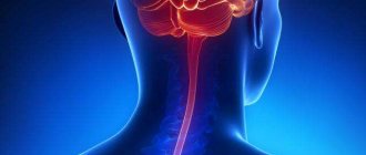
The scalene muscles are 3 large muscles that are located on both sides of the neck. With their upper edge they are attached to the lateral protrusions of the vertebrae (C2, C3, C4) of the cervical spine. With their lower edge they are attached to the 1st and 2nd ribs of the chest. Between the anterior and middle scalene muscles pass large blood vessels (subclavian artery and vein) and a bundle of nerve fibers (brachial nerve plexus).
Rice. 1. Anatomy of the neck - localization of the scalene muscles
Normally, muscles have elasticity, extensibility, are covered with “slippery” fascia and do not compress nerves and blood vessels. With pathological changes in the bone and muscle-tendon system, compression of these important organs of innervation and blood supply occurs, which causes the appearance of scalene muscle syndrome.
Depending on which muscle undergoes changes, a distinction is made between anterior scalene syndrome and posterior scalene syndrome. When nerves and blood vessels are compressed, neurovascular disorders are observed, accompanied by characteristic symptoms.
Symptoms of scalene muscle syndrome
Scalenus syndrome (SMS) may develop first as a myofascial pain syndrome. Due to overexertion or injury, the muscles spasm, structural changes occur in them, inflammation of the neck muscles develops, and trigger points (increased pain sensitivity) are formed. In this case, the main symptom of the pathology is pain. It radiates to the upper limb from the affected side, spreading from the shoulder joint to the tip of the phalanx of the ring or little finger. Often the soft tissues of the occipital part of the head and chest are involved in the pain process. The pain usually gets worse at night or when moving the head, upper limb, or taking a deep breath. The neck muscles hurt in different ways - from a mild nagging pain to a shooting, constant, burning pain.
Rice. 2. Map of the distribution of pain in scalene muscle syndrome
When a nerve bundle is compressed by a spasmodic muscle, the following manifestations are observed:
- a feeling of numbness, tingling, loss of sensitivity in the fourth and fifth fingers, as well as along the inner surface of the hand and forearm;
- swelling above the metacarpophalangeal joints of the II–V fingers and on the dorsum of the hand;
- stiffness of movements in the fingers, worsening in the morning.
When the great vessels are compressed, vascular disorders are observed:
- coldness of the extremity;
- cyanosis;
- numbness;
- swelling;
- disappearance of the pulse in the radial artery when raising the arm up and tilting the head to the affected side (Adson's test).
If lymphatic drainage is impaired, patients develop a swelling in the neck (Kovtunovich's pseudotumor) in the area of the supraclavicular fossa.
With prolonged disruption of innervation and blood supply, an imbalance of trophic processes occurs, which is accompanied by the following symptoms:
- damage to the nail plate – brittleness, unevenness;
- hair loss on the affected limb.
Long-term compression of nerve endings can lead to:
- hypertonicity of the neck muscles;
- heaviness, weakness in the hand;
- incomplete paralysis of a limb;
- atrophy of the muscles of the distal part of the arm.
As a rule, SLM is observed only on one side, much less often on both. The most dangerous complications occur when there is pain in the left neck.
Clinical picture
Signs and symptoms of SVA vary from patient to patient depending on the location of the nerve and/or vessel involved, which may include changes in sensation or mild pain.
Patients with SVA may experience pain or paresthesia in the face, neck, neck, shoulder, chest, and upper extremity. The patient may also complain of altered or absent sensation, weakness, fatigue, and a feeling of heaviness in the arm and hand. The skin may be marbled or pale. There may also be a change in the temperature of the limb.
These symptoms tend to worsen when the arm is placed above the head and rotated outward and the head is turned to the same or opposite side. Activities such as throwing a ball overhead, serving a tennis ball, painting a ceiling, driving a car, or typing can make symptoms worse.
When the superior brachial plexus nerves (C5, 6, 7) are involved, pain may occur in the lateral neck, extending to the ear and face, as well as the interscapular region posteriorly and the infraclavicular region anteriorly. The pain may move down the arm, in the projection of the radial nerve. There may also be headaches.
Patients with inferior brachial plexus nerve disease (C8, Th1) typically have symptoms that are localized to the anterior and posterior regions of the shoulder and extend down the ulnar side of the forearm to the hand, ring finger, and little finger.
There are four categories of SVA, and each has unique signs and symptoms. As a rule, SVA does not correspond to a dermatomal or myotomic pattern unless there is involvement of the nerve roots, which will be important in substantiating the diagnosis and planning treatment.
Arterial SVA
- Young man actively using his hands.
- Pain in hand.
- Pallor.
- Cold intolerance.
- Paresthesia.
- Symptoms appear spontaneously.
Venous SVA
- Young man actively using his hands.
- Cyanosis.
- Feeling of heaviness.
- Paresthesia of the fingers and hand (result of swelling).
- Swelling of the hand.
True SVA
- History of neck injury.
- Pain, paresthesia, numbness and/or weakness.
- Pain in the back of the head.
- Symptoms may occur during the day or at night.
- Impaired fine motor skills.
- Cold intolerance (possibly Raynaud's phenomenon).
- Objective weakness.
- Symptoms worsen during the day*.
Neurogenic SVA
- History of neck injury.
- Pain, paresthesia and a “feeling” of weakness.
- Pain in the back of the head.
- Nocturnal paresthesias, which often “wake up” the patient.
- Impaired fine motor skills.
- Cold intolerance (possibly Raynaud's phenomenon).
- Subjective weakness.
- Symptoms are worse at night**.
* - A patient who experiences symptoms throughout the day while in certain positions that result in increased tension or compression of the superior aperture. The most common aggravating postures are forward head position with prostration and depression of the shoulder girdle or overhead arm activity. These positions cause increased tension/compression, which can result in increased compression or irritation of the neurovascular bundle.
** - A patient who experiences symptoms at night, which may cause insomnia. This is due to the release of tension or compression of the superior aperture, resulting in the restoration of the perineural blood supply to the brachial plexus. This is used as an indicator of favorable outcome and resolution of symptoms.
Complications of scalene muscle syndrome
The most dangerous complications of SLM are:
- thrombosis of the subclavian artery;
- paresis and/or atrophy of the arm muscles;
- violation of the biomechanics of the neck and the whole body.
Large blood vessels pass between the anterior and middle scalene muscles - the subclavian artery and subclavian vein. Spinal vessels and arteries that supply blood to the brain depart from them. Therefore, thrombosis threatens to disrupt the blood supply to the spine, brain and body as a whole. Complaints that the neck is swollen on one side and there is a feeling of heaviness, pain, burning, and stiffness of movement are a signal for immediate hospitalization, surgical care and treatment.
A pinched nerve in the neck area leads to disruption of the innervation of the soft tissues of the hands, paresis and atrophy. If the consequences of atrophy and paralysis of the muscles of the limb are quite clear, then violations of the biomechanics of the neck need to be considered in more detail. When muscles are under prolonged tension, structural changes occur in the fibers - they compress, the muscles shorten and begin to “pull” the head forward. The curvature of the spinal column (lordosis) increases and a violation of the architectonics of the skeleton occurs. In this case, the muscles of the back of the neck and shoulder girdle are tense, and myofascial pain syndrome develops with the corresponding characteristics - trigger points and referred pain.
If the patient does not pay attention to the pain in the neck muscles and ignores treatment, then as a result of the action of compensatory mechanisms, curvature of the spinal column occurs in the lumbar and thoracic region. Changes in vector loads on the vertebrae cause deformation and protrusion of the intervertebral discs, the formation of hernias not only in the cervical region, but also in other areas of the spinal column.
Diagnostics
The main diagnostic difficulties are associated with the similarity of the clinical picture of SLM and vertebrogenic radicular syndrome, and the presence of signs of osteochondrosis in many patients. Incomplete physical examination of the patient leads to erroneous diagnosis of cervical radiculitis and incorrect prescription of treatment. Severe vascular abnormalities initially suggest scalene muscle syndrome. The list of examinations necessary to establish a diagnosis includes:
Examination by a neurologist. There is an antalgic position of the head, limitation of motor volume in the shoulder and cervical spine. Palpation reveals a decrease in the length of the scalene muscle, a high position of the upper rib, and its limited participation in respiratory movements. In the affected limb, hypotension, hyporeflexia, paresis, pain hypoesthesia of the radicular type, and decreased pulsation in the radial artery are detected. Tannotsi and Edson's tests are positive.
Diagnostics of SLM
Due to the variety of manifestations of the syndrome, variability in severity, and lack of characteristic signs, its diagnosis presents some difficulties.
An important diagnostic sign is the condition of the skin, muscles, hair and nail plate. Feeling the neck reveals hypertonicity of the anterior scalene muscle and pain when pressed. A test for spasm is carried out - with maximum rotation in the direction of the lesion and strong pressing of the chin to the collarbone, the spasmed muscle is tensioned and trigger points are activated.
Another indicative test is the Adson test. On the affected side, the patient should raise the limb upward and tilt the head towards this shoulder. With SLM, the pulse in the arm will not be palpable. Specialists at the Manual Medicine Clinic “Galia Ignatieva MD” conduct physical examinations - examination, palpation, skin sensitivity tests of individual parts on the hand. During physical examinations, the doctor may measure temperature and pulse pressure in different arms and in different positions.
In addition to visual examination and physical examinations, the Manual Medicine Clinic “Galia Ignatieva MD” prescribes instrumental methods:
- electromyography (EMG), which allows you to assess the anatomical and functional characteristics of nerves and muscles;
- radiography - to identify anatomical abnormalities, the presence of an additional rib in the upper part of the chest;
- MRI – to visualize the condition of soft tissues, localization of compression of nerves and blood vessels;
- CT – for layer-by-layer visualization of changes in bone tissue;
- Ultrasound, Doppler mapping, angiography - to determine the presence of arterial stenosis and the speed of blood flow in them.
Specialists can also prescribe laboratory tests to identify inflammatory processes, assess blood sugar and hormone levels, which help in the differential diagnosis of SLM.
In the initial stage of SLM, instrumental diagnostic methods are uninformative and can give a false answer. Therefore, the most reliable method for diagnosing the syndrome is manual muscle testing. It allows you to conduct a comprehensive assessment of the condition and physiology, dynamics of movements, and identify the causes of disorders more quickly and informatively than other diagnostic methods.
Main reasons:
Trauma – A traumatic episode may cause damage to the bone or soft tissue at the thoracic outlet. Most people with SLM have a history of one or another episode of an accident, injury at work or at home. Congenital anomalies, such as an extra rib or a tight ligament connecting the spinal column to a rib, can reduce the costoclavicular space.
Poor posture - sagging shoulders or excessive forward tilt of the head can put compression on the area where nerves and blood vessels exit the chest.
Frequent, repetitive movements can wear down the tissues and lead to SLM. An example would be movements associated with raising the arm (the collarbone lowers and the costoclavicular gap decreases), such as swimming, baseball, tennis, weightlifting.
Other causes: Weight gain (pregnancy or obesity), overdeveloped neck muscles (from weightlifting or martial arts), or holding your arms in one position for a long time (working on a computer) can put additional pressure on nerves and blood vessels. Diseases that impair nerve function, such as hypothyroidism and diabetes, may be predisposing factors for neurogenic SLM.
The scalene muscle group consists of three muscles:
- anterior scalene muscle;
- middle scalene muscle;
- posterior scalene muscle.
As a rule, they are attached to the sides of the cervical vertebrae and to the upper ribs. The scalene muscles are responsible for lateral movements of the head. They also move the neck, as well as the respiratory muscles, as they pull the ribs upward.
Symptoms:
- pain in the upper back (especially on the inside of the shoulder blade);
- neck pain;
- pain in the side of the face;
- pain in the upper chest;
- shoulder pain;
- pain in the hands;
- scalene muscle syndrome;
- lump in throat;
- hoarseness;
- temporomandibular joint syndrome.
Massaging trigger points located in the scalene muscle group will help get rid of such problems.
A complex pattern of pain caused by trigger points located in the anterior, middle and posterior scalene muscles. Some trigger points may only have one persistent area of referred pain.
Solid red shows the main zone of pain, grainy the zone of reflected pain.
A trigger point is a point that projects referred pain. J. Travel and Simons in the book “Myofascial pain and dysfunction. A Guide to Trigger Points,” described trigger points as hyperexcitable areas of localized muscle tension located in skeletal muscle and associated fascia.
The emergence and appearance of trigger points occurs during overexertion, prolonged and constant stress on muscles and muscle groups in the body.
Trigger points most often form where physical activity or postural stress causes significant mechanical stress or poor circulation.
These loads are associated with body position: raised shoulders, lowered and tense chest, hunched back, excessive arching in the lower back.
The presence and location of spasmodic muscles influence, through physical and emotional factors, a person’s posture, his ability to move and the ability to act in a physiologically optimal way, while other physiological parameters, blood supply, lymph flow and innervation are also affected.
Treatment of scalene syndrome
After conducting research and establishing an accurate diagnosis, the doctor at the Manual Medicine Clinic “Galia Ignatieva MD” develops an individual SLM therapy plan. Treatment of scalene muscle syndrome is most often carried out using conservative methods:
- medicinal;
- physiotherapy;
- reflexology;
- exercise therapy;
- manual therapy.
Scalenus syndrome, the treatment of which requires symptomatic therapy, involves the use of the following groups of drugs:
- NSAIDs – medications eliminate inflammation and relieve pain;
- muscle relaxants – relax muscles, eliminate hypertonicity, spasm and, as a result, pain;
- opioid analgesics – to eliminate severe pain that is unresponsive to other types of painkillers;
- drugs for neuropathic pain and restoration of nerve structure.
After the severity of the symptoms of SLM has been reduced, physiotherapy methods can be added to the complex effect:
- shock wave therapy;
- magnetic therapy;
- laser therapy;
- DDT – diadynamic therapy;
- electrotherapy;
- vibration impact;
- myostimulation;
- phonophoresis;
- massage.
Physiotherapy eliminates inflammation, pain, activates blood circulation in soft tissues and normalizes trophic processes. In combination with medication, the effectiveness of treatment increases.
Exercise therapy is one of the important elements of the treatment program. Individually selected exercises allow you to:
- improve posture;
- eliminate muscle hypertonicity;
- correctly distribute the load;
- strengthen the muscle corset;
- restore range of motion in the neck and upper limbs.
Drug blockades are prescribed for the purpose of differential diagnosis and treatment of SLM, but due to the proximity of the nerve plexus, the procedure must be performed by a specialist with extensive experience.
Rice. 3. Manual treatment of SLM.
If a nerve in the neck is pinched, a vertebrologist can tell you what to do. The effectiveness of manual therapy at the Clinic of Manual Medicine “Galia Ignatieva MD” in relieving pain, restoring the anatomical and physiological characteristics of structures, eliminating muscle blocks and clamps has been proven by numerous clinical studies.
The specialist not only eliminates the signs and consequences of the pathology, but also acts on the root cause of SLM, since manual therapy has virtually no contraindications, it can be prescribed to patients with drug allergies, children and the elderly. Gentle manual techniques will restore mobility to the arm and neck, activate blood circulation, eliminate congestion, normalize metabolic processes and help remove metabolic products.
The use of surgical treatment is indicated only in exceptional cases - when there is a threat of arterial thrombosis, damage to nerve endings, the presence of an additional rib, etc. When the left neck hurts, the surgeon decides how to treat the anomaly.
Diagnosis and treatment of anterior scalene muscle syndrome at ON CLINIC in Ryazan
During a face-to-face consultation, a neurologist collects an anamnesis of the disease and identifies the reasons that triggered its development. Treatment for patients diagnosed with Natzfinger syndrome consists primarily of relieving them of all bothersome symptoms, namely pain and loss of sensation. The goal of therapy is to restore the motor functions of the upper limb, as well as bring its muscles and blood vessels to a normal state.
If the disease has gone too far, then the only effective way to treat it is surgery.
Prevention of SLM
Specific methods for preventing SLM have not been developed. To prevent the occurrence of pathology, it is necessary to eliminate the causes of SLM:
- avoid hypothermia;
- eat rationally and control body weight;
- lead an active lifestyle - physical inactivity is one of the factors in the development of pathology;
- promptly identify and treat pathologies of the spinal column and musculotendinous system;
- avoid overstraining the muscles of the neck and shoulder girdle;
- train correctly;
- avoid prolonged fixed posture;
- the workplace must be ergonomic.
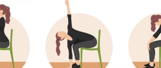
Anyone who has to spend a long time at a computer, desk, or operator console should know how to relieve neck muscle spasms with the help of special exercises. Examples of exercises can be found in specialized literature or shown by a specialist at the Manual Medicine Clinic “Galia Ignatieva MD”.
Manual therapy plays a great role in preventing relapse of SLM. A vertebrologist will show you how to relax your neck muscles. The patient can perform simple exercises and self-massage at home. In difficult cases, the help of a professional with extensive experience is necessary.