Basal ganglia system
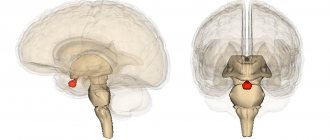
3D image - active brain, side view.
3D image - active brain, bottom view.
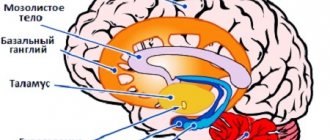
The basal ganglia are a complex of large structures located in the central part of the brain surrounding the central limbic system.
The basal ganglia are involved in the integration of feelings, thoughts and movements, as well as in regulating motor function. The basal ganglia set our body's regime, whether it operates in a state of rest or the level of anxiety. In addition, they help modulate motivation and are likely to contribute to feelings of pleasure and ecstasy. Let's look at each of these functions in more detail.
So, in the basal ganglia there is a process of integration of feelings, thoughts and movements. That is why, when you get excited, you jump up, when you get nervous, you tremble, when you get scared, you freeze in place, or you stand there, unable to utter a word when your boss scolds you. The basal ganglia are responsible for the gradual integration of emotions, thoughts and movements, so when overloaded they simply turn off. One of my patients suffered severe burns in a traffic accident. When he lay on the asphalt, engulfed in flames, those who were nearby froze in horror and could not take a single step to help him. For many years he could not get rid of the unpleasant feeling and understand why no one moved to help him. He was tormented by thoughts: “Did they really care? Am I really unworthy of help? For many years this man lived with the physical pain of his injuries and the mental pain of the indifference he faced. With what relief he learned that it was not a matter of indifference. The emotional shock of the very sight of the accident placed undue stress on the basal ganglia of eyewitnesses. As a result, they simply lost the ability to move, despite the fact that they most likely wanted to help.
When the basal ganglia are overactive (as is the case in people with high anxiety), the likelihood that a stressful situation will be too much for that person increases, and the person and his thoughts will freeze. For people with reduced activity in the basal ganglia (as in people with attention deficit disorder), it is a stressful situation that can push them into action. It is these people who are the first to arrive at the scene of an incident and react quite fearlessly to a stressful situation. For example, one of my friends who has ADD acts much faster in critical situations than I do. (As I mentioned in Chapter 2, I have a congenital overactivity of the basal ganglia.) I remember one day he and I were getting ready to leave a cafe and stood at the cash register to pay our check when suddenly the woman standing in front of us collapsed to the ground. My friend quickly began to help her, but I myself froze motionless from surprise and stress. At the same time, I myself am a doctor, but my friend is not! Previously, due to my inability to react quickly in such moments, I was constantly tormented by feelings of guilt. Subsequently, I realized with relief that my brain, or more precisely, the activity of the basal ganglia, did not allow me to act quickly in critical situations.
The basal ganglia are also responsible for fine motor skills, coordination, writing, etc. Let's look again at attention deficit disorder as an example. Many ADD patients, both children and adults, have terrible handwriting. The process of writing itself is difficult for them. Many adults and teens with ADD prefer to write in print because it does not require long, continuous, fluid movement. Many people with ADD complain that they have difficulty formulating their thoughts and putting them on paper. We know that medications that help with ADD are the psychostimulants Ritalin, Dexedrine or Adderall, which stimulate the basal ganglia to produce the neurotransmitter dopamine. After taking these medications, there is often a significant improvement in handwriting and the ability to express one's thoughts in writing. In addition, patients themselves say that taking these drugs generally improves their motor coordination. As an example, here are two samples of handwriting from the same patient, Tommy (14 years old), before and after treatment.
You can better understand what motor functions the basal ganglia are responsible for by looking at two diseases: Parkinson's disease and Tourette's syndrome. Parkinson's disease is caused by a deficiency of dopamine in the basal ganglia. It is manifested by hand tremors, muscle rigidity, difficulty moving, poor facial expressions, awkwardness and decreased motor activity. Often, the use of dopamine-stimulating drugs such as I-dopa significantly relieves these symptoms, allowing patients to move more easily and smoothly. The basal ganglia are also responsible for suppressing unwanted motor activity. When this part of the brain becomes abnormal, the risk of developing Tourette's syndrome, which is a combination of motor and vocal tics, increases. We will return to consider this condition later.
Based on the brain images obtained, we see that the basal ganglia are responsible for controlling the body's resting state or anxiety levels. Increased activity in the basal ganglia is often associated with anxiety, tension, wariness, and increased fear. Lack of basal ganglia activity causes problems related to motivation, energy, and ability to function.
Interestingly, among the most highly motivated individuals (such as CEOs) who underwent scanning, we noted high activity in this part of the brain. Thus, we can assume that in some cases, increased activity of the basal ganglia stimulates motivation, due to which these people become the “drivers” of society. Let's say my mother, who, like me, has slightly increased activity in the basal ganglia, can be a little anxious at times, but she is also a very active woman. She plays golf four or five times a week, has raised seven children without any apparent stress, and is always busy doing things for others. I believe that by using the excess energy generated by the increased activity of the basal ganglia in this way, it prevents the development of anxiety.
Another interesting finding is that the basal ganglia appears to be involved in controlling feelings of pleasure. At the Brookhaven National Laboratory in New York, a team led by Nora Volkova examined brain images to understand which centers were affected by cocaine and Ritalin. Both substances are absorbed primarily by the basal ganglia. Cocaine develops addiction, but Ritalin, in doses prescribed for ADD, does not. This study allowed us to confidently answer the question of why this happens. Cocaine greatly increases the uptake of dopamine by brain tissue. The concentration of this substance rapidly increases, and then just as rapidly decreases. A person who uses cocaine gets a wave of pleasure, but then it goes away, and then he (or she) wants to relive the feeling. Unlike cocaine, Ritalin, which also provides an influx of additional dopamine into the basal ganglia, acts more mildly, and after its use, dopamine levels decrease more slowly. Volkova's group hypothesized that cocaine cravings are triggered and perpetuated by activation of the basal ganglia. Ritalin, in turn, increases motivation, the ability to concentrate and focus for long periods of time, but does not cause the “coming” and desire to take more of it. (We do not consider cases where a person uses this drug in doses significantly higher than those used in clinical practice.) Thus, addiction does not occur. In fact, when I prescribe this drug to teenagers with ADD, I often find that they simply forget to take it.
Intense love affects the brain much like cocaine, releasing a powerful hit of dopamine in the basal ganglia. Love actually produces a distinct physical effect. I once scanned the brain of my friend Bill shortly after he met his lover. He was, as they say, head over heels in love with her. After the third date, which they spent on the ocean shore in each other’s arms, he came to my work to tell me about his new love. He was so happy that his state resembled euphoria. And it just so happened that while Bill was sitting with me, our technician came to me and said that we had an extra dose of the isotope left, and therefore, if I need to do an unscheduled scan, then there is such an opportunity. I already had a picture of my friend's brain that I took for the control group, which contains pictures of normal brain function. And then I decided to take a second shot of him. What I saw shocked me, without exaggeration. His brain looked as if he was under the influence of a large dose of cocaine. Activity in both the left and right basal ganglia was extremely high, almost like during a seizure. Love has the same powerful effect on the brain as drugs.
Basal ganglia: functions, norm and pathology
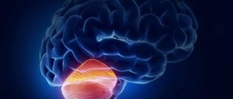
In order for the human body to carry out complex motor programs every day, there are specialized structures of the brain, such as the cerebellum and basal ganglia, which are in close connection with areas of the cerebral cortex.
While the cerebellum ensures the synchronization of movements and their instantaneous compliance with requirements, the basal ganglia allow the body to plan complex motor programs, and most importantly, to implement them. In addition to the regulation of movements, the subcortical nuclei are involved in the cognitive activity of the brain, and therefore in the formation of emotions. These are the ones that will be discussed in this article.
To better understand the mechanisms of normal and pathological processes, it is first worth considering the structure of the subcortical nuclei: their location and the formation of numerous connections with the thalamus and cortical areas. From an anatomical point of view, the basal ganglia include the caudate nucleus, putamen, globus pallidus and pelvis. These four nuclei make up the striatum. However, the often used concept of “striatum” includes only two formations - the putamen and the caudate nucleus. These formations are located mainly lateral to the thalamus and occupy most of the internal regions of the cerebral hemispheres.
All information that enters the basal ganglia in the form of signals is distributed independently of each other along special parallel information processing pathways. These pathways form functional circles, which are also independent and include different regions of the cortex. Therefore, the specific role of the nuclei is determined precisely by that area of the cortex that is located in the same functional circle with the nucleus.
The basal ganglia complex receives motor excitatory signals from the premotor areas of the cortex, processes them and again returns them to the cortex, but to the primary motor area. The “intention” for movement arises in the premotor cortex, from where the striatum receives motor commands. Thanks to this connection, the basal ganglia are able to “turn on” motor behavioral programs even before they occur. At the same time, the programs themselves are already laid down in the ganglia, and they only have to make a decision - what action to resort to and whether to resort to it at all. The execution of complex motor acts triggered by the cortex is ensured using a direct nerve pathway. Its function is as follows: the basal ganglia facilitate actions dictated by the cortex and suppress unnecessary accompanying ones.
All information that comes to the basal ganglia is collected from several regions of the cortex. Sensitive fibers form excitatory glutamatergic synapses with striatal neurons, which are combined into functional modules that process information using the same type of mechanisms. Since motor and sensory fibers differ in the nature of the information they transmit using an impulse, the modules to which the fibers are directed will be different. For this reason, bundles of fibers heading to different modules form separate stripes, from which the striatum got its name (Latin corpus striatum - striatum).
Neurons of the striatum, in turn, form GABAergic synapses with cells of the inner segment of the globus pallidus and part of the substantia nigra (the first sequential inhibitory switching). Neurons of these structures form inhibitory GABAergic synapses on the anterior and ventral nuclei of the thalamus (second sequential inhibitory switching), which leads to constant inhibition of the passage of excitation from the thalamus to the cortex. The processes of neurons in the thalamic nuclei, which are directed to the cortex, form the main efferent glutamatergic pathways. Therefore, when the internal segment of the globus pallidus and part of the substantia nigra are suppressed, the excitatory effect of the thalamus on the cortex increases, which makes it easier to perform the movement.
Since the transfer of information between brain structures is ensured by chemical receptors, the issue of constant synthesis of the mediator is especially important. The role of the supplier of biologically active substances in this case is played by the black substance, which received its name because of the pigment - neuromelanin, which gives it the appropriate color. The substantia nigra produces dopamine, which works as an excitatory neurotransmitter and also serves as an important part of the brain's reward system.
Excitatory dopamine receptors of type D1 are localized on the neurons of the direct pathway, due to which chemical signals from the negative situation excite this pathway. The phasic and tonic components of human movements depend on the ratio of dopamine and glutamate concentrations. With the predominance of dopamine, the phasic, fast component of movement is strengthened, with the predominance of glutamate, acetylcholine and GABA - the tonic, slow component. Along with the strengthening of the phasic component, the speed of movements also increases, but there is a decrease in tone.
To maintain the normal functioning of the motor systems, it is necessary to maintain both the anatomical integrity of the pathways and maintain a certain level of the transmitter. Accordingly, damage to structures or excess/lack of neurotransmitter entails serious consequences. When the basal ganglia are damaged, motor activity disorders occur - dyskinesia (hypokinesis or hyperkinesis) and changes in muscle tone (hypotonia or muscle rigidity). With functional disorders of the globus pallidus, spontaneous and often constant wave-like movements of the hand, arm, neck or face are observed. Such movements are called athetosis.
Damage to the subthalamic nucleus (also related to the basal ganglia) leads to sweeping movements of the entire limb. This condition is called hemiballismus. Multiple small lesions in the putamen lead to rapid twitching in the hands, face and other parts of the body, which is called chorea. Lesions of the substantia nigra lead to a widespread and extremely severe disease associated with akinesia and tremor. This disease is known as Parkinson's disease.
The main clinical manifestations of Parkinson's disease are hypokinesia and muscle rigidity. Hypokinesia manifests itself in a very slow performance of active motor actions: the beginning of a motor act is difficult, there is no cooperative movement of the upper limbs - synkinesis, they are motionless when walking (acheirokinesis). Muscular rigidity is a kind of resistance to passive movements; it appears not only in the initial phase of movement, but also in all subsequent phases of muscle stretching. The limb seems to freeze in the position that is given to it.
In addition to the above-described manifestations, a mask-like face is also observed - amicable, with a fixed gaze, rare blinking, sometimes absent for several minutes, and pale gesticulation. Hypokinesia and rigidity can be observed in isolation, but they are often accompanied by hyperkinesis in the form of tremor of the fingers (like counting coins), the chin area and lower extremities. Despite the fact that tremor is one of the clinical signs of parkinsonism, its pathogenesis remains unclear. Although from a neurological point of view, unlike other motor signs, tremor has precise electrophysiological characteristics (frequency, phase and power).
The goal of various clinical trials is to find the relationship between tremors and dopamine. However, it was found that changes in tremor characteristics in response to the action of dopaminergic drugs are quite variable. These unique features of tremor and new neuroimaging techniques are driving new research into this pathology.
The article by Helmich RC, devoted to the cerebral basis of the occurrence of parkinsonian tremor, discusses in detail the hypotheses of its occurrence due to increased interaction between the subcortical nuclei and the cerebellar-thalamo-cortical circuit. Typically, the strengthening of these connections is caused by an increase in the number of dopaminergic receptors in the nuclei due to the influence of various factors (for example, psychological stress).
The authors of the article also discuss in detail models that help understand the pathogenesis of tremor. One example is the “dimmer-switch” model, according to which cerebral activity associated with tremor in Parkinsonism first arises in the basal ganglia and then increases and spreads to the cerebellar-thalamo-cortical circuit (spread is ensured by the activation of excitatory synapses).
In addition to the well-known function of the basal ganglia, which is to control motor activity, there are also less studied, but no less interesting “responsibilities” of these subcortical structures.
In their paper, Lukas Maurer and colleagues proposed a concept that describes disturbances in “brain circuits” to explain a variety of neuropsychiatric diseases. The diseases are characterized by pathological changes in the structure of neural networks, including changes in oscillatory signaling of cortical-subcortical circuits in the basal ganglia system. Some of these circuits play a significant role in maintaining the body's energy balance. Therefore, the article focuses on the relationship between obesity and changes in oscillatory signaling of limbic corticobasal circuits.
The scientists performed repeated electrophysiological recordings of action potentials on the membranes of neurons in this corticobasal circuit. The test subjects were live rats under urethane anesthesia that were fed a HFD (high fat diet) for four weeks prior to the study. Potentials were recorded both in the absence of an external stimulus and under the influence of glucose. Analysis of the findings demonstrates increased beta activity in the NAC nucleus accumbens, associated with decreased coherence between this nucleus and the cortex in animals on a HFD.
Thus, it can be concluded that spontaneous beta activity is strongly correlated with endocrine indicators of obesity. However, the glucose challenge increased beta activity in those animals not on the HFD. Moreover, intraventricular administration of insulin also increased NAC activity. Thanks to the study, it was possible to confirm the hypotheses about the presence of a correlation between the limbic corticobasal loop, obesity and serum insulin levels. Therefore, insulin resistance and obesity can be considered as consequences of oscillatory disturbances in the cells of the limbic corticobasal circuit.
This circuit plays a role in the brain that is responsible for processing information related to food intake and reward. In addition, it is inextricably linked with the hypothalamic regions of the midbrain, which regulate the homeostatic functions of the body. In a comparison of the two patients, the obese individual had increased activity in corticobasal structures during food anticipation and decreased activity when receiving a reward. In accordance with this, obesity can be considered as a neuropsychiatric disorder similar to drug addiction, since it is also based on addiction, only in this case it is food addiction.
Having considered two completely different functions of the basal ganglia, we can conclude how complex and intricate the nature of any brain structure is. But it is precisely this complexity that will continue to attract a person seeking to learn new things.
Sources:
- Helmich RC The cerebral basis of Parkinsonian tremor: A network perspective //Movement Disorders. – 2021.
- Maurer L. et al. High-fat diet-induced obesity and insulin resistance are characterized by differential beta oscillatory signaling of the limbic cortico-basal ganglia loop //Scientific Reports. – 2021. – T. 7. – No. 1. – P. 15555.
- Erofeev N. Physiology of the central nervous system: textbook. – Litres, 2021.
- Guyton A.K., Hall D.E. Medical physiology. – Logobook. ru, 2008.
The role of the basal ganglia and cerebellum in the regulation of movements.
The constituent components of the basal ganglia are the caudate nucleus and putamen, which together form the striatum (neostriatum), the globus pallidus (pallidum), and the fence (claustrum).
The basal ganglia system is connected to the substantia nigra of the midbrain and the subthalamic nucleus. The input to the basal ganglia system is from the association cortex to the neurons of the striatum. The cortex has an activating effect on the striatum (mediator glutamic acid). The striatum is connected to the globus pallidus. From the globus pallidus, information enters the ventrolateral thalamus (motor nuclei of the thalamus), and from them to the motor cortex. The striatum has a inhibitory effect on the globus pallidus. The globus pallidus is a repository of programs for slow stereotypies of the axial muscles. The substantia nigra has an inhibitory effect on the striatum (transmitter dopamine). Thus, the basal ganglia system does not receive afferent input from skeletal muscle receptors and is not directly connected to skeletal muscle.
Functions of the basal ganglia
1. Based on basic programs, they participate in the formation of slow stereotypical movements.
2. Engage in motivational behavior.
3. Participate in the change of the sleep-wake cycle of the cortex.
4. Participate in the formation of conditioned reflexes.
Basal ganglia dysfunction
1. Akinesia - it is difficult to start a movement and it is difficult to finish it, rigidity - long-term maintenance of a posture, resting tremor, face in the form of a mask, constrained gait.
2. Hyperkinesis - obsessive uncontrolled movements.
Postnotonic reflexes according to Magnus.
Studies of the role of the brain stem in the regulation of muscle tone and body posture were carried out on thalamic cats, i.e., transection was carried out above the level of the diencephalon. Receptors of the vestibular apparatus, proprioceptors of muscles, joints and ligaments of the neck area, and mechanoreceptors of the skin are involved in the occurrence of reflexes. Signals from these receptors arrive to the nuclei of the brain stem - the red nucleus, Deiters nucleus, and the nuclei of the reticular formation.
1. Static reflexes are a reflex redistribution of muscle tone when the position of the head relative to the body changes. Observed during movements without acceleration, i.e. standing, sitting, lying down. Function: maintaining balance when changing the position of the head in space. They are divided into two groups: position reflexes (posture-tonic) and straightening reflexes.
a) Position reflexes: cervical tonic - when the position of the head changes and the receptors in the neck are excited, the tone of the muscles of the fore and hind limbs changes:
- turning the head to the side leads to an increase in muscle tone of the extensors on the side of rotation and the flexors on the contralateral side;
- when the head is tilted down, the tone of the flexors of the forelimbs and extensors of the hind limbs increases;
- when the head is tilted back, the forelimbs straighten (increased tone of the extensors), the hind limbs bend (increased tone of the flexors);
- in the supine position, the extensors are activated, the flexors are inhibited;
- in the prone position, the flexors are activated and the extensors are inhibited.
b) Rectifying reflexes appear when the body posture changes. Developed sequentially:
- position the head with the crown up, eyes along the horizon;
- straightening the body;
— installation of limbs (redistribution of tone).
2. Statokinetic reflexes develop under the action of angular accelerations. Rotation of the torso or head leads to activation of the semicircular canal receptors and rotation of the head and eyes in the direction opposite to the rotation. They are important for maintaining orientation in space.
Under the action of linear accelerations, the receptors of the otolithic apparatus are activated. The influence of this afferentation can be seen in the so-called elevator reflexes:
a) when the elevator moves down, the tone of the extensors increases;
b) when the elevator moves upward, the tone of the flexors increases.
These reflexes are involved in maintaining balance when running and jumping.