Our specialists
Tarasova Svetlana Vitalievna
Expert No. 1 in the treatment of headaches and migraines. Head of the Center for the Treatment of Pain and Multiple Sclerosis.
Somnologist.
Epileptologist. Botulinum therapist. The doctor is a neurologist of the highest category. Physiotherapist. Doctor of Medical Sciences.
Experience: 23 years.Derevianko Leonid Sergeevich
Head of the Center for Diagnostics and Treatment of Sleep Disorders.
The doctor is a neurologist of the highest category. Vertebrologist. Somnologist. Epileptologist. Botulinum therapist. Physiotherapist. Experience: 23 years.
Bezgina Elena Vladimirovna
The doctor is a neurologist of the highest category. Botulinum therapist. Physiotherapist. Experience: 24 years.
Palagin Maxim Anatolievich
The doctor is a neurologist. Somnologist. Epileptologist. Botulinum therapist. Physiotherapist. Experience: 6 years.
Mizonov Sergey Vladimirovich
The doctor is a neurologist. Chiropractor. Osteopath. Physiotherapist. Experience: 8 years.
Drozdova Lyubov Vladimirovna
The doctor is a neurologist. Vertebroneurologist. Ozone therapist. Physiotherapist. Experience: 17 years.
Zhuravleva Nadezhda Vladimirovna
Head of the center for diagnosis and treatment of myasthenia gravis.
The doctor is a neurologist of the highest category. Physiotherapist. Experience: 16 years.
Causes of hemifacial spasm
There are several causes of hemifacial spasm. Typically, hemispasm is caused by compression of the facial nerve root by a vessel (most often, this is the anterior inferior cerebellar artery, but compression of the root by the posterior inferior cerebellar artery, superior cerebellar artery, vertebral artery, labyrinthine artery, basilar artery, and also branches of the anterior inferior cerebellar artery is possible). Less commonly, compression by vascular malformations or veins is possible. The cause of hemifacial spasm can sometimes be benign tumors or cysts in the cerebellopontine angle, foci of demyelination in multiple sclerosis, adhesions, bone deformities, as well as tumors of the 4th ventricle, brain stem, and cerebellum.
Read also
Hyperhidrosis
Hyperhidrosis is a condition that is manifested by excessive, inadequate sweating due to increased activity of the sweat glands.
It happens that a person sweats so profusely that his clothes get wet very quickly, from his hands… Read more
Spasmodic torticollis (cervical dystonia)
This is a chronic disease that occurs as a result of impaired muscle tone in the neck and is manifested by incorrect head position or forced rotation of the neck muscles. According to statistics, this disease...
More details
Torsion dystonia
This is a chronic nervous disease that manifests itself in the form of uncontrolled tonic muscle contractions, which can result in pathological postures and hyperkinesis. Torsion dystonia…
More details
Blepharospasm
Blepharospasm is a chronic disease characterized by involuntary squinting of the eyelids due to increased tone of the orbicularis oculi muscles. Most often, the causes of blepharospasm are extremely difficult to establish or...
More details
Treatment of spasticity
Spasticity is an excessive increase in muscle tone, the cause of this condition can be a stroke, traumatic brain injury, spinal injury, spinal cord problems, neuroinfection, multiple…
More details
Facial hemispasm
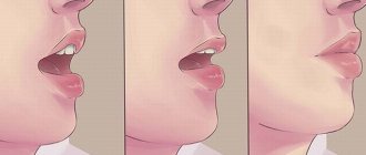
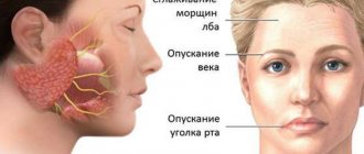
Facial hemispasm (hemifacial spasm, Brissot's disease) is a symptom complex of unilateral hyperactive dysfunction of the facial nerve, which develops as a result of compression of the facial nerve root in the area of its exit from the brain stem. It is characterized by the appearance of moderate and intermittent painless convulsions (tonic or clonic) in the orbicularis oculi muscle, gradually progressing in frequency and strength of contractions with the gradual involvement of all facial muscles. The duration of the attack can reach several hours. As the disease develops, a tonic half-mask appears on half the face: narrowing of the palpebral fissure, tightening of the nasolabial fold, tension of the subcutaneous muscle of the neck. Sometimes the attack is interrupted by pressing on the brow ridge or other part of the face. There are typical hemifacial spasms (starts in the orbicularis oculi muscle and then involves all the underlying facial muscles of half the face) and atypical (starts from the muscles of the cheek and gradually involves all the overlying facial muscles). Hemispasm can be combined with trigeminal neuralgia, and unilateral dysfunction of the auditory nerve is possible.
The prevalence of the disease is 8 patients with hemifacial spasm per 1 million population. Women are 2 times more likely to get sick.
Pathogenesis
Typically, the cause of facial muscle twitching is compression of the facial nerve root by a vessel. Most often this is the anterior inferior cerebellar artery, less often - other intracranial arteries. In isolated cases, compression by vascular malformations or veins is possible. With hemifacial spasm, the vessel compresses the complex of the facial and auditory nerves. Rare causes of hemifacial spasm are considered to be benign tumors or cysts of the cerebellopontine angle, multiple sclerosis, cicatricial adhesions of various origins, and bone deformities of the temporal bone pyramid. An even rarer cause of hemifacial spasm is Chiari malformation.
Diagnostics
The diagnosis of hemifacial spasm is made based on the patient’s complaints and the clinical picture of the disease. Hemifacial spasm usually begins from the lower eyelid on one side of the face, and as the disease progresses, it gradually spreads to the muscles of the cheek, chin and neck; At the beginning of the disease, convulsions are clonic, then they acquire a tonic-clonic character.
The value of instrumental research methods is insignificant, and only allows one to exclude or confirm the presence of a space-occupying formation of the cerebellopontine angle: the area in the cranial cavity where the root of the facial nerve is located. MRI allows us to identify the close location of the vessel and the facial nerve, while the neurovascular conflict in patients suffering from facial hemispasm is not always detected.
Treatment
Traditionally, vascular and anticonvulsant drugs are used, the effect of which is observed in approximately 20% of patients. At the onset of the disease, it is often possible to conduct courses of acupuncture, which in some cases provide a short-term positive effect.
Recently, symptomatic treatment through regular (2-3 times a year) injection of botulinum toxin into the facial muscles has become increasingly common. The therapeutic effect of botulinum toxin injections of varying severity lasts up to 18 weeks, so patients need to repeat them. In this case, the time intervals between injections, during which there are no twitches of the facial muscles, can gradually decrease.
If conservative treatment of hemifacial spasm is ineffective, surgical treatment is used: vascular decompression of the facial nerve root using microsurgical techniques, which in most cases eliminates the cause of the disease and achieves significant relief of symptoms or recovery.
Treatment of clonic hemifacial spasm
Treatment is prescribed only after confirmation of the diagnosis by a medical specialist. Anticonvulsants, muscle relaxants, antidepressants, botulinum toxin injections are indicated. Psychotherapy, electrical stimulation, massage, radiotherapy are carried out; if compression of the facial nerve is detected, surgical treatment is performed.
| Injection points for botulinum toxin preparations |
The clinical effect appears within 7 days, the maximum effect within 2 weeks.
Essential drugs
There are contraindications. Specialist consultation is required.
- Botox (muscle relaxant). Dosage regimen: superficially 15-50 units intramuscularly into the orbicularis oculi muscle. Total dose for 12 weeks. should not exceed 100 units.
- Dysport (muscle relaxant). Dosage regimen: subcutaneously 40-125 units into the junction between the preseptal and orbital parts of both the inferior and superior orbital muscles of each eye. Administration of the drug should be repeated every 8 weeks. or depending on the clinical situation.
- Amitriptyline (sedative, antidepressant drug). Dosage regimen: taken orally, without chewing, immediately after meals at a dose of 25-100 mg/day. (at night). After achieving a therapeutic effect, switch to the minimum effective dose of 10-50 mg/day.
- Carbamazepine (anticonvulsant). Dosage regimen: orally at an initial dose of 0.1 g for 4-5 days, then the dose is increased to 0.4-1.2 g/day. After 3-4 weeks. the dose is reduced to 0.1-0.2 g per day.
The main causes of bruxism in adults and jaw strain
- psycho-emotional reasons: nervous overstrain, prolonged stress, difficult experiences of situations,
- dental reasons: prolonged absence of teeth, malocclusion, diseases of the temporomandibular joint, complete or almost complete absence of teeth, inconvenient and poorly made crowns and dentures, fillings that are too high, injuries to the dental system,
- disorders of the musculoskeletal system: injuries and curvature of the spine, poor posture, asymmetry in the work of the core muscles,
- neurological causes: epilepsy, birth trauma, brain and central nervous system damage,
- other reasons: taking certain medications, smoking, abuse of caffeine-containing drinks.
“We always take a comprehensive approach to solving problems of hypertonicity of facial muscles. First of all, we look for causes from the maxillofacial apparatus - we carry out a full diagnosis and examination, evaluate the functioning of the masticatory muscles, identify the patient’s characteristic habits and collect a medical history. If necessary, we refer the patient to specialized specialists - a neurologist, psychotherapist, etc. It is very important to cure hypertonicity - at least so that the patient in the future can safely begin high-quality dental restoration: implantation, prosthetics or treatment.”
Aida Vladimirovna Jutova, implant surgeon, periodontist, work experience of more than 9 years make an appointment
Diagnostics
The diagnosis is made on the basis of complaints, anamnesis, clinical data and additional examination methods by a neurologist. In case of hearing impairment, consultation with a related specialist - an otorhinolaryngologist is required.
Additional research methods include:
Electromyography. Registers an abnormal muscle response to stimulation of the branches of the facial nerve.- Magnetic resonance imaging of the brain. Allows you to identify pathological formations.
- Cerebral MR angiography. With its help, you can trace the topography of anatomical structures relative to each other, in particular this concerns neurovascular conflict.
- Differential diagnosis is carried out with idiopathic blepharospasm, the main difference of which is the displacement of the eyebrow closer to the orbit when the eyelids close. HFS is also differentiated from psychogenic spasm, facial myokymia and paroxysms of focal epilepsy.
Clinical picture
The typical form of HFS begins with a spastic contraction of the muscle located near the eye. The patient notes an increase in the number of blinks, and after some time the nerve impulses spread to the remaining muscles of the corresponding half of the face. The squeezing of the eyes can be so intense and frequent that a person sometimes loses the ability to see with that eye. In the atypical form, the process begins with contraction of the lips and cheeks, then the orbital and frontal areas are involved. The pathology does not cause pain.
Attacks of spastic contractions can occur suddenly, without previous factors. But still, in most cases they are provoked by stress and neuropsychic overstrain.
Unlike other neurological disorders, hemifacial spasm does not go away during sleep or at rest. Babinski's sign is the main symptom accompanying HFS: the closure of the eyelids on the affected side occurs simultaneously with the raising of the eyebrow.
Hemispasm of the facial nerve is a chronic process, as a result of which a persistent decrease in its function develops. Neuralgia is observed in 5% of patients, and hearing loss is observed in 15%.
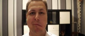
What are the symptoms of hemifacial spasm?
The twitching usually starts in the eye. At first they arise and go away on their own. Gradually they become stronger and become permanent, involving more and more muscles on one side of the face. Most often these are the muscles of the jaw and mouth. The corner of the mouth may be pulled up during permanent spasm. The left side is involved more often than the right. Naturally, constantly closing your eyes can affect your vision. The spasm can get worse when the person is nervous or tired, and it always improves when lying down.
Symptoms of hypertonicity of the masticatory muscles and bruxism
- the chewing surfaces of the teeth become worn and flat (the cutting edges disappear),
- tooth enamel chips, wedge-shaped defects appear, fillings fly out, teeth quickly decay, become loose, gums hurt,
- you experience discomfort when opening your mouth, while eating or yawning - there are clicks, crunching sounds,
- you or your loved ones notice that you are grinding your teeth - the characteristic grinding appears mainly at night (the so-called “night bruxism”), when it is difficult for a person to control himself during sleep,
- cramps the jaw - you cannot fully open or close your mouth, the jaw seems to be blocked,
- it is difficult for you to keep your mouth open for a long time, overexertion and pain appear during bruxism,
- when talking or chewing, you feel that your facial muscles are “clogged”, tired,
- when you are angry, tense or just working in concentration - your mouth is tightly closed, your lips are pursed, your teeth are closed, you are literally clenching your jaw,
- there are problems with your bite – your teeth don’t fit together correctly, your jaws are misaligned relative to each other, causing you to bite your cheeks or tongue,
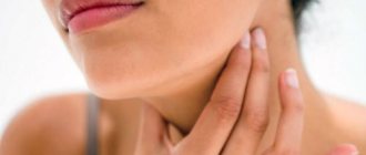
- after concentrated work or under stress, you often have a pressing sensation in your temples, pain and dizziness,
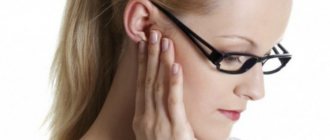
- you notice ringing and noise in your ears,
- Over time, the lower half of your face has become visually square, heavy,
- there are problems with posture - you are slouched, your lower back hurts, your neck is stiff, one shoulder is higher than the other.
If you find one or more signs in yourself, then this is a reason to consult a specialist for medical help. And the first step is a comprehensive diagnosis and search for the causes of hypertension.
We approach the treatment of bruxism comprehensively! We carry out in-depth diagnostics, find the exact cause of facial muscle spasm and eliminate it using advanced methods - safely and with high results.
Free consultation
Treatment
Treatment of hemifacial spasm is carried out comprehensively. Drug therapy and surgical interventions are used. In rare cases, pharmacological drugs cause side effects, and operations cause relapse.
Therapeutic treatment includes the prescription of antiepileptic drugs, but the results of long-term use of medications have not been well studied. Botulinum toxin injections are preferred; in the early stages of the disease they are successful in 75% of cases.
Among the surgical techniques are:
Removal of tumor and tumor-like formations. It is carried out at the level of microsurgery. The duration of the operation depends on the volume of the tumor and its location.- Interventions on pathologically altered vessels. An aneurysm of an artery or vein is eliminated by closing its neck, that is, clipping. This method helps not only to eliminate HFS, but also to prevent rupture of the vessel wall with subsequent hemorrhage.
- Microvascular decompression. Surgery is often performed in the presence of neurovascular conflict. A Teflon protector is installed between the artery and nerve in contact. In rare cases, such an operation may damage nearby nerves, but over time, their functions are restored. Relapse after microvascular decompression is observed in 15-20% of cases.