Binswanger's disease – is one of the subtypes of discirculatory encephalopathy - a large group of neurological diseases caused by chronic hypoxia of brain tissue. Usually develops against the background of long-term progressive hypertension and atherosclerosis. The disease is accompanied by various clinical symptoms: deterioration of cognitive functions, motor and muscle disorders. The disease can be treated only in the early stages, while pathological changes in the nerve tissue of the brain are reversible. In the later stages of the disease, therapy is reduced to maintaining basic functions, round-the-clock care and monitoring the patient’s condition. Doctors at the Leto mental health center will conduct a comprehensive examination of the patient and prescribe the correct treatment. If necessary, it is possible to be hospitalized in the hospital of our clinic, where the person is guaranteed due attention from doctors and medical staff, comfort and care from a doctor.
Etiology and pathogenesis
In more than 95% of cases, the underlying cause of Binswanger's disease is hypertension. Although it still remains a mystery to neurologists why some people suffer from progressive symptoms of encephalopathy, while others do not have any changes in intelligence, motor coordination, etc. until old age.
It is believed that the risk of developing such a disease is especially high if:
- sudden changes in blood pressure (BP);
- frequent hypertensive crises;
- episodes of hypertension during night rest (normally, pressure should be at least 10% lower than physiological);
- hereditary predisposition (stroke, chronic hypertension, cognitive disorders in close relatives);
- changes in the rheological properties of blood (increased levels of hematocrit, fibrinogen, blood viscosity, which leads to increased platelet aggregation on the inner surface of the vascular endothelium and narrowing of the lumen of cerebral vessels);
- old age, age-related cerebral amyloidosis;
- diabetes mellitus, which is especially difficult to correct with medication;
- overweight, alcohol abuse, drug use;
- vasculitis (inflammatory process in the vascular endothelium) of an autoimmune or infectious nature;
- mechanical compression of cerebral vessels by a tumor, compression due to spinal curvature, osteochondrosis;
- hematological pathologies (congenital or acquired).
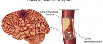
The pathogenesis of Binswanger's disease is based on atherosclerotic damage to the vascular network supplying blood to the white matter of the brain. Periodic fluctuations in blood pressure lead to cerebral circulation disorders and lacunar infarctions - small (up to 15 mm) foci of necrosis.
Publications in the media
This article provides brief information about a number of diseases that do not form separate articles. See also Various syndromes.
Alexander's disease (203450, r) is a severe neurodegenerative disease of the central nervous system manifested in children by a lag in psychomotor development, convulsive seizures, paralysis, megaloencephaly in combination with extensive leukodystrophy (decay of myelin), especially of the frontal lobes, and progressive hydrocephalus. Symptoms are similar to Canavan disease. In both disorders, marked changes in astrocytes are observed. Rosenthal fibers are commonly found in astrocytomas, optic nerve gliomas, and chronic reactive gliosis, but are particularly prominent in Alexander disease. Laboratory: Rosenthal fibers (homogeneous eosinophilic masses that form elongated narrow fibers up to 30 µm in length) are scattered throughout the cortex and white matter of the brain, most numerous perivascularly and in the subependymal areas. Synonyms: Rosenthal disease, Rosenthal leukodystrophy. Note. Hypoproconvertinemia (227500, hemocoagulation factor VII deficiency) is also known as Alexander WS. ICD-10. G31.8 Other specified degenerative diseases of the nervous system.
Balo disease (periaxial concentric encephalitis) is a demyelinating disease, clinically similar to Schilder’s disease (central paralysis and paresis, hyperkinesis, epileptiform seizures, visual disturbances, mental disorders), but distinguished by the presence of concentrically located ring-shaped areas of altered nervous tissue in the white matter of the brain. ICD-10. G37.5 Concentric sclerosis [Balo].
Binswanger disease is an organic dementia that develops due to cerebral atherosclerosis and cerebral hypertension, small focal lesions, recurrent edema of the white matter, secondary demyelination “Binswanger encephalopathy” Binswanger atherosclerotic encephalopathy “Binswanger syndrome.” ICD-10. F01.8 Other vascular dementia.
Vincent's disease is an acute, sometimes recurrent pathological lesion of the gums, characterized by ulceration and necrosis of the gingival margin and destruction of the gingival papillae; usually a consequence of the symbiosis of the spindle-shaped rod (Plaut-Vincent) Fusobacterium fusiforme and the spirochete (Vincent) Treponema vincentii “ulcerative gingivitis” ulcerative membranous gingivitis. ICD-10. A69.0 Other infections caused by spirochetes; necrotizing ulcerative stomatitis.
Granulomatous chronic disease - congenital (forms and genes: • autosomal, cytochrome b negative form: CYBA, 233690, 16q24; • deficiency of neutrophil cytosolic factor NCF 1: NCF1, 233700, 7q11.23; • deficiency of neutrophil cytosolic factor NCF 2: NCF2, 233710, 1q25; • X linked, CYBB, CGD, 306400, Xp21.1) impaired digestion of phagocytosed bacteria by polymorphonuclear leukocytes, leading to decreased resistance to infections (see also hereditary agranulocytosis) “Congenital dysphagocytosis. ICD-10. D83.0 General variable immunodeficiency with predominant abnormalities in the number and functional activity of B cells.
Darier's disease is a hereditary dermatosis (*124200, 12q23–q24.1, DAR gene, Â), characterized by a disorder of the keratinization process of the dyskeratosis type. Clinical picture: multiple miliary follicular hyperkeratotic papules, grouped into large plaques with a warty or papillomatous surface; mental retardation, possible development of psychoses and mood disorders. Synonyms: Daria vegetans follicular psorospermosis, follicular dyskeratosis, follicular ichthyosis, keratosis follicular vegetans. ICD-10. L87.0 Keratosis follicular and parafollicular, penetrating the skin [Kierle disease].
Decompression sickness is a symptom complex caused by the release (from physiological fluids) of nitrogen bubbles (dissolved at high pressure); occurs with a sudden decrease in pressure, characterized by headache, pain in the arms, legs, joints and epigastrium, itching of the skin, dizziness, shortness of breath, cough, suffocation, vomiting, weakness, in severe cases - paralysis, peripheral circulatory collapse. Treatment: • 100% oxygen through a mask • fluid replacement (avoid the use of 5% glucose and hypotonic solutions in cases of spinal cord damage) • prompt referral to the hyperbaric department. ICD-10. T70.3 Caisson sickness [decompression sickness].
Demyelinating diseases are a group of diseases morphologically characterized by demyelination, i.e. destruction of the myelin sheaths of nerve fibers; etiology is unclear (multiple sclerosis, acute disseminated encephalomyelitis, acute hemorrhagic leukoencephalitis, adrenoleukodystrophy, neuromyelitis optica), congenital metabolic disorders (aminoaciduria, Tay-Sachs disease, Guillain-Barré syndrome, etc.), acquired disorders (trauma, ischemia, toxic effects). ICD-10. G35–G37 Demyelinating diseases of the central nervous system • Acute disseminated encephalomyelitis is an acute demyelinating disease of the central nervous system that occurs after a viral, less often bacterial infection (post-infectious encephalomyelitis), vaccination (post-vaccination encephalomyelitis - smallpox, rabies vaccines) or spontaneously and is characterized by the formation of perivenular foci of inflammation and demyelination with variable general and local symptoms; Treatment mainly consists of GC therapy. ICD-10. G04.0 Acute disseminated encephalitis • Acute hemorrhagic leukoencephalitis is an acute demyelinating disease characterized by the formation of perivenular foci of demyelination, inflammatory infiltration, vasculitis, necrosis of the vessel walls, fibrin deposition and numerous hemorrhages in the white matter of the hemispheres, spinal cord trunk and spinal cord; the clinical picture resembles acute disseminated encephalomyelitis; prognosis is usually poor, death occurs within 2–4 days; treatment mainly involves GC therapy. ICD-10. G36.1 Acute and subacute hemorrhagic leukoencephalitis • Opticomyelitis - a type of acute disseminated encephalomyelitis with predominant damage to the optic nerves and spinal cord (thoracic, less often cervical segments); clinically manifested by blindness, central scotoma, focal symptoms reminiscent of multiple sclerosis; the disease occurs spontaneously, after infectious diseases or as a sign of multiple sclerosis; treatment consists mainly of GC therapy. Synonyms • Devika disease • Devika syndrome. ICD-10. G36.0 Neuromyelitis optica (Devic's disease).
Dandy-Walker disease (*304340, Xq25–q27, r) - developmental anomalies of the fourth ventricle in the form of cerebellar hypoplasia (Dandy-Walker malformation, see article), hydrocephalus, atresia of the foramen of Luschka and Magendie. Characterized by mental retardation, spasticity, choreoathetosis, convulsive seizures, hearing defects, numerous and varied symptoms due to defects in the development of the brain, facial and trunk skeleton, and internal organs. The disease often ends in death in the newborn period. ICD-10 • Q03.1 Atresia of the foramina of Magendie and Luschka • Q04.8 Other specified congenital anomalies of the brain.
Dercum disease. Multiple painful lipomas and/or foci of diffuse fat deposition are formed in the subcutaneous tissue; it occurs mainly in women (5:1) in menopause, accompanied by adynamia, asthenia, and depression. Synonyms • Adiposis dolorosa • Adiposalgia • Dercum syndrome • Lipalgia • Painful lipomatosis • Painful obesity. ICD-10. E88.2 Lipomatosis, not elsewhere classified.
Dyggwe–Melchior–Clausen disease (*223800, r). Clinical picture: disproportionate dwarfism, protruding chin, microcephaly, protruding sternum, barrel chest, emphasized lordosis, osteochondrodysplasia, platyspondyly, claw-shaped hand, general limitation of joint mobility, subluxation of the hips, gait disturbances, mental retardation (not always), spinal cord compression . Laboratory data: hyperexcretion of mucopolysaccharides in the urine. ICD-10. Q87.1 Syndromes of congenital anomalies manifesting predominantly as dwarfism.
Seitelberger disease (neuraxonal infantile dystrophy, *256600, r) is clinically similar to Hallervorden-Spatz disease. Distinctive signs are keratitis, urinary retention, primary hypothyroidism, diabetes insipidus and pathology of the thermoregulatory center, the presence of spherical bodies within the hypothalamic-pituitary region and the Auerbach plexus. ICD-10. G31.8 Other specified degenerative diseases of the nervous system.
Calve's disease is osteochondropathy of the vertebral body. The lower thoracic and upper lumbar vertebrae are most often affected. Clinical picture: back pain that disappears at rest and appears during physical activity, tension in the back muscles, protrusion of the spinous process of the affected vertebra is often observed. X-ray picture: uniform flattening of the vertebral body, sometimes with a small wedge anteriorly. In the later stages of the disease, if no treatment has been carried out, only a spotted shadow of the vertebra is revealed on radiographs. Treatment lasts several years • Complete unloading of the spine (bed rest, reclination), physiotherapy, massage, exercise therapy • Sanatorium-resort treatment (staying in a bone-tuberculosis sanatorium is recommended), vitamin therapy, ultraviolet irradiation, wearing a corset. ICD-10. M43.0 Spondylolysis.
Caroli's disease (*263200, mutation of the PKHD1 gene [600643, fibrocystin, Â]). Clinically: congenital polycystic dilatation of the intrahepatic bile ducts, cholangitis, liver abscesses, epigastric pain, vomiting, hepatomegaly, bleeding from varices of the esophagus and stomach, portal hypertension, association with polycystic kidney disease, repeated episodes of hyperthermia. Diagnostics. CT for visualization of bile ducts. Treatment • Antibacterial therapy to reduce the frequency and intensity of episodes of cholangitis • Liver transplantation. ICD-10. Q44.5 Congenital anomalies of the gallbladder, bile ducts and liver—Other congenital anomalies of the bile ducts.
Keller I disease is osteochondropathy of the navicular bone of the foot. The predominant age is 3–10 years. The predominant gender is male. Clinical picture • The disease is usually bilateral, lasts for 1 year • Pain in the tarsus, aggravated by pressure, as well as at night, sometimes swelling • Lameness, the child walks with support on the outer arch of the foot. The X-ray picture shows a decrease in the bone core, fragmentation and flattening of the scaphoid bone, widening of the joint space. Treatment is conservative: wearing a plaster boot, physiotherapy, massage, exercise therapy. ICD-10. M92.6 Juvenile osteochondrosis of the tarsus.
Keller II disease - osteochondropathy of the heads of the II and III metatarsal bones. The predominant age is 10–20 years. The predominant gender is female. Clinical picture • The disease progresses slowly, lasts several years and often ends with the appearance of deforming arthrosis • Pain at the base of the 2nd and 3rd toes occurs spontaneously and intensifies while walking • Swelling is often detected in the initial stages. X-ray picture - compaction, spotty pattern, flattening of the heads of the metatarsal bones, widening of the joint space. Treatment • Providing rest for the limb - immobilization, wearing orthopedic shoes • Surgical treatment - resection of the head of the metatarsal bone (arthroplasty). ICD-10. M92.7 Juvenile osteochondrosis of the metatarsus.
Koenig's disease is a limited aseptic necrosis of the articular epiphysis. The necrotic area is rejected into the joint cavity in the form of an articular mouse. The knee joint is most often affected (necrosis of the internal femoral condyle), less often the elbow, hip, and ankle joints. The predominant age is 15–30 years. The predominant gender is male. Clinical picture. There are two stages of the disease: • Stage I - intermittent pain, discomfort. Radiologically, in the area of the internal condyle of the femur, a focus of clearing with a bone body located inside is revealed • Stage II - clinic of entrapment, blockade of the knee joint. The limb remains in a fixed position, often in a flexion position. Free fluid is detected in the joint cavity. Radiographs show an “empty niche”, a foreign body in the joint cavity. treatment • In the initial stages, resection of the focus of osteonecrosis is recommended • If a fragment is rejected into the joint cavity - removal of the foreign body • In the postoperative period - immobilization with a plaster splint, physiotherapy (UHF in an oligothermic dosage, Bernard currents, from 6-7 days - exercise therapy). Synonym: Dissecting osteochondrosis of the hip and knee joints. ICD-10. M93.2 Osteochondritis dissecans.
Kienbeck's disease is osteochondropathy of the lunate bone of the hand. The risk factor is chronic microtrauma. The predominant age is 20–40 years • The predominant gender is female. Clinical picture. Pain in the area of the lunate bone, sharply intensifying with pressure and movements in the wrist joint; edema. X-ray picture. Induration, spotty pattern and reduction in size of the lunate. Treatment • Prolonged immobilization, physical therapy • Surgical treatment (removal of the lunate bone) is indicated when conservative treatment is ineffective and pain continues. ICD-10. M92.2 Juvenile osteochondrosis of the hand.
Maple syrup disease (leucinosis) is a severe hereditary disease (acidosis, vomiting, hypoglycemia, seizures, mental and physical retardation, specific smell of urine). The incidence of the disease is approximately 1:300,000. Biochemistry: several defects in the subunits of the mitochondrial enzyme complex, branched-chain a-ketoacid decarboxylase (EC 1.2.4.4, forms and genes) leading to the development of the disease are known: • type Ia: BCKDHA, MSUD1, 248600, 19q13 .1 q13.2 • type Ib: BCKDHB, E1B, 248611, 6p22 p21 • type II: DBT, BCATE2, 248610, 1p31). Thiamine is effective in some forms. ICD-10. E71.0 Maple syrup disease.
Canavan disease (*271900, EC 3.5.1.15, mutations in the ASPA aspartoacylase [aminoacylase 2] gene, 17pter p13, r) manifests itself in early childhood with increasing paralysis and blindness; megaloencephaly, severe demyelination and formation of cavities in the cerebral hemispheres. Clinically: atony of the neck muscles, hyperextension of the joints of the legs and arms, severe retardation in intellectual development, megaloencephaly, blindness with onset in infancy, death usually in the first 18 months. Laboratory findings: spongy degeneration of white matter, pathology of astrocyte mitochondria, increased levels of acetylaspartate in urine, cerebrospinal fluid and blood plasma. Synonyms: spongy white matter degeneration of the brain, Canavan–Van Bogaert–Bertrand disease, spongiform degeneration of the central nervous system. ICD-10. G37.8 Other specified demyelinating diseases of the central nervous system.
Kümmel's disease is post-traumatic osteochondropathy of the vertebrae. Clinical picture. There are three stages of the disease: • I – 5–8 days after injury: pain in the area of the affected vertebra • II – several months: asymptomatic period • III – persistent pain in the area of the affected vertebra, kyphosis. X-ray: wedge-shaped decrease in vertebral height. Treatment: unloading of the spine, warm baths, back massage, exercise therapy, Bernard currents, electrophoresis with calcium chloride, mud applications, wearing a corset. Synonym: traumatic spondylitis. ICD-10. M48.3 Traumatic spondylopathy
Lafora's disease - myoclonus epilepsy (254780, MELF locus, 6q23 q25), starting at 11–18 years of age; progressive mental disorders, EEG changes in the posterior parts of the brain, PAS positive inclusions in the brain, myocardium, liver, skin (myoclonic bodies, or Lafora bodies); death occurs within 10 years from the onset of the disease. ICD-10. G40.8 Other specified forms of epilepsy.
Leva disease - blockade of the His bundle branch in patients with healthy myocardium and coronary arteries; the result of fibrosis or calcification of the conduction system and involvement of the membranous septum, apex of the muscular septum, mitral and aortic valve rings; polymorphic and polytopic extrasystoles, fainting; Several forms are described. ICD-10. I44.6 Other and unspecified bundle blockades.
Lenegra disease is dystrophic and sclerotic changes in the conduction system of the heart of unknown etiology in the absence of damage to the rest of the myocardium and coronary arteries, manifested by disturbances in atrioventricular and intraventricular conduction. ICD-10. I45.8 Other specified conduction disorders.
Lee's disease is a hereditary (À, r, Â) demyelinating disease with progressive degeneration of the white matter of the brain and spinal cord; manifested by pyramidal and extrapyramidal disorders, cranial nerve dysfunction, and myocardial damage. ICD-10. G37.8 Other specified demyelinating diseases of the central nervous system.
Letterer–Siwe disease (*246400, r) is an acute progressive histiocytosis in young children, characterized by fever, hemorrhagic purpura, enlarged lymph nodes, infiltration in the lungs, hepatosplenomegaly with jaundice, pancytopenia; in the red bone marrow, liver and spleen there are a significant number of histiocytes “Reticulohistiocytosis is non-lipoid. ICD-10. C96.0 Letterer–Siwe disease.
Lindau disease (*193300, 3p26–p25, VHL gene defect, B) - multiple retinal hemangiomas in combination with hemangioma or hemangioblastoma of the cerebellum and the wall of the fourth ventricle of the brain (sometimes with damage to the spinal cord); cysts or hamartomas of the kidney, adrenal gland and other organs are possible. Clinical picture • Headache • Progressive visual impairment • Unilateral ataxia • Dizziness • Retinal detachment • Papilledema • Retinal hemangiomas • Subarachnoid hemorrhages • Polycythemia • Hemangiomas of the lungs, adrenal glands, cerebellum, spinal cord, liver • Malignant tumors of the pancreas, hemangioblastoma, pheochromocytoma, hypernephroma, epididymal tumor • Multiple renal cysts • Hypercalcemia and hypokalemia in pheochromocytoma. Treatment • Surgical • Radiation therapy. Synonyms • Cerebroretinal angiomatosis • Angioblastomatosis • Angiophakomatosis of the retina and cerebellum • Von Hippel–Lindau disease • Von Hippel–Lindau syndrome. ICD-10. D18.0 Hemangioma of any location. Note. Mutation of the VHL gene also leads to the development of kidney carcinoma.
Leiner's disease (227100, r) is a combination of generalized erythroderma, diarrhea, delayed physical development, increased susceptibility to infections due to hypoplasia of lymphoid tissue, which leads to death in early childhood.
Marchiafava–Binjami disease is a degenerative process in the corpus callosum that occurs primarily in alcoholism. Pathogenesis is progressive degeneration of the corpus callosum. Characteristic signs: progressive dementia, confusion, hallucinations, tremors, rigidity and convulsions. The course is usually chronic and progressive. Synonyms: Marchiafava syndrome, encephalopathy with demyelination of the corpus callosum. ICD-10. G37.1 Central demyelination of the corpus callosum.
Merzbacher-Peliceus disease is a hereditary (312080, PLP, PMD [lipophilin], Xq22] degenerative disease of the brain; it is based on progressive sclerosis of the white matter of the frontal lobes and vasomotor disorders. Types • Classic - nystagmus and tremor appear in the first months of life, delayed motor development, sometimes choreoathetosis, spasticity, seizures, death at an early age • Type 2 - death within months and tens of months after birth • Type 3 - transitional form, death within the first decade • Type 4 - adult form Clinically : nystagmus , optic nerve atrophy, neonatal hypotonia, ataxia, spastic paraparesis, dementia, muscle rigidity and tremor, microcephaly, respiratory distress syndrome. Synonyms: diffuse familial cerebral sclerosis, sudanophilic leukodystrophy, Pelizaeus-Merzbacher leukodystrophy. ICD-10. G37.8 Others specified demyelinating diseases of the central nervous system Note: Mutations in the lipophilin gene also lead to the development of spastic paraplegia type 2 (312920).
Machado-Joseph disease (*109150, 14q24.3–q32, MJD gene [SCA3], Â). Clinically: ataxia, parkinsonism, dystonia, muscle fasciculations, absence of normal leg reflexes, cerebellar tremor, extensor plantar reflexes, exophthalmos, limitation of eye movements, nystagmus, muscle atrophy, diabetes, onset after 40 years. Laboratory: neuronal death and gliosis in the substantia nigra, pontine nuclei, vestibular and cranial nerve nuclei, Clark's columns and anterior horns of the spinal cord, abnormalities on the electrooculogram. Synonyms: spinocerebellar ataxia type 3, spinocerebellar atrophy type 3, Azores disease, spinopontine atrophy, nigro-spino-dentate degeneration. Note: Machado and Joseph are the names of people from the Azores, in whose offspring the disease was first described. ICD-10. G23.2 Striatonigral degeneration.
Pyla's disease (*265900, r). Clinically: metaphyseal dysplasia, valgus deformity of the knee joints, Erlenmeyer flask-shaped femurs, moderate involvement of the skull bones, abnormally wide proximal part of the humerus, ulna and radius. ICD-10. Q78.5 Metaphyseal dysplasia.
Parry-Romberg disease (progressive hemiatrophy of the face) is a gradually developing atrophy of all tissues of one half of the face, homolateral atrophy of the vocal cord, larynx, tongue, hair loss (including eyelashes and eyebrows). Usually occurs between the ages of 10 and 20 years. Treatment: plastic reconstructive surgery; skin grafting; subcutaneous fat grafting; for pain resembling that of trigeminal neuralgia, carbamazepine is prescribed. Synonyms: Romberg's disease, Romberg's syndrome.
Periodic disease is a hereditary disease (249100, 16p13, r) observed in Armenians and Jews. Clinically: short-term and recurrent attacks of fever with pain in the abdomen, chest and joints; erythema similar to that of erysipelas; the course is relapsing. A complication is amyloidosis, which develops even in the absence of the described attacks. Treatment: colchicine. Synonyms: familial paroxysmal polyserositis, Janeway-Mosenthal paroxysmal syndrome, periodic peritonitis, Reimann's disease, Segal-Kattan-Mamu syndrome. ICD-10. E85.0 Hereditary familial amyloidosis without neuropathy.
Peak disease (172,700). Total presenile dementia with “disintegration” of speech is a rare progressive destructive disease of the brain; Clinically similar to Alzheimer's disease, it occurs with disturbances in logical thinking and perception, apathy, amnesia, aphasia, and extrapyramidal disorders. Pathologically: cortical atrophy is limited to the frontal and temporal lobes, unusual inclusions in neurons. The course is usually chronic and progressive. Synonyms: limited presenile brain atrophy, Pick syndrome. ICD-10. G31.0+ F02.0* Limited cerebral atrophy + dementia.
Pulsating muscles 1 disease (*600332; 1q41, RMD1 gene, Â). Myotonia with muscle hypertrophy, different from Thomsen's congenital myotonia (160800). Clinically: myotonia, muscle hypertrophy, cramps, pain, rigidity, rotational muscle contractions (in severe cases). ICD-10. G71.1 Myotonic disorders.
Refsum disease is a peroxisomal storage disease (accumulation of phytanic acid). Genetic classification • Juvenile form (*266510, PEX1, r) • Classic form (*266500, PHYH, PAHX, 602026, 10pter–p11.2, r) • Refsum disease with pipecolatacidemia (*600964, r). Clinically: retinitis pigmentosa, miosis, ptosis, ataxia, anosmia, deafness, demyelinating polyneuropathy, increased protein content in the cerebrospinal fluid, ECG changes, ichthyosis; in the juvenile form, mental retardation is pronounced, facial dysmorphism (flat face), hepatomegaly, steatorrhea, and osteoporosis are characteristic. A diet excluding phytanic acid and plasmapheresis stop the progression of neurological disorders. Synonym: phytanic acid storage disease. ICD-10. G60.1 Refsum disease
Family Byler disease (*211600, 18q21–q22, FIC1, r gene defect) is a progressive familial intrahepatic cholestasis type 1. Clinically: large, loose, foul-smelling stools, episodes of jaundice (possibly associated with infection), hepatosplenomegaly, Kayser-Fleischer ring, growth retardation, death between 17 months and 8 years of age. ICD-10. Q44.7 Other congenital liver abnormalities.
Silverscheld's disease is a chondrodysplasia characterized by shortening of the proximal and middle segments of the limbs, thickening of the epiphyses, curvature of the spine, dwarfism, clubfoot and saddle nose. ICD-10. Q77.8 Other osteochondrodysplasia with growth defects of long bones and spinal column.
Tangier disease (*205400, post-translational modification defect, r) is an inherited deficiency of HDL in combination with a low content of apolipoprotein A1 (synthesis of the latter is not impaired), accumulation of foam cells containing cholesteryl esters, enlargement and pronounced hyperemia of the tonsils, hepatosplenomegaly, lymphadenopathy, hypercholesterolemia. Other clinical manifestations • Cardiac - increased risk of coronary artery disease • Neurological •• recurrent neuropathy •• loss of pain and temperature sensitivity in the distal extremities • Muscular - progressive muscle weakness • Ophthalmological •• decreased visual acuity •• incomplete closure of the eyelids •• keratopathy. Synonym: Analphalipoproteinemia. Note. Tangier, an island in the Chesapeake Bay (Virginia, USA), where the disease was first recorded. ICD-10. E78.6 Lipoprotein deficiency
von Graefe disease is a series of inherited nosological units in the form of a slowly progressive bilateral myopathy affecting the muscles of the eyeball. Clinically: progressive ptosis, bilateral external ophthalmoplegia, weakness of the muscles of the face, pharynx, tongue, shoulder girdle. Synonyms: Graefe myopathy, progressive external chronic ophthalmoplegia, ophthalmoplegic myopathy. ICD-10. H49.4 Progressive external ophthalmoplegia.
Hallervorden–Spatz disease (*234200, 20p13–p12.3, defect of the NBIA1, r gene) is a pathological process of demyelination of nerve fibers connecting the striatum and the globus pallidus. Clinically: aphasia, movement disorders, chorea, athetosis, dystonia, muscle rigidity, mental retardation, hyperkinesis, gait disturbance, dysphagia, convulsions, torticollis, tremor, dental deformation, facial grimaces, optic atrophy, nystagmus, retinitis pigmentosa, blepharospasm, hyperpigmentation skin, bedsores, aspiration pneumonia. Laboratory: pathology of the basal ganglia on MRI. Treatment: levodopa, tryptophan, large doses of vitamins. ICD-10. G23.0 Hallervorden–Spatz disease.
Hand–Schüller–Christian disease is histiocytosis, characterized by the formation in the bones, skin, lymph nodes, bone marrow and internal organs of foci of cell proliferation (presumably sedentary macrophages) with an increased content of cholesteryl esters in the cytoplasm; clinically manifested by diabetes insipidus, exophthalmos, and foci of bone destruction (especially the skull). Synonyms: Schüller's disease, Schüller's syndrome, histiocytosis X, lipoid granulomatosis, Christian-Schüller's disease, lipogranulomatosis. Note. The disease was first described in 1865 and 1876 by the English pathologist Thomas Smith; Hand A. in 1893 described polyuria in combination, as he believed, with tuberculous bone lesions; Schüller A. in 1915 described two cases of damage to the skull bones in Hand–Schüller–Christian disease; Christian G. in 1919 provided a complete description of Hand–Schüller–Christian disease. ICD-10. D76.0 Langerhans cell histiocytosis, not elsewhere classified.
Chagas disease is an acute, subacute or chronic form of trypanosomiasis (pathogen: Trypanosoma cruzi). Acute forms develop after bites from vectors—predatory bugs of the genus Triatoma. Epidemiology: endemic areas - Central and South America. Clinical picture: at the site of pathogen penetration there is a skin tumor (shagoma), often on the face with regional enlargement of the lymph nodes; 1–3 weeks after the bite - injection of the sclera, swelling of the eyelids; fever; the chronic form is characteristic only of adults, who were usually ill in childhood (manifested by degenerative changes in internal organs). Research methods: microscopy of blood smears, serological methods. Treatment: symptomatic. Synonyms: South American trypanosomiasis, Chagas-Cruz disease. ICD-10. B57 Chagas disease.
Scheuermann–Mau disease is osteochondropathy of the apophyses (growth zones) of the vertebrae, leading to curvature of the spine (juvenile kyphosis), one of the most common diseases of the spine in children. Clinically: fatigue, thoracic kyphosis, pain, aggravated by straightening the spine and applying pressure. X-ray: wedge-shaped deformation of the vertebrae, disturbances of growth plates. Treatment: bed rest on a hard bed with a shield, in the acute stage - a plaster bed, exercise therapy to strengthen the muscles of the back and abdomen, physiotherapy, electrophoresis with calcium chloride, cocarboxylase, ozokerite and mud applications. ICD-10. M42.0 Juvenile osteochondrosis of the spine.
Clinical manifestations
Lacunar infarctions arising against the background of Binswanger's disease are accompanied by:
- deterioration of tactile sensitivity and motor skills of the right or left hand;
- attacks of dizziness, severe weakness and clouding of consciousness;
- headache.
The listed symptoms are reversible. As a rule, clinical manifestations gradually increase over 3–6 days and then decline. Sometimes lacunar infarctions are practically asymptomatic and unnoticed by the person himself.
The danger is posed by repeated focal lacunar infarctions, which lead to symptoms typical of Binswanger's disease. This:
- A gradual increase in cognitive dysfunction up to the development of dementia, which is accompanied by memory loss, personality degradation , the formation of delusional ideas (usually persecution, harm, hypochondriacal content), hallucinosis, replacement of true memories with false ones, etc.
- Walking disorders. In the initial stages of the disease, a shuffling, unsteady gait and swaying when walking are noted. As the condition worsens, such pathological changes progress to the point of complete inability to walk or even stand. In this case, the sensitivity of the body and limbs can be preserved, and the functions of the cerebellum are not lost.
- Caused by neurological pathology, periodic urinary incontinence , which in the later stages of the disease is transformed into a total loss of control over urination and defecation.
- Emotional-volitional disorders: apathy, indifference, coldness towards other family members, deterioration of intellectual abilities, thirst for knowledge, etc.
- Speech and hearing impairments due to pathology of the innervation of the muscles of the palate, vocal cords, and tongue.
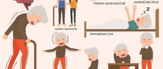
Initial symptoms of pathology: when to see a doctor
- memory deterioration: at first a person cannot remember and reproduce recent events and information, but gradually the patient begins to forget what happened much earlier;
- frequent falls;
- sleep disorders;
- headache, accompanied by ringing and noise in the ears, blurred vision;
- weakness, decreased performance, increased fatigue, intolerance to physical and mental stress;
- imperative urge to urinate, involuntary loss of urine;
You should also pay attention to excessive irritability, mood swings (but depression, despondency, melancholy usually predominate), distractibility, and forgetfulness. Such manifestations are especially dangerous with concomitant hypertension.
Forms of vascular dementia
The brain provides us with conscious existence, being responsible for mental, emotional, and adaptive processes. Its substance is strictly structured. Each of its departments is responsible for its functions.
However, any part of the brain can undergo destructive changes, as a result of which a certain type of activity is disrupted. Based on this, several forms of the disease are distinguished, differing not only in location, but also in the caliber of the affected vessels.
Dysmnestic or lacunar dementia occurs against the background of destruction of small-diameter vessels. As a result, multiple infarct foci appear in the thickness of the white and gray matter. This is the most classic variant of the course of the disease, in which all pathological manifestations are not clearly expressed. There is a measured decrease in intellectual abilities, mild memory impairment, and slight slowness of psychomotor skills.
The multi-infarction form is accompanied by damage to vessels of medium diameter, and usually develops in non-acute pathological processes. Its manifestations are insignificant and go unnoticed for a long time even by the patient himself. Its course is gradual. The disorders first progress and then freeze at a certain stage until the next micro-stroke. Among the symptoms of this type of disease, cognitive impairment comes to the fore. Neurological and emotional disorders gradually develop.
Subcortical vascular dementia is a disease of small vessels, against which atrophy of white matter cells occurs with the formation of ischemic areas. Scientists see the reason for this process in the accumulation of amyloid in the walls of the arteries, followed by its inflammation. The clinical picture of the disease is somewhat atypical. It can occur as Alzheimer's disease or as isolated dementia.
Autoimmune vasculitis, such as systemic lupus erythematosus and panarteritis, cause another form of the disease - cerebral vasculitis. It is expressed by dementia and confusion. As a rule, it affects patients over 50.
Mixed dementia combines two forms: vascular and atrophic, that is, Alzheimer's type. Therefore, in the picture of the disease one can observe symptoms of both vascular dementia and Alzheimer's disease, but the latter prevail over the former.
Diagnostics
The only reliable way to confirm the suspected diagnosis is to do neuroimaging using CT or MRI. The following changes are clearly visible on the tomogram results:
- leukoaraiosis: decreased density of the white matter of the brain;
- multiple foci of lacunar infarctions;
- expansion of the cerebral ventricles and subarachnoid space.
The following are recommended as auxiliary examination methods:
- Dopplerography and/or duplex scanning of cerebral vessels;
- electrocardiography and Echo-CG;
- daily blood pressure monitoring;
- general clinical blood and urine tests (lipid profile, coagulogram required).
Additionally, psychological testing is carried out to determine cognitive and intellectual functions, memory, and the ability to evaluate and analyze what is happening. It is also recommended to conduct research to identify markers of hereditary diseases that can cause similar symptoms.
Treatment
Patients need supervision by cardiologists, neurologists, psychiatrists, endocrinologists, therapists, and all specialists.
In case of Binswanger's disease, two goals are set before treatment - to improve the patient's quality of life and to stop the progression of encephalopathy. Patients are hospitalized, but the main treatment is carried out on an outpatient basis.
Drug treatment is based on restoring normal blood pressure and restoring neurocognitive functions. Galantamine, Memantine and Donepezil are used for this. The emotional sphere is restored with the help of antidepressants.
- Treatment is prescribed based on the stage, degree and etiology of the disease. The therapy is aimed at etiological and symptomatic factors. When selecting medications, the emotional state, stage of the underlying disease, and the patient’s well-being are taken into account.
- An important factor in correcting the patient’s condition is control of arterial hypertension. The selection of antihypertensive therapy is very important, since a sharp decrease in pressure and its surges should not be allowed. The pressure should not drop more than 120/95 mm. rt. Art. as this can lead to a sharp deterioration in the patient's condition.
- Patients with atherosclerotic encephalopathy are prescribed anticoagulants and drugs that improve brain trophism. In severe depressive states, the patient is treated by psychiatrists, prescribing nootropic drugs and antidepressants.
- Statins, drugs that lower blood cholesterol levels, are used in the treatment of such patients. In the later stages of the disease, paliotropic therapy is used. The main task of the attending physician during this period is to alleviate the patient’s condition.
Cost of services
| CONSULTATIONS OF SPECIALISTS | |
| Initial consultation with a psychiatrist (60 min.) | 6,000 rub. |
| Repeated consultation | 5,000 rub. |
| Consultation with a psychiatrist-narcologist (60 min.) | 5,000 rub. |
| Consultation with a psychologist | 3,500 rub. |
| Consultation with Gromova E.V. (50 minutes) | 12,000 rub. |
| PSYCHOTHERAPY | |
| Psychotherapy (session) | 7,000 rub. |
| Psychotherapy (5 sessions) | 30,000 rub. |
| Psychotherapy (10 sessions) | 60,000 rub. |
| Group psychotherapy (3-7 people) | 3,500 rub. |
| Psychotherapy session with E.V. Gromova (50 minutes) | 12,000 rub. |
| TREATMENT IN A HOSPITAL | |
| Ward for 4 persons | 10,000 rub./day |
| Ward for 3 persons | 13,000 rub./day |
| Ward 1 bed VIP | 23,000 rub./day |
| Individual post | 5,000 rub. |
| PETE | 15,000 rub./day |
This list does not contain all prices for services provided by our clinic. The full price list can be found on the “Prices” , or by calling: 8(969)060-93-93. Initial consultation is FREE!
Description of the pathology
Binsvangr's disease is named after a scientist who lived in the second half of the 19th century and studied brain pathologies. Then Alzheimer continued to study his discoveries. For a long time, this pathology was not recognized in medicine, but everything changed dramatically with the advent of MRI and CT scans of the brain. Thanks to modern research, it has become possible to trace the slightest deviations in the structure of the organ.
Progressive vascular leukoencephalopathy (synonymous with the main diagnosis) develops against the background of a prolonged increase in blood pressure in the vessels of the brain. In such patients, a change in the amount of white matter is clearly visible, which leads to the development of dementia and loss of coordination of movements.
The disease is difficult to treat, especially in severe cases. Most patients with this diagnosis receive a disability group. Today, studies continue to study the causes of Binswanger's disease.
The disease is characterized by sleep disturbances (difficulty falling asleep, restless, often interrupted sleep), increased irritability, forgetfulness, and severe fatigue when performing usual work. Such violations are a reason for a detailed examination. Instrumental methods make it possible to identify characteristic signs and establish a diagnosis with 99% accuracy.
A CT scan shows the presence of bilateral leukoaraiosis (damage to small blood vessels running in the white matter). MRI reveals numerous diffuse areas of high intensity (T2 mode) with a diameter of about 2 cm in the white matter. An MRI study shows structural changes in tissues characteristic of Binswanger's disease:
- Decreased density of white medulla, mainly in the area of the lateral ventricles.
- The presence of multiple post-infarction cysts of small diameter.
- Brain edema of a focal or diffuse nature.
- Foci of microhemorrhage.
- Increased volume and size of the ventricles.
To identify the nature of disorders in the circulatory system, instrumental diagnostics are carried out using MR angiography and Doppler ultrasound. Electroencephalography shows the characteristics of the bioelectrical activity of parts of the brain (level of convulsive readiness, predisposition to epileptic seizures). During the examination by a neurologist, the patient's neurological status is determined. Diagnostic criteria:
- Dementia is the main, obligatory symptom of the disease.
- History of vascular pathologies, signs of a systemic (autoimmune) type of vascular disease.
- Signs of vascular pathology of the brain with focal neurological symptoms.
- Neurological disorders characteristic of damage to subcortical (subcortical) brain structures - motor disorders, changes in gait, paratonia (uncontrolled resistance to passive movements), urinary incontinence.
In this case, there is no severe dementia and diffusely located foci of partial necrosis localized in cortical structures (according to CT, MRI). A blood test (general, biochemical) is prescribed, the levels of glucose, lipids, creatinine, and urea are determined. Using an electrocardiogram, the characteristics of the heart (heart rate, number of contractions) are revealed. The degree of cognitive impairment is determined using the MMSE scale. A consultation with an ophthalmologist, cardiologist, or therapist is indicated.
Treatment of Binswanger's disease in medical
First of all, antithrombotic and antihypertensive therapy is selected. Systolic blood pressure must be kept within 130–150 mm Hg. Acetylsalicylic acid is considered the safest drug for normalizing the rheological properties of blood. Aspirin, which is familiar to many, can cause complications in the digestive tract, so enteric-coated tablet forms are recommended at a dose of 1 mg per 1 kg of weight.
- Drugs are prescribed to: improve antioxidant function;
- lipid profile correction;
- maintaining, and ideally restoring, cognitive functions;
- stimulation of cerebral circulation and protection of nerve cells from hypoxia;
- correction of urinary function, improvement of gait coordination.
Important!
Failure to achieve results from drug therapy is an indication for surgical intervention for vascular bypass surgery.
Preventive non-drug therapy
Knowing the possible risk factors, certain rules of prevention should be followed by all patients with hypertension. First of all you need to:
- Adhere to a strict diet , which is especially important if you are overweight. Since it is quite difficult to correctly create a diet on your own and calculate the calorie content of dishes, we recommend consulting a professional nutritionist. The specialist will fully write out the menu, taking into account not only the patient’s health condition, but also his wishes, answer all questions and be constantly in touch.
- Regular blood pressure monitoring. The most accurate readings are provided by a mechanical tonometer, but such a device is not suitable for independent use. And if there is no one to help the patient, we advise you to acquire an electronic device. The measurement results must be recorded in a special diary.
- Refusal (or at least strict limitation) from caffeine-containing drinks , strong tea, and alcohol.
- To quit smoking , if you cannot overcome the craving for nicotine on your own, we will invite you to a consultation with a narcologist.
- Stick to a daily routine , rest more, and engage in feasible physical activity.
- Massage of the cervical spine to eliminate occlusion and improve blood circulation.
As a rule, outpatient medication and physical treatment are sufficient to correct the main symptoms of the disease. But if necessary (for example, with the progression of cognitive dysfunction, the inability to fully care for the patient), our doctors offer hospitalization in a hospital equipped with everything necessary. To make an appointment or call a doctor at home for a consultation, call us at 8(969)060-93-93 at any time. With this pathology, even a day of delay can be fatal!
Duration and quality of life of patients
The disease is progressive, the patient can be brought to a more or less normal quality of life.
It is necessary to assess the patient’s adequacy, find out how independent and able to work he can be. For obvious reasons, it is better not to leave patients with this disease alone or to completely secure the area where they will be staying for a long time.
It is advisable to foresee all dangerous situations that arise on the path of a sick person. More often than not, these patients are suspended from driving.
If a person develops a tendency to wander and regularly leaves home, then all safety options must be provided.
Find the answer
Are you having any problem? Enter “Symptom” or “Name of the disease” into the form, press Enter and you will find out all the treatment for this problem or disease.
The relatives of such a patient must understand the responsibility they have and know the complexity of the disease.
The disease worsens the quality of life and significantly shortens it.
In terms of prognosis, it is unfavorable, but there are bright flashes of improvement, which after a short time still return to their original position.
Although with the right therapy you can prolong life by keeping your mind conscious.
Causes
The main prerequisite for the development of pathology is sclerotic changes in the elements of the circulatory system that supplies the white matter with blood. Causes:
- Long-term arterial hypertension (75-90% of cases).
- Amyloid angiopathy.
- Hereditary predisposition, in particular, autosomal dominant angiopathy, accompanied by leukoencephalopathy and infarctions of subcortical (subcortical) localization.
An elevated blood pressure value is considered to be 140/90 mm Hg. pillar and more. Risk factors that contribute to the development of pathology:
- Arterial hypotension (in elderly patients).
- Jumps in blood pressure associated with circadian rhythm disturbances (significant decrease or increase in values at night).
- History of diabetes mellitus.
- Pathologies of the cardiovascular system (rheumatic lesions, ischemic disease, pathological changes in heart rhythm) in the anamnesis.
- Excess weight.
The cause of the development of dementia is the disconnection of cortical-subcortical neural connections, which occurs as a result of damage to the subcortical white matter, dysfunction of the thalamus and basal ganglia.
Initial signs
- At the initial stage of its appearance, leukoencephalopathy of the brain has a hidden course. The person may appear to others as more distracted, awkward, or self-absorbed. In some cases, people experience increased tearfulness and irritability. They have problems with sleep - both falling asleep and intermittency of night rest.
- Additionally, muscle tone increases, which is manifested by convulsive twitching of individual muscle subgroups and eye movements. Speech difficulties arise - pronunciation and clarity of sounds suffer. Even short sentences and fame require some effort.
- Intellectual activity gradually deteriorates - the patient can still perform his usual work duties, but if it is necessary to learn new material, he has difficulties. Dementia at the initial stage is manifested by forgetfulness, as leukoencephalopathy progresses it will become more and more noticeable - the person loses his ability to work.
Symptoms
The clinical picture of Binswanger's disease is manifested by mental disorders. Most often these are disorders of the intellect and emotions. Intelligence gradually decreases, up to acquired dementia - dementia. In parallel, psychophysiological changes increase: memory decreases, attention is scattered.
The patient becomes hot-tempered and irritable. He finds it difficult to control his emotions. During the day, your mood often changes. Oddities are observed in behavior: unreasonable actions, socially unacceptable actions and emotional coldness.
As atherosclerotic encephalopathy progresses, neurological disorders are included in the clinical picture. The gait becomes unsteady, and it is difficult for the patient to maintain the posture. The gait disorder reaches the point where the patient begins to suddenly fall.
The pelvic innervation is disrupted. Patients cannot control urination and erectile dysfunction occurs. At the end of the disease, patients completely lose control of the pelvic organs: they urinate and perform bowel movements “for themselves.”
The clinical picture is complemented by pseudobulbar disorders. Swallowing problems occur, the voice becomes nasal, and speech loses clarity.