Painful conditions associated with disruption of the nervous system are common. But not all pathologies are considered a life sentence. There are a number of them in which the prognosis for recovery reaches 70% or higher. These include episyndrome.
Neither an adult nor a child is immune from such a violation. If an attack occurs for the first time, you should react to it correctly and think about what factors could have caused the deterioration. It is extremely important to take into account the entire medical history.
Causes
Key sources of problems in adults include:
- Post-traumatic consequences of damage to the cranial area.
- Tumor processes in the head region.
- Fainting or syncope as a consequence of a heart rhythm disorder.
- Parasitic lesion or abscess of the brain.
- Collapse.
- Hippocampal sclerosis.
If a seizure occurs in a child, symptomatic epilepsy is explained by the following effects:
- Oxygen starvation of the brain or asphyxia.
- Significant increase in body temperature.
- Ingestion of toxic substances into the body.
- Potassium-calcium imbalance.
- Hormonal imbalance.
- An infection dangerous to the head structures, meningitis.
Manifestations of the disease in young children and adolescents are much more pronounced than in adults. This feature is due to insufficient maturity and continued formation of the nervous system.
How does it manifest itself?
Dangerous signs in a child include the following external changes:
- Hoarseness, shortness of breath.
- Pallor of the skin.
- Headache of various origins.
- Uncharacteristic curvature of the joints of the limbs.
- Impaired consciousness.
- The appearance of foam on the lips.
- Convulsive contraction of the facial muscles, which gradually spreads to the entire body. A striking symptom of episyndrome is the rhythm of convulsions.
Such attacks are called focal. The type of disorder is determined by the location of the affected area. Complex cases with loss of consciousness characterize the episyndrome in children; among adults, a simple course is most often observed.
Forms of the disease
Symptomatic epilepsy is classified into frontal, temporal, parietal, and occipital. Depending on the affected area, an expansion of the clinical picture of the disease is noted.
Frontal form
Focal attacks are aggravated by the presence of the following symptoms:
- Motor disorders of the limbs, distortion of half the face.
- Sign of the supplementary motor cortex. It consists of a sharp tension in the arms and legs, and their attraction to the body. Spontaneous outbursts are possible.
- Opercular symptoms: drooling, chewing, smacking, rolling of the eyeballs.
- Adversive disorders with twitching of the head, its removal to the side.
The list is supplemented by partial attacks, including inadequate olfactory reactions, autonomic failure, and motor automatism. After two minutes, there is a transition from focal manifestations to generalized ones.
Temporal form
This variant of pathology is characterized by the following disorders:
- Auditory hallucinations - squeaking, noise, buzzing in the ears.
- Unreasonable sensations of the smell of gasoline, burnt rubber, paint.
- Visual impairment. The main symptom is incorrect display of the proportions of objects.
- Feeling of deja vu.
- Autonomic failure, which is manifested by rapid heartbeat, increased sweating, nausea, abdominal pain, fever.
- Obsessions.
- A sharp transition from an overly joyful state to a depressed one.
- Sleepwalking.
If there is an isolated disturbance of consciousness, a frozen gaze, wide open eyes, they speak of an amygdala-hippocampal lesion. With the lateral type of temporal lobe epilepsy, speech and hearing are additionally affected.
Parietal form
This variant of the episyndrome is distinguished by a number of manifestations:
- During anterior parietal attacks, certain parts of the body go numb and temporary parasthesia occurs.
- If the posterior areas are affected, impaired consciousness is accompanied by freezing with a gaze frozen at one point.
- During lower attacks, the clinical picture consists of dizziness and disorientation in space.
Occipital form
In addition to visual hallucinations, patients experience loss of visual fields and rapid blinking. Additionally, the eyeballs may twitch.
Pediatric absence epilepsy
The effectiveness of epilepsy treatment is determined primarily by adequate diagnosis of a specific form of epilepsy or epileptic syndrome. A lot depends on the form of epilepsy or epileptic syndrome, both in the prognosis of the course of the disease (preservation of intelligence, sensitivity to anticonvulsants) and in treatment tactics. It is the nosological diagnosis that dictates how quickly anticonvulsant therapy should be started, what drugs to use, what the doses of anticonvulsants should be and the duration of treatment [1].
Childhood absence epilepsy (CAE) is a form of idiopathic generalized epilepsy, manifested mainly by typical absence seizures with onset in childhood and the presence of a specific pattern on the EEG (generalized spike-wave activity with a frequency of 3 Hz) [2]. This is one of the most studied forms of epilepsy. Despite the widespread prevalence of DAE, as well as its long-term study, the diagnosis of DAE seems simple only at first glance. There are many unresolved questions regarding both the etiology and differential diagnosis and prognosis of this disease. There are many reviews on DAE in the domestic literature. First of all, these are chapters in large monographs on epilepsy, the authors of which are M. Yu. Nikanorova and K. Yu. Mukhin [3, 4]. However, it should be noted that most reviews are based on literature data and do not take into account the introduction of video-EEG monitoring into widespread clinical practice. Currently, in the process of preparing a new International Classification of Epilepsies and Epileptic Syndromes, epileptologists are trying to formulate more clear criteria for the diagnosis of DAE. The above allowed us to once again touch upon both known and little-studied aspects of this problem.
The 1989 International Classification of Epilepsies and Epileptic Syndromes provides the following criteria for the diagnosis of childhood absence epilepsy [2, 3, 4]:
- onset of epileptic seizures at the age of 6-7 years;
- genetic predisposition;
- higher prevalence in girls;
- frequent (several to multiple during the day) absence seizures;
- bilateral, synchronized symmetrical spike waves, usually at a frequency of 3 Hz, against a background of normal underlying bioelectrical activity on the EEG at the time of the attack;
- possible development of generalized tonic-clonic seizures (GTS) in adolescence.
Below we will focus on how these diagnostic criteria are modified in the light of modern knowledge about DAE.
In the general population of patients with epilepsy, childhood absence epilepsy occurs in 2–8% of cases [2]. DAE is thought to account for 10–12.3% of all epilepsies under 16 years of age. 6.3/100 thousand - 8.0/100,000 cases of the disease are registered annually in the population of children under 15 years of age. Between 60 and 70% of all patients are girls.
Childhood absence epilepsy belongs to the category of idiopathic, i.e. those in which there is no other cause of epilepsy other than a hereditary predisposition. The type of inheritance is not precisely established. Previously, an autosomal dominant mode of inheritance with age-dependent penetrance was assumed. Later, a hypothesis about polygenic inheritance was put forward. P. Loiseau et al. consider the most likely fact that the epileptic EEG pattern and predisposition to the occurrence of epilepsy are genetically determined, and DAE itself is multifactorial and is the result of the interaction of genetic and exogenous factors [2]. It has been established that hereditary burden of epilepsy is observed only in 17–20% of patients [2]. In such cases, relatives of the proband experience both typical absence seizures and generalized types of seizures. Most cases of DAE are sporadic. AV Delgado-Escueta et al. [5] provide several loci associated with DAE (Table 1).
The table shows that only a few families with the 8q24 locus meet the classical criteria for the diagnosis of DAE, and patients with the 1p or 4p locus most likely suffer from juvenile myoclonic epilepsy with an atypical course.
However, 75% of monozygotic twins whose siblings had DAE subsequently develop the same form of epilepsy. P. Loiseau et al. believe that the risk of developing epilepsy in a child whose parents suffer from DAE is only 6.8% [2].
Clinical manifestations
It is believed that epileptic seizures in DAE can begin between the ages of 4 and 10 years. The maximum number of cases of the disease occurs between 5 and 7 years of age. The onset of this form of epilepsy before the fourth year of life is unlikely, and after 10 years it is extremely rare [2].
Type of epileptic seizure. According to the International Classification of Epilepsy and Epileptic Syndromes of 1989, two types of generalized seizures are considered characteristic of DAE - typical absences and GTCS. Recent studies have shown that these two types of seizures cannot occur simultaneously in DAE. The onset of DAE is characterized by only typical absence seizures, and GTCS can be observed in adolescence (after 12 years), when absence seizures have already disappeared.
As you know, a typical absence seizure is a short (within a few seconds) generalized type of attack.
with a sudden start and end against a background of impaired consciousness. Typical absence seizures have their own clinical and EEG features in various absence epilepsies (see differential diagnosis). The child stops purposeful activity (talking, walking, eating), becomes motionless, and his gaze becomes absent. Mild automatisms are often observed, especially in the first seconds of an attack, as well as a clonic or tonic component of an attack. Possible pallor. Urination is extremely rare. The child never falls during the attack and resumes purposeful activity a few seconds after the end of the attack.
Absence is typical for DAE and has the following characteristics:
- factors provoking an attack - hyperventilation (in 100% of all cases), emotional stress (anger, fear, surprise, admiration, grief), intellectual factors (lack of interest, absent-mindedness);
- attacks may disappear or their frequency may decrease with physical and intellectual stress;
- the duration of the attack ranges from 4 to 20 s;
- high frequency of attacks (pycnoleptic course); it is difficult to establish the true frequency of attacks without video-EEG monitoring, but several tens to hundreds of attacks per day can be observed;
- attacks, as a rule, become more frequent after waking up or in the evening;
- consciousness is completely lost (after an attack - amnesia);
- automatisms (the complex nature of absence seizures) are often observed.
According to P. Loiseu et al. (2002), clinical criteria for excluding the diagnosis of DAE are [2]:
- simultaneous presence of absence seizures and GTCS in the clinical picture;
- incomplete impairment of consciousness or preservation of consciousness at the time of the attack;
- severe myoclonus of the eyelids, single or irregular myoclonus of the head, trunk or limbs during absence seizure.
The last statement is controversial, and not all authors agree with it. At the same time, all researchers agree that the presence of a myoclonic component of a typical absence seizure, as a rule, is significantly more often observed in DAE resistant to anticonvulsants.
Data on the frequency of generalized tonic-clonic seizures in adolescents with DAE are contradictory. It is believed that they are observed in 30–60% of all cases. Perhaps such a high percentage of convulsive seizures is due to the fact that researchers do not always strictly adhere to the criteria for the diagnosis of DAE, and some of the observed patients suffered from other absence epilepsies. GTCS begins 5–10 years after the onset of absence seizures. Researchers note the fact that attacks are rare and can be easily controlled with anticonvulsants [2]. Some patients may experience isolated attacks after lack of sleep or against the background of a stressful situation. Factors that increase the risk of developing GTCS in adolescence are the male gender of the patient, relatively late onset of epilepsy (at the age of 9–10 years) and late initiation of treatment (in 68% of cases) [2].
The neuropsychic status of most patients with childhood absence epilepsy remains normal. There are no intellectual impairments [3, 4]. However, hyperactivity and/or attention deficit disorder is observed in 30–50% of children, which is significantly higher than the population frequency of 5–7%. Most patients have learning difficulties [6], which, like behavioral characteristics, can be caused by various reasons: the attacks themselves, the influence of anticonvulsants, and psychological changes observed in the patients’ parents [2].
Absence status. In some children with DAE (in 7–24% of all cases), episodes of absence seizure status may occur. At the same time, the frequency of absence seizures increases, which leads to the development of a special condition, characterized primarily by varying degrees of impairment of consciousness: from mild drowsiness to stupor and lethargy. This condition lasts from one hour to several days and often ends in a generalized tonic-clonic seizure.
Electroencephalographic characteristics of DAE
Seizure EEG. Characterized by a bilateral, synchronous, symmetrical discharge of the “spike-slow wave” complexes. The frequency of paroxysmal EEG abnormalities increases significantly with hyperventilation and during sleep (during the transition from REM to NonREM phase). Both hyperventilation and waking EEG recordings after sleep deprivation can be used to provoke an epileptic pattern. It must be emphasized that various EEG variants of a typical absence in DAE occur much more often than a certain “ideal” averaged EEG pattern described in most epileptology manuals [7].
DAE is characterized by the following features of the spike-wave complex:
- in the structure of the “spike-slow wave” complex there can be from 1 to 2 spikes;
- During the first 2 s of the attack, only polyspikes (without slow waves) are possible. This onset is usually associated with the myoclonic component of absence seizure;
- the discharge frequency ranges from 2.7 to 4 Hz;
- there is a decrease in the discharge frequency towards the end of the attack (by 0.5–1 Hz);
- the amplitude of the complex is most pronounced in the fronto-central EEG leads. The amplitude of the spike may decrease towards the end of the attack, sometimes the spike completely disappears;
- Variable asymmetry of the discharge amplitude is possible, especially in patients undergoing treatment;
- the duration of the discharge is not less than 4, but not more than 20 s (on average 10–12 s). A complex lasting more than 5 s usually has clinical manifestations;
- The epileptic discharge begins abruptly and ends gradually.
P. Loiseau et al. (2002) believe that DAE is not characterized by fragmentation (rupture) of the discharge on the EEG, the presence of multiple spikes and photosensitivity. The last statement does not seem very correct. Photosensitivity (or photosensitivity) is the appearance of polyspike-slow wave complexes on the EEG in response to photostimulation. Photosensitivity appears to be inherited separately from DAE and can be observed with it, but with a frequency not exceeding that in the general pediatric population. It is believed that the peak frequency of the photoparoxysmal response occurs between the ages of 7 and 14 years - it is observed in 14% of boys and 6% of girls. After puberty, the incidence of photosensitivity in a population of healthy people is 1%. In any case, there appears to be no benefit in subjecting children with DAE to repeated photosensitivity tests if the first test is negative. In order to prohibit a child with DAE from watching TV or playing on the computer, it is necessary to prove not photosensitivity itself, but the fact that he has photogenic epilepsy (with the development of epileptic seizures in response to a specific provoking photogenic factor). A child in such a situation should undergo thorough testing to determine the nature of the photogenic provoking factor, and it can be very different (flickering sunlight, flashing artificial light at a disco, flickering TV or computer screen, etc.).
It is assumed that the generalized spike-slow wave discharge is genetically determined, but is inherited separately from DAE [7]. Therefore, it can occur in a variety of generalized forms of epilepsy and in healthy people. The hypothesis about the autosomal dominant mode of inheritance of this EEG pattern is currently considered untenable. It is necessary to take into account the data that a generalized spike-slow wave discharge can occur in approximately 1.5-2% of children without epilepsy. As a rule, these are healthy siblings of children with epilepsy. In these cases (in the absence of clinical seizures), discharges have no clinical significance, but reflect a genetic predisposition to the development of seizures. Treatment, according to H. Doose [7], is necessary only in cases where the frequency and severity of these EEG manifestations increases over time. Therefore, the diagnosis of DAE should be made not based on encephalographic data, but solely on the basis of the totality of the clinical and encephalographic picture.
Interictal EEG in DAE is usually described as “normal.” However, some variants of disturbances in basic bioelectrical activity are possible. Quite often, parieto-occipital delta rhythms are observed, which decrease when the eyes are opened [7]. This phenomenon is regarded as a favorable prognostic symptom—such patients are less likely to experience GTCS in the future. If there is a diffuse slowdown in the main bioelectrical activity, then first of all we must exclude an overdose of drugs and the presence of valproate encephalopathy. A slowdown may also occur with inadequate treatment of DAE with phenytoin.
The EEG during absence seizures shows continuous generalized epileptic activity, often, but not always, similar to that characteristic of a single typical absence seizure.
Differential diagnosis
Differential diagnosis must be made with other forms of epilepsy, which are characterized by typical absence seizures. In addition to DAE, the “continuum” of absence epilepsies includes the following forms of epilepsies:
- juvenile absence epilepsy;
- juvenile myoclonic epilepsy;
- epilepsy with myoclonic absence seizures;
- perioral myoclonus with absence seizures;
- myoclonus of the eyelids with absence seizures;
- absence epilepsy of early childhood.
All of the above forms of epilepsy are characterized by typical absence seizures, but their clinical features, as well as the characteristics of the course, vary depending on the form of epilepsy. Absence in these forms of epilepsy can be either the only type of seizure or combined with other types of generalized seizures (GTX, myoclonus), and can be either short (6–10 s) or long (about 30 s). It can occur with either complete or partial impairment of consciousness [8, 9]. The degree of severity of automatisms and motor components of absence also varies in individual forms of epilepsy (Table 2).
A rather difficult diagnostic situation arises in cases where typical absence seizures begin before the age of 4 years. Typically, if absence seizures occur in early childhood, they are different from those with DAE. Absence seizures in this case have a spanioleptic course, can be combined with other types of seizures (myoclonus, GTCS), and are resistant to anticonvulsants. Children with this type of absence seizure usually have neurological disorders and suffer from mental retardation. Absence epilepsy of early childhood, described by H. Doose et al. in 1965 and included in the International Classification of Epilepsies and Epileptic Syndromes in 1989, is currently not considered nosologically independent. Typical absence seizures have also been described in the structure of symptomatic epilepsy with a variety of brain injuries - arteriovenous malformations, tumors, meningitis, etc. [2]. In this situation, it is still unclear whether typical absence seizures are a consequence of brain damage or an accidental combination of brain damage and idiopathic form of epilepsy.
Certain diagnostic difficulties may arise during the differential diagnosis of a typical absence seizure and a complex focal seizure. As a rule, a frontal focal seizure may clinically look like an absence seizure, since it can cause rapid spread of the attack to both hemispheres. However, the EEG shows focal changes that precede the appearance of spike-wave complexes.
Evolution and forecast of current
The average duration of absence seizures with DAE is 6.6 years, i.e. e. absence seizures disappear at the age of 10–14 years [2]. The disappearance of absence seizures does not always mean recovery from epilepsy, as GTCS may occur during puberty (see above). In 6% of all cases of DAE, absence seizures persist in adults. They become rare, sometimes occur against the background of provoking factors (lack of sleep, menstruation), and their clinical manifestations are less pronounced [2].
Patients with DAE require anticonvulsants, since epileptic seizures are very common and can cause cognitive impairment. In addition, the possibility of developing a non-convulsive status must be taken into account. The goal of treatment for DAE is to achieve clinical-encephalographic remission, i.e. a stable (at least two years) absence of clinical attacks and normalization of the EEG. According to various authors, clinical and laboratory remission against the background of anticonvulsants is possible in 75–90% of all cases [2, 3, 4].
Epileptic seizures in DAE are well suppressed by both broad-spectrum anticonvulsants (sodium valproate, lamotrigine) and anti-absence drugs (ethosuximide). On the other hand, epileptic seizures in DAE are prone to aggravation (increased frequency of attacks and development of status epilepticus) when carbamazepine or vigabatrin are prescribed. Some drugs (phenytoin and phenobarbital) are not effective in treating absence seizures [10].
It is believed that sodium valproate (Depakine, Convulex, Convulsofin) and ethosuximide (Suxilep) are equally effective in the treatment of DAE, and they are the first choice drugs [1, 2, 3, 4, 10, 11]. For DAE without generalized convulsive seizures, the basic drug for monotherapy is ethosuximide (suxilep), which is prescribed according to the following scheme: for children under 6 years of age, start with 10–15 mg/kg (no more than 250 mg/day) with a gradual increase in dose every 4–7 days until remission of attacks is achieved or the first signs of intolerance to the drug appear. In children over 6 years of age, you can start with 250 mg/kg, the maintenance dose is 15–30 mg/kg/day.
The possibility of achieving remission with monotherapy with sodium valproate and ethosuximide is 70-75%, with lamotrigine (Lamictal) - 50-60% [2]. In addition to its slightly lower effectiveness, lamotrigine has another drawback - a slow rate of administration. When using sodium valproate, preference should be given to long-acting dosage forms [11], namely Depakine Chrono. Double use of Depakine Chrono during the day ensures a stable concentration of the drug in the blood, and therefore its effectiveness and reduces the risk of side effects. In addition, a convenient dosage (2 times a day) makes it easier for the patient to comply with the drug regimen.
A small proportion of patients (10 to 15% of all cases of DAE) achieve remission when prescribed low doses of Depakine Chrono (10–15 mg/kg body weight per day). If remission is not achieved, then the dose should be increased to 20–30 mg/kg body weight per day. If necessary, increase to 40 mg/kg body weight per day, provided the drug is well tolerated. Monotherapy cannot be considered ineffective until the maximum tolerated dose of anticonvulsants has been reached. If the maximum well-tolerated doses of depakine chrono are not effective, alternative monotherapy with ethosuximide (up to 20 mg/kg body weight per day) or lamotrigine (up to 10 mg/kg body weight per day) is possible [2, 11].
If sequential monotherapies are ineffective, the use of 2 anticonvulsants is indicated [1, 2, 3, 4]. The combination of sodium valproate and ethosuximide has been proven to be more effective than monotherapy with these drugs. It is also suggested that sodium valproate and lamotrigine have a synergistic effect [1]. Therefore, this combination is possible in the treatment of DAE. The dose of lamotrigine, if combined with sodium valproate, should be reduced to 5 mg/kg body weight per day.
Resistance to anticonvulsants is observed in 5–10% of all cases of DAE. Even in the presence of such resistance, a significant reduction in the number of attacks under the influence of anticonvulsants is usually possible.
Cancellation of anticonvulsants is possible 1.5-2 years after the cessation of epileptic seizures. The optimal duration of anticonvulsant treatment for DAE is still not known; it can only be determined by conducting long-term prospective multicenter studies on this issue. The condition for discontinuation of anticonvulsants is also the normalization of the EEG. It is believed that the abolition of anticonvulsants should be carried out gradually (over 4-8 weeks) in order to prevent relapses. If repeated GTCS occurs during puberty, this serves as a reason for re-prescribing anticonvulsants.
Epileptology is a branch of neurology that is characterized by extremely rapid development. Our ideas about the etiology and course of various epilepsies and epileptic syndromes are constantly changing, and the criteria for their diagnosis are also being revised. We hope that the new classification of epilepsies and epileptic syndromes, which is currently being created, will take into account all the latest data regarding DAE.
For questions regarding literature, please contact the editor.
E. D. Belousova, Candidate of Medical Sciences A. Yu. Ermakov, Candidate of Medical Sciences, Moscow Research Institute of Pediatrics and Pediatric Surgery, Ministry of Health of the Russian Federation, Moscow
Episyndrome and epilepsy, what is the difference
In epilepsy with chronic damage to the nervous system, the factor that provokes seizures is increased discharges in the brain neurons. To which the body reacts with simple or complex seizures, convulsive damage to both the local area and all muscles. The high probability of death is due to hypoxia and accompanying acidosis developing against the background of increased activity. As a result of respiratory arrhythmia, the brain begins to swell, microcirculation is disrupted, which leads to a coma.
The difference between epilepsy and episyndrome is that the second option is not characterized by a chronic course. The source of the problem is age-related vascular changes, injuries, tumors, and brain abscesses. If the attack was isolated, the prognosis is extremely favorable, long-term remission occurs in at least 70% of patients, subject to adequate therapy and prevention.
Introduction
Classification is the division of a set of objects on a certain basis into types, which are further divided into subspecies, etc.
The basis of division can be significant (natural classification), for example, table D.I. Mendeleev, and unimportant (auxiliary classification), for example, an alphabetical catalog.
The main purpose of classification is to determine the place of objects in the system and establish connections between them.
The nature of the classification depends on its purpose. For example, from the point of view of a housewife who has not read the works of Darwin and Lamarck, all fish existing on our planet are divided into edible and inedible.
For a doctor, classification should primarily have practical significance. Thanks to it, he should receive specific diagnostic and treatment algorithms.
Currently, the main classifications in epileptology are the following: ICD-10 (International Statistical Classification of Diseases and Related Health Problems) and ILAE 2017 (International League Against Epilepsy Classification).
Diagnosis of the disease
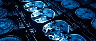
If the final diagnosis of the patient’s condition indicates “episyndrome,” the research process is just beginning and it is too early to talk about the final verdict. Two methods are considered the most informative:
- Based on the use of X-rays.
- Using a strong magnetic field.
Both techniques help to recognize the presence or absence of brain lesions. As an additional study, an encephalogram is prescribed to record the attacks themselves and detect their location.
During diagnosis, a number of patients with episyndrome are not diagnosed with pathological disorders, while periodic seizures recur. This condition is considered as presumably symptomatic epilepsy.
Causes
The root cause of alcoholic epilepsy is often organic damage to brain cells as a result of a “toxic attack.” This is preceded by a long “experience” of abuse or periodic use of surrogate alcohol.
But there are also risk factors that can cause an attack, regardless of the duration of alcoholism:
- History of traumatic brain injury;
- Cerebrovascular pathologies;
- Compounded hereditary history (epilepsy);
- Neuroinfections.
Alcohol seizures are considered to be quite rare and the most severe symptom of chronic alcoholism.
It is believed that a pathological symptom appears with a continuous history of alcoholism of at least 7–10 years. But there are known cases of seizures occurring after 1–2 years of systematic abuse.
Chronic alcoholism causes metabolic disorders in the central nervous system. Intracellular disorders are characterized by a failure in the transport of calcium and chlorine, as well as dysfunction of gamma-aminobutyric acid receptors. Pathological metabolic effects are temporary and disappear completely after alcohol withdrawal.
Clinical and neurophysiological features of epilepsy in alcohol abusers / V. O. Generalov, T. R. Sadykov, A. I. Fedin, Yu. V. Kazakova, L. P. Mishnyakova, E. V. Amcheslavskaya - Bulletin of the Russian State Medical University. - No. 5 - 2009. - 45 - 46 p.
Treatment options
The choice of treatment tactics is based on several factors, including age, probable cause, and individual course of the attack. Adult patients are offered the following regimen:
- For long-term use, medicinal herbs with a relaxing effect are prescribed. These are wild rosemary, violet, oregano, celandine, linden.
- Plan a diet with limited consumption of liquids, pickles, and smoked foods.
- Prescribed drugs with anticonvulsant and tranquilizing effects. Their purpose is to reduce the frequency of attacks and muscle relaxation, respectively.
Treatment of children involves the following stages:
- Organizing a diet with an emphasis on fatty foods and reducing the amount of protein. Thanks to this diet, lipids become the body's only energy resource. The reduction in the number of seizures occurs due to the active work of internal structures on the formation of ketone bodies.
- Restoration of calcium-potassium balance.
- Herbal medicine using the medicinal plants mentioned above.
The main goal of treatment is to relieve the underlying cause. Only in this case it is possible to eliminate disability, get an effective result and a normal, fulfilling life.
Treatment
An epileptic seizure can occur at any stage of alcoholism. It is impossible to cope with the disease on your own. A generalized attack of alcoholic epilepsy is a life-threatening condition. Therefore, treatment is carried out only in a hospital setting.
There is no specific therapy for alcoholic epilepsy. Drug treatment is carried out to maintain the functioning of vital organs and systems. And also to reduce the risk of secondary complications. The main condition for a favorable prognosis is complete abstinence from alcohol.
Symptomatic therapy:
- Stopping a convulsive attack. Systematic use of anticonvulsants to increase the seizure threshold;
- Prevention of cerebral edema and cardiovascular pathologies - forcing diuresis;
- Muscle relaxants;
- Maintaining water and electrolyte balance;
- Rehabilitation therapy – vitamins, nootropics.
After stabilization of the condition, it is recommended to undergo comprehensive rehabilitation in specialized institutions. With timely treatment of alcoholism, the prognosis is usually favorable.
One of the most important stages of alcohol addiction rehabilitation is psychotherapeutic correction. Its tasks and specific techniques change at each stage of the treatment process. It is especially important that the patient acknowledges his problem and the need for treatment.
Fundamentals of narcology: textbook. Benefit /M. M. Burkin, S. V. Goranskaya. – Petrozavodsk. : Karelia, 2002. – 71 – 73 p.
First aid during an attack
In case of a repeated attack or the initial manifestation of the disease, it is sometimes possible to do without an ambulance. If the patient’s consciousness is preserved, they sit him down and ask him about his general well-being. Subject to an adequate assessment, serious measures may not be applied. When there is an unconscious state, adhere to the sequence below:
- Place the adult or child on his back with his head slightly raised (you can use folded clothes). This precaution will help prevent blows and cranial injuries in the event of convulsions.
- A handkerchief is placed in the mouth to prevent tongue biting.
- If there is excessive salivation, turn the head to the side to avoid choking.
- You need to be prepared for a possible one-minute apnea. The situation is under control when breathing resumes after a couple of seconds.
Errors in providing first aid include bringing a person with a seizure to consciousness, stopping a seizure with mechanical forces. Such measures will not give the expected result (just like forcibly opening clenched teeth).
The duration of one attack is on average 3 minutes, during which the main goal of first aid is to exclude injury, maintain the correct position of the tongue, and prevent hypoxia. If you feel better, you can’t get up immediately - you need to maintain a 10-minute interval. Calling an ambulance is justified in the following situations:
- The patient is a woman expecting a baby.
- The attack occurred in an elderly person or a child.
- The duration of the seizures exceeds 3 minutes.
- Unconsciousness persists after 10 minutes.
Symptoms
An attack of true alcoholic epilepsy is a harbinger of alcohol withdrawal. That is, a seizure always occurs as a result of alcohol withdrawal after prolonged or periodic ethanol intoxication. The pathological condition develops several days after withdrawal or against the background of a sharp decrease in the daily dose of alcohol.
During an alcoholic seizure, the clinical picture is homogeneous. But the severity of symptoms and the nature of manifestations may differ.
| Alcoholism stage | Probability of occurrence |
| I | 17 – 20 % |
| II–III | 60 – 64 % |
| III | 17 – 19 % |
An attack of alcoholic epilepsy occurs suddenly and without warning. Sometimes there is an atypical scenario of a non-convulsive seizure. A person falls into a kind of stupor, after which a disorder of speech and thinking occurs. The moment of the attack is completely amnesic.
Narcologists noted that in a state of alcoholic intoxication the convulsive threshold is high, so seizures do not occur. But with the formation of withdrawal syndrome, the convulsive threshold becomes significantly lower. Before an attack, anxiety increases, and visual or auditory hallucinations may occur. In isolated cases, before fainting, victims feel a second of numbness in the leg. The attack lasts on average 3 minutes and in most patients occurs while awake.
A severe form of epileptic seizure is considered to be a recurrence of a seizure after a short time interval - a transition to epileptic status.
After an epileptic seizure, the victim regains consciousness and feels overwhelmed. Sleep sets in for a few hours. In some patients, a full-blown attack does not occur. A day after stopping alcohol, only burning pain in the muscles occurs.