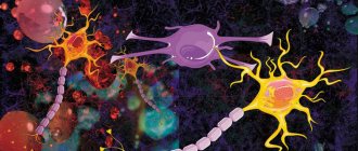
Oligodendrocytes
, or
oligodendrogliocytes
- neuroglial cells. This is the most numerous group of glial cells [1]. Oligodendrocytes are localized in the central nervous system.
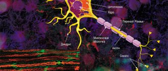
Oligodendrocytes are oval-shaped cells with processes. Their main function is the myelination of CNS axons. Each oligodendrogliocyte has many processes, each of which wraps itself around part of an axon. As a result, one oligodendrocyte is connected to several neurons. This ensures isolation of the axon, and, as a consequence, the possibility of rapid saltatory conduction of nerve impulses (along the nodes of Ranvier, remaining between isolated areas).
Oligodendrocytes also perform a trophic function in relation to neurons, taking an active part in their metabolism.
A type of oligodendrocyte in the peripheral nervous system is Schwann cells.
The breakdown of myelin sheaths leads to disruption of signal conduction, this process is called demyelination and causes a number of diseases, these can be both autoimmune diseases and genetic ones, and it can also cause senile dementia.
Oligodendrocytes also provide a depot function: they synthesize and store cholesterol necessary for the construction and renewal of the membrane, and also share it with neurons for the same purposes.
Thanks to the myelin sheaths, neuron restoration is possible, provided that the body is not damaged. They preserve the “tunnels” through which new fiber will grow.
The heir of the great histologist and brain phagocytes
The term “microglia” was introduced by a student of the famous histologist Santiago Ramon y Cajal, Pio del Rio Gortega, back in the 1920s, when he divided the glial cells of the brain into macro- and microglia.
Subsequently, they began to be called Gortega cells, and so the scientist forever wrote his name in history. Pio del Rio Gortega (1882-1945).
Credit: Wikipedia
Pio del Rio-Gortega first introduced the concept of microglia as a specific cellular element of the central nervous system on June 1, 1920 in the article La microglía y su transformación en células en bastoncito y cuerpos granuloadiposos.
- Functions[ | code]
- Synaptic connections
- Morphological components of the reflex arc.
- Microscopic structure of a neuron. Classification of neurons.
- Structure and functions of neuroglia (astrocytes, oligodendrocytes, microglia, ependyma). Relationships between neurons and neuroglia.
- Neuron structure
- Cell membrane
- Damaged wrapper
- Pathologies
- What is neuroglia
Gortega's first work
He later developed these ideas in the chapter Microglia
, written for the book that later became iconic -
Cytology and Cellular Pathology of the Nervous System
(Cytology and Cellular Pathology of the Nervous System). It was published under the editorship of Wilder Penfield in 1932. In this chapter, Rio-Gortega talked about the fact that microglia migrate to the central nervous system in the early stages of embryogenesis and in the developing brain there is a temporary form of it - amoeboid. It, actively phagocytizing (absorbing foreign inclusions), performs a protective function in the early postpartum period, when the blood-brain barrier is not yet fully developed and substances from the blood easily enter the brain. Moreover, such cells have a greater ability to migrate and multiply.
Origin and development of microglia. Credit: public domain
It should be noted that Gortega had predecessors and he was not the discoverer of Gortega cells. It is still unclear who was first. It seems that in 1878, Karl Frohmann identified cellular changes in certain areas of the brain and spinal cord of a 22-year-old patient who died of multiple sclerosis. We know for sure that at the end of the 19th and beginning of the 20th centuries these cells were seen and sketched by Franz Nissl, Alois Alzheimer and Ludwig Merzbacher. And Gortega’s teacher himself, Santiago Ramon y Cajal, also made his mark in this field.
Sketches by Alois Alzheimer It is now known that microglia are fundamentally different from the bone marrow-derived monocytes/macrophages that are often found in peripheral tissues. The difference is that its cells originate from primitive macrophages, which originate from the wall of the yolk sac, during embryogenesis (8th week of embryonic development) and enter the brain rudiment through the circulatory system.
These precursors surround the neuroepithelium of the developing brain around the 9th week of embryonic development and on the 64th day enter the neuroepithelium, beginning to populate the tissue of the central nervous system. Indeed, microgliocytes at this stage of development have an amoeboid rather than a branching shape.
It is interesting that the scientist back in the 20s of the 20th century in the chapter “ Microglia
“wrote that migrating along the choroid plexus that entwines the neural tube and the pathways of the white medulla, microglial cells penetrate into all parts of the central nervous system. In the formed brain, they are similar in appearance to astrocytes (macroglia cells), and in this form they can be distinguished by a body and many processes that are not intertwined with each other (this state of microglia is known today as “resting” microglia). That is, the idea of them has not changed much after decades.
Microglia completely populate the central nervous system only by day 28 of postnatal development. The development and survival of microglia depends on several factors, including the transcription factor PU.1 as well as CSF1R.
In the already formed brain, microglial cells are distributed evenly in all its parts and, with rare exceptions, show little variability. But as soon as a pathological process occurs, these cells, when activated, acquire an amoeboid shape, which is characteristic of them in the early stages of embryogenesis.
Del Rio-Gortega introduced his postulates based on research published in a series of papers between 1919 and 1927, in which he used an improved technique of silver impregnation (a special stain invented by Camillo Golgi, for which he shared with Ramón y) to label microglial cells. Kahalem received the Nobel Prize). The imaging technique was tedious and quite time-consuming, but it was worth it - high-quality and clear images of microglial cells were obtained.
In addition to the term microglia, del Rio-Gortega also coined the term microgliocyte. In neuroscience textbooks published after World War II, these cells were already called microglial cells. The name, although slightly changed to microglial cells, remains relevant to this day.
What is needed for oligodendrocyte maturation?
Oligodendrocytes are a type of neuroglial cells that cover the processes of neurons with a myelin sheath within the central nervous system. Like other neuroglial cells (with the exception of microglia), oligodendrocytes are formed from the mesoderm in the embryonic period, but the molecular pathways responsible for their differentiation are poorly understood. According to a recent study published in the journal Neuron, differentiation of oligodendrocytes requires that some of their mRNAs undergo a special modification known as m6A methylation (that is, the addition of a methyl group to the sixth nitrogen atom of adenosine, which is part of the mRNA).
It turned out that this modification is necessary for the correct splicing of the mRNA encoding the protein neurofascin 155, an important factor in the development of neuroglial cells.
The molecular mechanisms underlying cell differentiation and specialization are perhaps the most confusing area of cell and developmental biology. The development and maturation of many types of cells, which have been relatively well studied in the mature state, remain a blank spot to this day. The reason for this injustice is that most often the differentiation of cells from unspecialized progenitor cells is accompanied by simultaneous changes in the expression levels of many genes, which cannot be tracked individually.
The situation began to change in recent years, when, due to the rapid development of high-throughput sequencing, it became possible to obtain information about all RNA molecules present in a cell at a given time (this RNA profile of a cell is called a transcriptome). Now we can not only monitor quantitative changes in the level of expression of certain genes during the differentiation process, but also identify special modifications in mRNA molecules that they undergo after transcription.
One of the most common post-transcriptional modifications of mRNA in mammalian cells is the covalent addition of a methyl group (-CH3) to the N6 nitrogen atom of the adenosine nitrogenous base, known as m6A-methylation. Such a seemingly insignificant change can have enormous consequences for further stages of protein biosynthesis. The role of m6A methylation has already been shown in many processes associated with the development of the body, and the maturation of oligodendrocytes is one of them.
The authors of the study were able to demonstrate that during the maturation of oligodendrocytes, dynamic changes occur in the frequency of occurrence of m6A methylation in a number of transcripts. It turned out that when the METTL14 protein, which is responsible for introducing this modification, was “turned off,” the number of oligodendrocytes sharply decreased, as did the level of myelination of axons in the central nervous system, although the number of oligodendrocyte precursor cells turned out to be unchanged.
It turned out that inactivation of METTL14 disrupts the postmitotic development of oligodendrocytes, and it affects the transcriptomes of oligodendrocyte progenitor cells and oligodendrocytes themselves differently. At the molecular level, disturbances in m6A methylation cause failures in the process of splicing (excision of certain nucleotides) of many transcripts, including neurofascin 155 protein transcripts.
This isoform of neurofascin is specific for neuroglial cells and is necessary for their proper maturation. However, there are still many unexplored aspects in the molecular differentiation of neuroglial cells, including oligodendrocytes.
Text: Elizaveta Minina
m6A mRNA Methylation Is Essential for Oligodendrocyte Maturation and CNS Myelination by Xu, H., Dzhashiashvili, Y., Shah, A., Kunjamma, R.B., Weng, Y., Elbaz, B., … Popko, B. in Neuron. (2019)
doi:10.1016/j.neuron.2019.12.013
Functions[ | code]
Oligodendrocytes are closely associated with nerve cells, and like other glial cells, oligodendrocytes provide scaffolding as well as trophic support to neurons by producing glial-derived neurotrophic factor (GDNF), brain-derived neurotrophic factor (BDNF), and insulin-like growth factor-1 (IGF-1). . Additionally, the mammalian nervous system relies heavily on myelin sheaths to reduce ion leakage and reduce cell membrane capacitance. Myelin also increases the speed of nerve impulses when action potentials jump along nodes of Ranvier between Schwann cells (in the peripheral nervous system) or oligodendrocytes (in the central nervous system). In addition, the impulse velocity of a myelinated axon increases proportionally to its diameter, whereas the impulse velocity of unmyelinated fibers increases only as the square root of the axon diameter. The thickness of the insulation should be proportional to the diameter of the fiber. The optimal ratio for the maximum speed of impulse conduction is 0.6, divided by the diameter of the entire fiber (including the myelin sheath).
Satellite oligodendrocytes are functionally different from other oligodendrocytes. They are not attached to neurons and therefore do not serve an insulating role. They regulate the composition of extracellular fluid. Satellite oligodendrocytes are considered part of the gray matter, whereas myelinating oligodendrocytes are part of the white matter.
Important Neuroplasticity of the brain: how to develop the most important organ
Myelination is an important factor influencing intelligence. Neuroscientist Vincent J. Schmithorst has suggested that there is a relationship between the volume of white matter in the brain and intelligence. People with more white matter have higher IQ scores. A study conducted by Janice M. Juraska on rats found that rats raised in an enriched environment had a greater degree of myelination in their corpus callosum.
Function
There are two types of oligodendrocytes, each of them differs in its place of primary localization and the functions it performs. Among them are:
- myelinating;
- satellite.
Along with other glial cells, olidendrocytes have a close connection with the entire nervous system. For neurons, they provide support, also performing the function of trophic support for the production of glial trophic factors, as well as brain neurotrophic factors and insulin-like growth factors. Throughout the nervous system of every mammal there is a strong dependence on myelin structure. Due to the myelin sheath covering the nerve structures, the ionic component leaks, and the capacitance of cell membranes decreases.
It is due to the structure of the myelin sheath that the speed of transmission of the nerve impulse increases, in cases where spasmodic propagation of action potentials occurs in the nodes of Ranvier, as well as in the spaces located between areas of Schwann cells at the level of the peripheral parts of the nervous system), as well as oligodendrocytes ( in the region of the central nervous system). The increase in the speed of propagation of the nerve impulse will be a value dependent on the thickness of the axon carrying out the transmission.
There are also other types of oligodendrocytes that are satellite. They differ significantly in their function. They are not attached to the structure of other neurons, and therefore do not play an insulating role. Due to satellite oligodendrocytes, the composition of extracellular fluid components is regulated. Unlike myelinating oligodendrocytes, satellite cells belong to the gray matter of the brain, which performs a different functional role.
Synaptic connections
The formation of neural networks is based on electrical excitation, which consists of two processes:
- the launch of electrical excitation from the energy of external influences occurs due to the special sensitivity of the membranes located on the dendrites;
- triggering cellular activity based on the received signal and influencing other structural units of the nervous system.
Neurons are connected to each other through special structures - synapses. They consist of presynaptic and postsynaptic membranes, between which there is a synaptic cleft filled with fluid.
By the nature of their action, synapses can be excitatory or inhibitory. Signal transmission can be chemical or electrical.
In the first case, neurotransmitters are synthesized on the presynaptic membrane, which arrive at the receptors of the postsynaptic membrane of another cell from special vesicles - vesicles. After their influence, ions of a certain type can massively enter a neighboring neuron. This occurs through potassium and sodium channels. In the normal state, they are closed, there are negatively charged ions inside the cell, and positively charged ones outside. Consequently, a voltage difference is formed across the shell. This is the resting potential. After positively charged ions enter, an action potential occurs - a nerve impulse.
Cell balance is restored with the help of specialized proteins - potassium-sodium pumps.
Properties of chemical synapses:
- excitation is carried out only in one direction;
- the presence of a delay of 0.5 to 2 ms during signal transmission, associated with the duration of the processes of mediator release, its transmission, interaction with the receptor and the formation of an action potential;
- fatigue may occur due to depletion of the transmitter supply or the appearance of persistent depolarization of the membrane;
- high sensitivity to poisons, drugs and other biologically active substances.
Electrical transmission is characterized by a narrow synaptic gap and reduced resistance between membranes. In this case, the potential created on the presynaptic membrane causes excitation to spread to the postsynaptic membrane.
Properties of electrical synapses:
- the speed of information transfer is higher than in chemical synapses;
- Both one-way and two-way signal transmission (in the opposite direction) is possible.
There are also mixed synapses, in which excitation can be transmitted both with the help of neurotransmitters and with the help of electrical impulses.
Memory includes the storage and reproduction of received information. As a result of training, so-called memory traces remain, and their sets form engrams - “records”. The neural mechanism is as follows: certain impulses pass through the chain many times, structural and biochemical changes are formed in the synapses. This process is called consolidation. Repeated use of the same contacts creates specific proteins - these are memory traces.
Morphological components of the reflex arc.
reflex arc
- several interconnected neurons, ensures the organ’s response to irritation. consists of three links: afferent (receptor), associative (insert) and effector. the intercalary may be absent.
Autonomous (vegetative) arc: the afferent link is represented by a pseudounipolar neuron. its body lies in the spinal ganglion, and the dendrites form endings with tissues, organs, vessels, and glands. the axon enters the spinal cord as part of the dorsal roots, goes into the lateral horns of the gray matter, where it forms synapses with the intercalary link. interneurons are multipolar, their dendrites are located in the lateral horns of the gray matter, and the axon through the anterior roots rushes to the autonomic ganglion, where it ends in a synapse with the effector link. the neurons of this link are also multipolar, and the axon is directed to the cells of the working organs (smooth muscle, glands, heart)
somatic arc: everything is the same with the receptor link, only the dendrites are in contact with the skin and skeletal muscles. The axons go to the dorsal horns of the gray body, where the interneurons are located. some collaterals pass into the anterior horns, where they contact motor neurons. the intercalary link differs only in location (posterior horns). the neurons of the effector link lie in the spinal ganglia, from where, as part of the mixed nerve, they go to the skeletal muscles, forming motor plaques on them.
Origin
Oligodendroglia develop from oligodendrocyte progenitor cells.
Most oligodendrocytes develop during embryogenesis and early infancy from the periventricular germinal areas.
Cells of the subventricular zone migrate from the germinal zones to populate the developing white and gray matter, where they differentiate and mature into myelin-forming oligodendroglia.
However, it remains to be seen whether all oligodendroglial progenitors undergo this sequence of events. It has been suggested that some of them undergo apoptosis, that is, they self-destruct, while others fail to differentiate into mature oligodendroglia and persist as adult oligodendroglial progenitors. Notably, the oligodendrocyte population originating in the subventricular zone can be dramatically expanded by administration of epidermal growth factor (EGF).
Now the origin of oligodendrocytes is actively discussed by scientists.
At birth, myelination is widespread only in certain regions of the brain, and it continues until the age of 25-30 years.
Microscopic structure of a neuron. Classification of neurons.
neuron body
- nucleus and nearby cytoplasm + any grEPR, AG, etc. There are a lot of receptors on the plasmalemma. the nucleus is large, with 2-3 nucleoli; Barr’s body lives near the nucleus in women. The plasmalemma is capable of conducting impulses and has na/k pumps (they say that it is not sodium that is pumped in, but calcium). The grEPR forms clumps, which is why the whole thing is called a tigroid (Nissl body). they disintegrate with prolonged irritation or damage to the neuron. everything else is uninteresting. many mitochondria, developed AG, active endosomes... cytoskeleton - MCT (neurotubes), microfilaments, PF (neurofilaments). neurophile and neurotubules are connected by bridges, forming neurofibrils.
dendrite
. conducts impulses from neuron to neuron through axo-dendritic synapses located in the area of protrusions of the cytoplasm - dendritic spines. There are many dendrites, they are short and highly branched. neurotubules and neurofilaments carry out dendritic transport.
axon
. the process is long and extends from the axon hillock on the body of the neuron (where the impulse is generated). most of it is covered with glia. The cytoplasm of the axon is even named in a special way - axoplasm. it, of course, contains MKT and PF, as well as gLEPR, AG tanks, mitochondria and all that. axons can have perpendicular branches - collaterals. at the end, the axon often breaks up into small branches (telodendria), everyone knows how it ends.
the movement of substances along the axon is carried out due to dynein and kinesin, there is retrograde (from the axon to the body), and there is anterograde (from the body to the axon)
Important Diseases of the pituitary gland and hypothalamic-pituitary system
classification
- morphological
unipolar (amacrine neurons of the retina and interglomerular neurons of the olfactory bulb)
- bipolar (have an axon and a dendrite. These also include pseudounipolar)
- multipolar (axon and several dendrites)
functional
- sensitive (afferent)
- motor (efferent)
- associative (insert)
biochemical
- cholinergic (mediator - acetylcholine)
- adrenergic (transmitter norepinephrine)
- serotonergic (everything is clear)
- dopaminergic
- GABAergic (gamma-aminobutyric acid)
- purinergic (ATP and derivatives)
- peptidergic (mediators - substance P, enkephalins, endorphins and a bunch of other neuropeptides)
Neuron structure
Each structure in the human body consists of specific tissues inherent to the organ or system. In nervous tissue - neuron (neurocyte, nerve, neuron, nerve fiber). What are brain neurons? This is a structural and functional unit of nervous tissue that is part of the brain. In addition to the anatomical definition of a neuron, there is also a functional one - it is a cell excited by electrical impulses, capable of processing, storing and transmitting information to other neurons using chemical and electrical signals.
The structure of a nerve cell is not as complex as that of specific cells of other tissues; it also determines its function. A neurocyte consists of a body (another name is soma), and processes - axon and dendrite. Each element of a neuron performs its own function. The soma is surrounded by a layer of fatty tissue, allowing only fat-soluble substances to pass through. Inside the body there is a nucleus and other organelles: ribosomes, endoplasmic reticulum and others.
In addition to the neurons themselves, the following cells predominate in the brain, namely: glial cells. They are often called brain glue for their function: glia serve as a support function for neurons, providing an environment for them. Glial tissue provides nerve tissue with the ability to regenerate, nourish, and assist in the creation of nerve impulses.
The number of neurons in the brain has always interested researchers in the field of neurophysiology. Thus, the number of nerve cells varied from 14 billion to 100. Recent studies by Brazilian specialists revealed that the number of neurons averages 86 billion cells.
Processes
The tools in the hands of a neuron are the processes, thanks to which the neuron is able to perform its function as a transmitter and storer of information. It is the processes that form a wide nerve network, which allows the human psyche to reveal itself in all its glory. There is a myth that a person’s mental abilities depend on the number of neurons or on the weight of the brain, but this is not so: those people whose fields and subfields of the brain are highly developed (several times more) become geniuses. Due to this, fields responsible for certain functions will be able to perform these functions more creatively and quickly.
Axon
An axon is a long extension of a neuron that transmits nerve impulses from the nerve soma to other similar cells or organs innervated by a specific part of the nerve column. Nature has endowed vertebrates with a bonus - myelin fiber, the structure of which contains Schwann cells, between which there are small empty areas - nodes of Ranvier. Along them, like a ladder, nerve impulses jump from one area to another. This structure makes it possible to speed up the transmission of information several times (up to about 100 meters per second). The speed of movement of an electrical impulse along a fiber that does not have myelin is on average 2-3 meters per second.
Dendrites
Another type of nerve cell extension is dendrites. Unlike the long and solid axon, the dendrite is a short and branched structure. This process is not involved in transmitting information, but only in receiving it. Thus, excitation reaches the neuron body using short dendritic branches. The complexity of information that a dendrite is capable of receiving is determined by its synapses (specific nerve receptors), namely its surface diameter. Dendrites, thanks to the huge number of their spines, are capable of establishing hundreds of thousands of contacts with other cells.
Metabolism in a neuron
A distinctive feature of nerve cells is their metabolism. Metabolism in the neurocyte is distinguished by its high speed and predominance of aerobic (oxygen-based) processes. This feature of the cell is explained by the fact that the work of the brain is extremely energy-intensive, and its need for oxygen is great. Although the brain weighs only 2% of the body's weight, its oxygen consumption is approximately 46 ml/min, which is 25% of the body's total consumption.
The main source of energy for brain tissue, in addition to oxygen, is glucose, where it undergoes complex biochemical transformations. Ultimately, large amounts of energy are released from the sugar compounds. Thus, the question of how to improve neural connections in the brain can be answered: eat foods containing glucose compounds.
Literature
Alexei Verkhratsky, Vladimir Parpura. Introduction to Neuroglia. Morgan & Claypool, 2014.
| This is a draft article on neurobiology. You can help the project by adding to it. |
| Afferent nerve/Sensory neuron | GSA GVA SSA SVA Nerve fibers (Muscle spindles (Ia), Neurotendon spindle (Ib), II or Aβ fibers, III or Aδ fibers, IV or C fibers) |
| Efferent nerve/Motor neuron | GSE GVE SVE Upper motor neuron Lower motor neuron (α motor neurons, γ motor neurons) |
| Synapse | Chemical synapse Neuromuscular synapse Ephaps (Electrical synapse) Neuropil Synaptic vesicle |
| Touch receptor | Meissner's corpuscle Merkel's corpuscle Pacini's corpuscle Ruffini's corpuscle Neuromuscular spindle Free nerve ending Olfactory neuron Photoreceptor cells Hair cells Taste bud |
| Neuroglia | Astrocytes (Radial glia) Oligodendrocytes Ependymal cells (Tanycytes) Microglia |
| Myelin (White matter) | CNS: Oligodendrocytes PNS: Schwann cells (Neurolemma · Node of Ranvier/Internodal segment · Myelin notch) |
| Connective tissue | Epineurium Perineurium Endoneurium Nerve fiber bundles Meninges: dura mater, arachnoid membrane, pia mater |
Cell membrane
Thanks to the membrane, the cell has its potential. When it is transmitted along the chain, excitable tissue is innervated. Contact between the membranes of neighboring neurons occurs at synapses. Maintaining a constant internal environment is an important component of the life of any cell. And the membrane subtly regulates the concentration of molecules and charged ions in the cytoplasm. At the same time, they are transported in the required quantities for metabolic reactions to occur at an optimal level.
Important: Main symptoms and clinical picture of Parkinson's disease
The main component of the human or other mammal's brain is the neuron (also known as neuron). It is these cells that form the nervous tissue. The presence of neurons helps to adapt to environmental conditions, feel, and think. With their help, a signal is transmitted to the desired area of the body. Neurotransmitters are used for this purpose. Knowing the structure of a neuron and its features, one can understand the essence of many diseases and processes in brain tissue.
Damaged wrapper
Figure 5. Sensory impairment of the polyneuritic type. The name "sock-glove" is due to the fact that the anatomical areas corresponding to nerve damage are similar to the areas covered by these items of clothing.
It seems to me that the following rule is quite suitable for the human body: if there is an organ, then there must be a disease associated with it. In principle, this rule can be extended to molecular processes: if there is a process, there are also diseases associated with disruption of this process. In the case of myelin, these are demyelinating diseases. There are quite a few of them, but I will talk in more detail about two - Guillain-Barré syndrome and multiple sclerosis. In these disorders, damage to myelin leads to disruption of adequate signal transmission along the nerves, which causes the symptoms of the disease.
Guillain-Barré syndrome (GBS) is a disease of the peripheral nervous system in which the myelin sheath formed by Schwann cells is destroyed. GBS is a classic autoimmune disease. As a rule, it is preceded by an infection (often caused by a microbe). The presence of various pathogens in the human body triggers autoimmune damage to the myelin of nerve fibers by T- and B-lymphocytes. Clinically, this is manifested by muscle weakness, sensory impairment of the “socks-gloves” type (polyneuritic type) (Fig. 5). In the future, muscle weakness can increase until complete paralysis of the limbs and damage to the trunk muscles. Damages to the sensitive nervous system can also be varied: from a decrease in the ability to distinguish one’s own movements (impaired deep sensitivity) to severe pain. In severe forms of GBS, the main danger is the loss of the ability to breathe independently, requiring connection to a mechanical ventilation device (ALV). To treat GBS, plasmapheresis (purification of plasma from harmful antibodies) and intravenous infusions of human immunoglobulin preparations are currently used to normalize the immune response. In most cases, treatment leads to lasting recovery.
Figure 6. Lesions of the central nervous system in multiple sclerosis appear as white plaques on MRI.
Figure 7. Depending on the location of damage to the nervous system in multiple sclerosis, there may be different symptoms: from tremor and ataxia with damage to the cerebellum to emotional disorders with localization of lesions in the frontal lobes.
Pathologies
Damage to oligodendrocytes is observed in demyelinating diseases such as multiple sclerosis and various leukodystrophies. Oligodendrocyte dysfunction may also be associated with the pathophysiology of schizophrenia and bipolar disorder.
Injuries to the body, such as spinal cord injuries, can also cause demyelination. Cerebral palsy (sometimes developing from periventricular leukomalacia, which is local or widespread aseptic necrosis of the white matter of the cerebral hemispheres and occurs most often in premature infants) is mainly congenital or caused by damage to the newly formed brain (trauma to brain structures during fetal development and childbirth). In cerebral palsy, spinal cord injury, stroke and possibly multiple sclerosis, oligodendrocytes are thought to be damaged by excessive release of the neurotransmitter glutamate. Damage has also been shown to be mediated by NMDA glutamate receptors.
Oligodendrocytes are also susceptible to infection by human polyomavirus (JC virus), which causes progressive multifocal leukoencephalopathy (PML), a condition that specifically affects the white matter, usually in immunocompromised patients.
Tumors of oligodendroglia are called oligodendrogliomas. The chemotherapeutic agent fluorouracil (5-FU) causes oligodendrocyte damage in mice, leading to both acute acute central nervous system damage and delayed central nervous system degeneration that gradually worsens over time.
Clinical significance[edit]
See also: Myelin § Dysmyelination and demyelinating diseases
Diseases that result in oligodendrocyte damage include demyelinating diseases such as multiple sclerosis and various leukodystrophies. Trauma to the body, such as spinal cord injury, can also cause demyelination. Immature oligodendrocytes, which increase in number during mid-gestation, are more vulnerable to hypoxic injury and are involved in periventricular leukomalacia. [25] It is largely a congenital condition associated with damage to the newly formed brain and can therefore lead to cerebral palsy. In cerebral palsy, spinal cord injury, stroke and possibly multiple sclerosis, oligodendrocytes are thought to be damaged due to excessive release of the neurotransmitter, glutamate. [26] Damage has also been shown to be mediated by N-methyl-D-aspartate receptors. [26] Oligodendrocyte dysfunction may also be involved in the pathophysiology of schizophrenia and bipolar disorder. [27]
Oligodendrocytes are also susceptible to infection by JC virus, which causes progressive multifocal leukoencephalopathy (PML), a condition that specifically affects the white matter, typically in immunocompromised patients. Tumors of oligodendrocytes are called oligodendrogliomas. The chemotherapeutic agent fluorouracil (5-FU) causes oligodendrocyte damage in mice, resulting in both acute central nervous system (CNS) damage and progressive worsening of delayed CNS degeneration. [28] [29]