Author
: Grachev Ilya Illarionovich
Editor
: Efremov Mikhail Mikhailovich
Date of publication: 05/13/2014 Date of update: 04/12/2021 All doctors of the clinic
- Knee pain due to coronavirus and ARVI
- Diagnostics
The knees are more susceptible to injury and various diseases than other joints. A significant proportion of orthopedist-traumatologist and rheumatologist patients complain of severe knee pain. From this article you will learn what to do if you suddenly experience severe pain. She is being treated, the main thing is to seek medical help with her problems in time. They will always help you at the medical office (Moscow).
What exactly hurts in the knee?
The knee joint is the most complex joint, as it bears the main load. Structure: three bones (femur, tibia and patella form a single trochlear joint, consisting of two interconnected joints: femorotibial (tibiofemoral) and femoral-patellofemoral (patellofemoral).
The patella is a flat sesamoid (additional in the joint) bone that is attached to the head of the femur, sliding in its concave groove and acting as a block. Structural features: the anterior surface of the patella is covered with periosteum, the posterior surface, connecting to the femur, is covered with hyaline cartilage. The patella is strengthened by ligaments: main and lateral – vertical (upper and lower) and horizontal (lateral – internal and external).
O transmits the force of the quadriceps femoris muscle to the musculoskeletal formations of the lower leg, ensuring extension of the lower leg at the knee joint. The surface of the joint-forming bones is covered with cartilage, which acts as a shock absorber. Additional shock absorbers that protect the joint from injury are two crescent-shaped cartilaginous menisci located between the femur and tibia. The joint is held in its correct position by ligaments, tendons and the surrounding capsule.
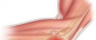
Structure of the knee joint
Injuries and diseases affect various joint tissues. Not all of them can get sick. Thus, cartilage tissue does not have nerve endings and therefore can be destroyed imperceptibly and painlessly. But the ligaments and synovial membrane have many nerve endings and in case of injury or inflammatory processes they immediately begin to react, which manifests itself in the form of severe pain. With significant destruction of articular cartilage, pain may be associated with the involvement of the periosteum, the outer layer of bone that has good innervation, in the process.
What to do if you have severe knee pain
Intense knee pain may appear suddenly or develop gradually. In any case, it often becomes unbearable. If severe pain occurs, you need to calm down and immediately seek medical help. All this can be treated, specialists will be able to help even with advanced disease. If you can’t see a doctor at the moment, you can temporarily relieve the pain on your own.
But you should remember that if you have pain in the knee joint, this is a temporary measure; you still need to seek medical help; you cannot do without it. And it’s better not to delay.
How to treat knee and leg pain at home
To alleviate your condition with severe pain in the knee joint, you can take the following emergency measures:
- Perform pain relief using tablets:
- Nise (nimesulide)
– for severe pain, take 1 tablet 100 mg; an effective modern medicine, belongs to the group of non-steroidal anti-inflammatory drugs (NSAIDs) of the latest generation and has a minimum of side effects, including no negative effect on the gastrointestinal tract (GIT); in case of an acute inflammatory process, it will relieve not only pain, but also fever, and will also reduce tissue swelling if the knee is swollen; - Analgin
– you can take a 500 mg tablet once; also relieves inflammation, swelling and pain. You should not abuse this drug, as with frequent use it can cause severe blood disorders. - Pentalgin
is a combined pain reliever that contains two anti-inflammatory painkillers (paracetamol and naproxen), an anti-vasospasm agent (drotaverine) and the antihistamine pheniramine - it will help if the knee is swollen. Take one tablet (no more than 3 - 4 tablets per day). - Use external painkillers (ointments, gels). The most effective:
- emulgel Voltaren
, active ingredient – diclofenac; The peculiarity of the drug is that, due to its structure, it is instantly absorbed into the skin, penetrates its deep layers, relieves inflammation, swelling and pain; the effect lasts for several hours; the ointment is applied to the skin in the knee area twice a day and lightly rubbed in; - Menovazin
is an alcohol solution for external use with a combined composition; benzocaine and procaine have a local analgesic effect, menthol enhances this effect by dilating skin blood vessels and a feeling of pleasant cold, relieving inflammation and heat; the solution is rubbed into the inflamed tissue, which immediately causes relief; - Dimexide gel
is an analgesic and anti-inflammatory agent that is applied to the skin 1 – 2 times a day; A feature of the gel is its rapid healing effect; in addition, it suppresses the intra-articular proliferation of connective tissue, thereby preventing the development of limb immobility; has contraindications: severe diseases of the kidneys, liver, cardiovascular system, pregnancy, breastfeeding. - Injections. If there is no effect from tablets and external agents, the medicine is administered as an intramuscular or subcutaneous injection:
- Diclofenac in solution for injection
- available in ampoules of 3 ml (25 mg per 1 ml); a total of 75 mg per ampoule - a single dose for intramuscular administration; the drug is very effective, quickly helps if the knee hurts and is swollen, but has side effects, therefore it is contraindicated for erosive and ulcerative lesions of the gastrointestinal tract; - Analgin in solution for injection
- available in ampoules of 2 ml (250 mg in 1 ml); a total of 500 mg in an ampoule - a single dose for an adult; Frequent use is not recommended, as it has many side effects.
All of the drugs listed can be purchased at a pharmacy without a prescription.
For chronic pain in the knee joints, when there is no significant inflammation, you can perform physical exercises that strengthen the muscular-ligamentous system, improve blood circulation and metabolism. Systematic training leads to a gradual decrease in pain, despite the fact that the legs are pretty crunchy.
An approximate set of exercises for knee pain:
- Fixed knee. Lie on your back, bend one leg at the knee, lift it and hold it in this state for a minute; the second leg is motionless at this time; straighten and lower your leg very slowly, rest for 10 seconds and repeat the exercise with the other leg; repeat 10 times;
- Double leg bending with knees near the face. Lie on your back, bend your legs at the hips and knees, fix the latter near your face and hold it like that for a minute. Then slowly straighten and lower your legs, rest for 10 seconds and repeat; do 5–6 approaches, gradually increasing the load.
Exercises for knee pain
Acute pain in the knee requires a state of rest; all physical activity and training are contraindicated.
In case of acute pain in the knee joint, accompanied by swelling and redness of the skin, a violation of the general condition, fever, you cannot move the leg, it must be kept at rest. And only after the inflammation begins to subside, you can move first to passive exercises (performed by an assistant) and then to active exercises (performed by the patient himself).
What not to do if you have pain
For chronic pain, the following should not be allowed:
- bruised knee - you can avoid this by arranging your life in such a way as to reduce the risk of injury to a minimum;
- excess body weight is an additional burden on the knees, especially in the elderly;
- heavy physical activity, jumping, strength sports; You should also not run;
- any intoxication, so you need to get rid of bad habits (smoking, alcohol abuse), treat all chronic diseases and foci of infection;
- wearing uncomfortable tight shoes, high-heeled shoes;
- stress, lack of sleep;
- sedentary lifestyle - you need to force yourself to move at certain intervals.
It is also impossible to apply warm compresses without a doctor’s prescription: in purulent and hemorrhagic (with intra-articular bleeding) processes, they can cause irreparable harm.
When you need to see a doctor urgently
If your knee hurts, urgent medical attention is required if the following symptoms appear:
- swelling, redness and tenderness in the knee combined with fever and general malaise;
- severe pain in the knee immediately after the injury or some time after it;
- gradual increase in pain intensity;
- periodically appearing pain after physical exertion, prolonged standing, sharp straightening of the leg;
- night pain and associated insomnia;
- if the pain in the knee is very strong, constant, the feeling that the inside of the patella hurts.
In any case, pain in the knee should be a reason to consult a doctor. There is no point in treating yourself: it may temporarily reduce or even eliminate pain, but it will not stop the progression of the disease and destruction of the joint. Treatment should be entrusted to a specialist.
Treatment
Conservative treatment
Conservative treatment for FMS includes pharmacologic agents (nonsteroidal anti-inflammatory drugs (NSAIDs), muscle relaxants, and medications for neuropathic pain), physical therapy, lifestyle changes, and psychotherapy.
Injections of local anesthetics, steroids, and botulinum toxin type A into the piriformis muscle can serve for both diagnosis and treatment. The practitioner should be familiar with variations in anatomy and the limitations of techniques based on anatomical landmarks. Recently, the ultrasound-guided injection method has been used. This technique has been shown to have both diagnostic and therapeutic value in the treatment of BMS.
FMS often becomes chronic, so pharmacological treatment is recommended for a short period of time.
Surgical intervention
Surgical interventions should be considered only when nonsurgical treatment has failed and symptoms become intractable and disabling. Classic indications for surgical treatment include abscesses, neoplasms, hematomas, and painful compression of the sciatic nerve vessels caused by gluteal varices.
Some authors have reported that surgical release of the piriformis muscle by cutting the tendon to free the sciatic nerve from compression results in immediate pain relief.
Sometimes the obturator internus muscle should be considered as a possible cause of pain in the area of innervation of the sciatic nerve. However, the diagnosis of obturator internus syndrome can only be made by ruling out other possible causes of gluteal pain, which is similar to how FMS is diagnosed. Surgical release of the obturator internus muscle can lead to both short-term and long-term pain relief in patients with retroacetabular pain syndrome and should be considered if conservative treatment has failed.
Postoperative management of the patient consists of partial weight reduction through the use of crutches for 2 weeks and selection of exercises. The above surgical approach has shown promising short-term results.
Treatment algorithm for retroacetabular pain syndrome:
Physical therapy
Although there are few recently published controlled studies that critically examine the effectiveness of noninvasive treatments, there are a number of treatment options for BMS that are well established in clinical practice.
Noninvasive treatments include physical therapy and lifestyle changes. According to Tonley et al., the most commonly reported physical therapy interventions include soft tissue mobilization, piriformis stretching, cryotherapy (hot packs or cold spray), and manual treatment of the lumbosacral spine.
In addition, Tonley describes an alternative approach to the treatment of BMS. Treatment focused on functional "Therapeutic Hip Exercises" exercises aimed at strengthening the hip extensors, abductors, and external rotators and correcting abnormal movement patterns. Despite the positive results (complete elimination of pain in the lower back, cessation of pain in the buttock and thigh), further research is required, since this protocol was tested in only one patient.
In order not to miss anything interesting, subscribe to our Telegram channel.
To achieve a 60-70% improvement, the patient usually undergoes 2-3 treatments per week for 2-3 months.
- Start with ultrasound treatment: 2.0-2.5 W/cm2 for 10-14 minutes. Apply the ultrasound gel using wide strokes in a longitudinal direction along the piriformis muscle from the connecting tendon to the lateral edge of the greater sciatic foramen. The patient should be in the contralateral lateral decubitus position and in the FAIR position (flexion, adduction, internal rotation).
- Before stretching the piriformis muscle, treat the same area with hot compresses or a cold spray for 10 minutes. Using hot and cold before stretching is very helpful in reducing pain.
- After this, move on to stretching the piriformis muscle, which can be performed in a variety of ways. It is important not to push down, but to direct the pressure along the surface (tangentially) to the ipsilateral shoulder (Fischman et al. (2002), level of evidence A2). When pressing downward, the sciatic nerve is compressed along the tendinous edge of the superior gemellus muscle. Another way to stretch the muscle is the FAIR position. The patient is in the supine position with the hip flexed, adducted and internally rotated. The patient then moves the foot on the affected side across and over the knee of the unaffected leg. The specialist can increase the stretch by using muscle-energetic techniques.
- After stretching the piriformis muscle, you can perform myofascial release of the lumbar muscles, as well as do Mackenzie exercises.
- Because FMS can occur when a tight piriformis muscle is forced to do the work of other large muscles (such as the gluteus maximus or gluteus medius), an alternative approach to treating FMS using a strengthening program for the hip muscles (especially weak gluteal muscles) with relearning of the movement may help. in pain relief.
Your therapist can also give you some tips to avoid worsening your symptoms.
- Avoid sitting for long periods of time; Standing and walking are required every 20 minutes.
- Make frequent stops as you move to stand and stretch.
- Prevent injury to the gluteal region and avoid further aggravating actions.
- Daily stretching is recommended to prevent recurrence of BMS.
Home exercises
The patient can also do some exercises at home.
- Rolling from side to side with bending and straightening of the knees, lying on one side of the body.
- Rotate from side to side while standing with relaxed arms for 1 minute every few hours.
- “Riding a bicycle” while lying on your back.
- Knee curls - up to 6 repetitions every few hours.
- Taking a warm bath.
What to do for severe pain in the knee of various types
Painful sensations in the knee can vary in nature and duration among people. They can be constantly aching or develop only under certain loads, at night, etc. For example, some pathological processes are characterized by pain in the knee when bending, others - pain in the knee when walking, etc. Only a specialist can figure out what exactly hurts and why, and how to help the patient.
Knee pain due to coronavirus and other viral diseases
Viral infections can cause joint inflammation. As a rule, these diseases develop against the background of an existing infection and pass without any consequences after its completion. Thus, with influenza and other acute respiratory viral infections with acute fever, both short-term joint and muscle pain and acute arthritis with inflammation and swelling of the knee joints may appear. Their course is favorable.
Coronavirus infection has its own characteristics: it occurs differently in each patient. Why this happens is unknown. During the disease, aching joint pain, swelling and redness sometimes appear - a sign of acute arthritis, but then they go away.
Arthritis, which begins approximately a month after coronavirus infection, is more dangerous. The fact is that it has a significant effect on the immune system. Malfunctions of the immune system lead to the development of autoimmune processes. This is especially dangerous for people who have close relatives suffering from chronic arthritis. Experts note a high risk of developing rheumatoid arthritis (RA) in such patients. The first sign of RA is stiffness of movement in the morning (difficulty bending the knee).
If arthritis appears after recovery from a viral infection, you should immediately contact a rheumatologist.
Severe pain under the knee
This may be a sign of the development of a Baker's cyst - a distended synovial bursa with fluid located in the popliteal area. The cyst and associated pain under the knee appear due to the fact that it is connected to the cavity of the knee joint and is filled with synovial fluid. In this case, the reverse flow of liquid is difficult for various reasons. Most often, the cyst develops against the background of bruises, arthrosis and arthritis of the knee.
A small cyst can go unnoticed for years. But with its significant size, the surrounding tissues begin to be compressed, causing pain under the knee, which intensifies with physical activity, including walking and running. Women get sick more often. Sometimes the cyst disappears on its own, but often it progresses and increases in volume, which can lead to rupture or suppuration.
If you have pain under your knee, it is better to consult a doctor as soon as possible. Conservative (removal of fluid from the cyst, injection of glucocorticoids into it) and surgical (removal of the cyst) treatment is carried out.
Severe pain in the left or right knee
Severe pain in combination with swelling and redness of the skin over the right or left knee usually indicates the development of an acute inflammatory process. This can be acute nonspecific arthritis (the process can turn purulent), reactive arthritis, which begins a few days after a urogenital or intestinal infection. Both diseases occur with similar symptoms; a correct diagnosis can only be established after a full examination.
The right or left knee can suffer from microtraumas, for example, in athletes or workers in certain professions who use one (usually the right) knee more.
It is important at an early stage not to use folk remedies, but to carry out the correct treatment, this will allow you to fully recover and forget about the pain forever. But even with advanced diseases, a specialist will always be able to provide assistance and relieve pain.
Crunching in joints - when to worry
Joint pain at rest
Knee and leg pain
Depending on the cause of the disease, its course and the presence of complications, severe knee pain may include:
- in the lower leg below the knee
- they are rare, this may indicate compression of the nerve branches innervating the muscles of the lower leg by the inflamed, swollen periarticular tissues in the knee area; pain under the knee in the back indicates compression of the branches of the sciatic nerve, and pain under the knee in the front indicates that the nerves innervating the anterior surface of the leg are affected; Such diseases are treated by a rheumatologist, but consultation with a neurologist is required; - above the knee, into the thigh
- often a sign of post-traumatic inflammatory process in the femoral-patellar joint; in this case, patellofemoral pain syndrome develops; the pain is strong, aching, sometimes twitching, aggravated by walking; an orthopedist-traumatologist and a rheumatologist will help you cope with pain; - on the side, on the inside
– pain can develop with injury and damage to the internal lateral articular ligament; pain on the side is strong, jerking, with hemarthrosis (intra-articular hemorrhage) - bursting, accompanied by imbalance of the joint, the whole leg crunches when moving; the same pain appears when the external collateral ligament is sprained; assistance will be provided by an orthopedist-traumatologist.
To eliminate pain, you need to accurately determine its cause, and this is impossible to do on your own. Need doctor's help.
Patella pain on the back, front and sides
Pain in the patella is almost always the result of acute or chronic injuries:
- pain behind the knee
- the causes are associated with damage to the femoral-patellar joint with the development of patellofemoral pain syndrome; the pain is aching, constant, aggravated by walking; - in the front
- such painful sensations are caused by superficial bruises of the knee or frequent prolonged stay on the knees with microtrauma to the kneecap; the pain is significant, as the periosteum, rich in nerve endings, is injured; - pain in the knee on the side -
when the horizontal internal or external patellar ligaments are ruptured or injured; injury can develop with frequent long-term microtrauma of the ligaments, for example, during jumping; accompanied by bleeding into the joint cavity (hemarthrosis); pain is strong, constant, accompanied by swelling; movements in the leg are impaired.
To prevent permanent dysfunction of the joint from developing at the site of injury, you should immediately seek help from an orthopedic traumatologist.
Knee pain radiating to the leg, heel, groin
Knee pain can radiate to the leg, groin, or back
The causes of radiating acute pain in the knee need to be understood. The reason may be:
- femoral nerve entrapment; the pain is acute, piercing in nature, begins sharply in the groin area, spreads along the anterior-inner surface of the thigh and knee, along nerve branches it can reach the inner edge of the foot and heel; sometimes the patient gets the impression that the knee hurts, but this is not the case;
- arthritis (gonarthritis) of various origins: swelling of the joint leads to pinched nerves and the spread of sharp pain in the knees up to the groin and down to the heel; in this case, a mainly aching pain is felt in the knee, which with sudden movements turns into acute pain with irradiation above and below the knee;
- bruise accompanied by hemarthrosis, ligament rupture, severe swelling and compression of nerves;
- bruise with a fracture or dislocation of the patella and imbalance of the joint; acute pain in the knee extends down the thigh to the groin, along the inner surface of the shin - lower, to the heel.
Severe knee pain of this nature requires immediate medical attention. No folk remedies will help, you need to call an ambulance.
Knee pain during extension and flexion
Most often, pain in the knee during flexion and extension, as well as when squatting, is a sign of tendonitis - an inflammatory process in the area of the tendon-ligamentous apparatus of the knee joint. It develops mainly in young men involved in sports and is a consequence of frequent repeated microtraumas associated with jumping and constant shaking of the limb. The first sign is the inability to straighten the knee painlessly.
The cause of painful flexion and extension of the knee can also be arthrosis - degenerative-dystrophic changes in the joint with the growth of connective and bone tissue that interferes with movement.
Only treatment of tendinitis or arthrosis helps relieve the patient from severe pain in the knee when flexing and extending.
Severe knee pain at night
Night pain is characteristic of inflammatory processes. This may be latent arthritis, periodically inflamed knee joint due to arthrosis (arthrosis-arthritis). During sleep, joint and periarticular tissues warm up, blood vessels dilate, which increases swelling.
The periarticular tissues swell, compress the nerve endings, and pain develops. If at the same time there is pain behind the knee, then this may be a sign of a Baker's cyst. How to get rid of swelling and night pain? Treatment of the underlying disease helps.
Knee pain when walking
Pain when moving, jogging, nagging pain under the knee are characteristic of degenerative-dystrophic processes in the knee - arthrosis, since during movement the articular surfaces are injured, completely or partially devoid of shock absorber cartilage. The periosteum, which has good innervation, suffers. Knee pain when descending stairs is also very common. After exercise, patients note that the pain in the knee continues to ache for some time. Over time, pain appears in the foot.
Long-term rehabilitation treatment with the use of chondroprotectors - medications that restore cartilage tissue - will help. In case of complete destruction of the joint - endoprosthetics.
Pain after exercise - squats, running, weight lifting
This indicates long-term microtrauma of the knee joints and the gradual formation of degenerative-dystrophic processes in them. Sometimes pain appears in one right or left knee. Joints crunch.
If there is severe pain in the knee after training, then the athlete needs rehabilitation treatment. Otherwise, there will be a gradual decrease in joint function, accompanied first by periodic and then constant pain.
Knee pain and crunching
Sharp, sudden pain in the knee and crunching (crackling) indicates a rupture of the meniscus - the cartilaginous shock-absorbing pads in the knee joint. This may be the result of injury or age-related wear and tear of the cartilage structures.
Severe joint pain may go away, but pain in the knee will remain when squatting, then dysfunction of the limb will gradually increase, accompanied first by pain during exercise, and then by constant pain, which intensifies when going down the stairs. Over time, the entire limb suffers and crunches, including the foot. Long-term rehabilitation treatment under the supervision of an orthopedist-traumatologist helps.
Knee pain and swelling
Pain accompanied by swelling always indicates the presence of an inflammatory process. It can be aseptic (without the presence of infection, usually after injury), infectious, infectious-allergic and autoimmune (with an allergy to one’s own tissues).
To get rid of such pain, you need to identify their cause and carry out treatment prescribed by a specialist.
Pinched nerve behind the knee
People engaged in office sedentary work are often diagnosed with a pinched nerve under the knee at the back - this pathology is always associated with a violation of the ergonomics of the workplace. an incorrectly selected office chair causes gross deformation of the nerve fiber and the formation of connective scar tissue around it.
In the area of the knee fossa, the sciatic nerve branches into the tibial and peroneal branches. This plexus is located very close to the surface of the skin. Any injury or prolonged compression provokes a pinching effect.
At the initial stage, a pinched nerve under the knee may manifest itself with the following clinical signs:
- pain in the lateral surfaces of the knee joint;
- a feeling of slight numbness in the area of the outer surface of the lower leg;
- decreased skin sensitivity;
- pain and tension in the area of the outer lateral surface of the foot;
- pain in the area of the talus of the heel when trying to step on the foot.
In a later period, the pain may completely disappear, but trophic changes in the tissue begin. This is due to the fact that the innervation of the muscular layer of the vascular wall is disrupted. There is an effect of expanding the lumen of blood and lymphatic vessels. Swelling of the legs and ankle joint begins. The skin becomes dry, flaky and pale. Then trophic ulcers may appear.
Vascular pathology develops secondarily: varicose veins of the lower extremities, thrombophlebitis, atherosclerosis, obliterating endarteritis, etc.
When the first signs of such pathologies appear, you should immediately seek medical help. don’t waste time, book a free appointment with a neurologist at our manual therapy clinic right now.
Injuries and diseases that cause knee pain
The most common pathologies accompanied by knee pain are:
- Closed and open knee injuries with intra-articular fractures, dislocations, meniscus tears. All of them lead to the development of inflammatory processes (arthritis), which then turn into degenerative-dystrophic processes (arthrosis) with the formation of constant pain and dysfunction of the knee. This is the most common cause of knee pain. Athletes are at risk. Even a minor injury to the knee can cause destruction of joint tissue cells, which leads to the development of an inflammatory process. With significant injuries, all these processes are pronounced and are accompanied by prolonged pain. Knee instability, which develops when the ligamentous apparatus is damaged, is of great importance. Over time, instability increases, nearby tissues are injured, which leads to increased pain in the knee joint, especially when descending stairs. In the absence of proper treatment, the process can be complicated by infection, become purulent, threatening the patient’s life, or become chronic with gradual progression and destruction of the joint, accompanied by severe pain.
- Arthritis of the knee joint is an inflammatory process of various origins, accompanied by an increase in body temperature and a disturbance in the general condition of the patient. Patients complain that the pain in the knee aches constantly. The disease can be infectious, infectious-allergic, autoimmune, metabolic in nature. The inflammatory process also occurs differently, depending on the cause that caused it (purulent, rheumatoid, psoriatic, gouty and other arthritis). Sometimes arthritis can be complicated by a Baker's cyst located in the popliteal region. Then there is pain behind the knee. Only adequate treatment prescribed by a traumatologist or rheumatologist will save you from joint destruction and disability.
- Arthrosis of the knee joint is a degenerative-dystrophic process in the joint that develops after injuries, acute and chronic inflammatory processes and against the background of age-related metabolic disorders in older people. There is destruction or thinning of the cartilage shock-absorbing tissue (menisci and the cartilaginous layer covering the articular surfaces of the bones). The destruction of the joint progresses slowly but steadily. I am concerned about pain and crunching when moving. In old age, this is the main cause of knee pain. The bones rub against each other, their structure is destroyed, the joint is deformed, all this is accompanied by severe pain.
- Cysts and tumors.
Characteristics/Clinical picture
- Patients with piriformis syndrome have a variety of symptoms, usually including persistent back pain, buttock pain, numbness, paresthesias, difficulty walking and other functional activities (eg, pain with sitting, squatting, standing, defecation, or pain that occurs during sexual intercourse).
- Patients may also experience pain in the buttock on the same side as the affected piriformis muscle, and in almost all cases, notice tenderness over the sciatic notch. Pain in the buttock may spread down the back of the thigh and lower leg.
- Patients with FMS may also suffer from leg swelling and sexual dysfunction.
- Pain may worsen with physical activity, prolonged sitting or walking, squatting, hip abduction and internal rotation, and any movements that increase tension in the piriformis muscle.
- Depending on the patient, pain may be relieved when the patient lies down, bends the knee, or walks. Some patients experience relief only when walking.
- FMS is not characterized by neurological disorders typical of radicular syndrome, such as decreased deep tendon reflexes and muscle weakness.
- The patient's leg can be shortened and externally rotated in the supine position. This external rotation may be a positive sign regarding damage to the piriformis muscle (the result of its contraction).
Differential diagnosis
FMS can “masquerade” as other common somatic disorders. These include:
- Thrombosis of the iliac vein.
- Bursitis of the greater trochanter of the femur.
- Painful sciatic nerve vascular compression syndrome caused by varicose veins of the gluteal veins.
- Intervertebral disc herniation.
- Postlaminectomy syndrome and/or coccydynia.
- Facet syndrome at the level of L4-5 or L5-S1.
- Unrecognized pelvic fractures.
- Undiagnosed kidney stones.
- Lumbosacral radiculopathy.
- Osteoarthrosis of the lumbosacral spine.
- Sacroiliac joint syndrome.
- Degenerative lesions of intervertebral discs.
- Compression fractures.
- Intra-articular damage to the hip joint: tears of the acetabular labrum, impingement syndrome of the hip joint.
- Lumbar spinal stenosis.
- Tumors, cysts.
- Gynecological reasons.
- Diseases such as appendicitis, pyelitis, hypernephroma, as well as diseases of the uterus, prostate and malignant neoplasms of the pelvic organs.
- Pseudoaneurysm of the inferior gluteal artery after gynecological surgery
- Sacroiliitis.
- Psychogenic disorders: physical fatigue, depression, frustration.
How to Treat Severe Knee Pain
Injuries and diseases of the knee often lead to the development of patellofemoral pain syndrome (PFPS), pain in the anterior part of the knee joint. Changes in the joint gradually destroy the patellar cartilage, contributing to the development of joint instability and constant severe pain in the knees. Only a course of rehabilitation treatment prescribed by a qualified specialist can help.
Diagnostics
CT and MRI for knee pain will help to study in detail the pathologies of hard and soft tissues
Knee pain has different causes and requires an individual approach to treatment. First of all, it is necessary to make a correct diagnosis, and this will require examination in a clinic. And only after this is treatment prescribed for knee pain.
Some diseases cannot be cured completely, but the doctor can eliminate the pain and stop the destruction of the joint.
To establish the causes of pain and diagnose the disease, the following examination is performed:
- Laboratory tests
- clinical, biochemical, immunological blood tests, if necessary - sampling and examination of intra-articular fluid (microscopy, culture on nutrient media to determine the sensitivity of the identified infection to antibiotics). - Instrumental studies
:- Ultrasound
– reveals pathology of soft tissues and the volume of intra-articular fluid; - X-ray
– changes in the bone tissue of the knee; - CT and MRI
– a more detailed study of the pathology of soft and hard tissues; - diagnostic arthroscopy
– appearance and changes in the synovium of the knee.
Methods for treating severe knee pain
Treatment measures are prescribed individually. Pain associated with a knee injury is treated by an orthopedic traumatologist; if the disease becomes chronic, consultation with a rheumatologist is necessary. If there is a suspicion of a purulent inflammatory process, then a surgeon will be better able to deal with the disease. All chronic inflammatory processes in the joints are treated by a rheumatologist, but specific infectious processes, for example, tuberculosis of the knee, are treated by a phthisiatrician or other specialist in close contact with a rheumatologist.
First of all, they try to eliminate pain as much as possible, using for this purpose both drug therapy (modern medications for knee pain, including ointments), and non-drug remedies for knee pain (manual therapy, massage, therapeutic exercises, reflexology courses and other traditional methods, folk remedies). To reduce the load on the joint and create rest, various immobilization methods are used (splints, splints), as well as taping - fixing the joint using special adhesive tapes.
In the medical department, in case of all acute joint pain, an emergency examination of the patient is carried out with simultaneous anesthesia. All modern and traditional methods of pain therapy are used in treatment. After this, according to indications, the patient is either hospitalized or prescribed a course of outpatient treatment followed by rehabilitation. Not a single patient with pain is left without our attention.
Survey
X-ray studies have limited use for diagnosing BMS. This type of research is used to exclude other pathological conditions; CT and MRI are used for this purpose.
Electromyography (EMG) may also be useful in the differential diagnosis of other possible pathologies, such as a herniated disc. Spinal nerve entrapment will result in changes in the EMG of the muscles closest to the piriformis muscle. However, in patients with FMS, EMG findings will be normal for the muscles closest to the piriformis muscle and abnormal for the muscles distal to it. Electromyographic studies that include active maneuvers such as the FAIR test (flexion, adduction, and internal rotation test) may have greater specificity and sensitivity than other available tests for diagnosing FMS.
Electrophysiological testing and blocks play an important role when the diagnosis is uncertain. Injections of anesthetics, steroids, and botulinum toxin type A into the piriformis muscle can serve both diagnostic and therapeutic purposes.
Rating scales
Roland-Morris questionnaire.
Objective examination
A thorough history and detailed neurological examination are important to make an accurate diagnosis.
Inspection
Patients with FMS may experience atrophy of the gluteal muscles, as well as shortening of the limb on the affected side. In chronic cases, muscle atrophy is observed in other muscles of the affected limb.
Palpation
The patient reports increased sensitivity during palpation of the greater sciatic notch, the sacroiliac joint area, or over the belly of the piriformis muscle. Spasm of the piriformis muscle can be detected with careful deep palpation.
With deep palpation in the gluteal region, soreness or sharp pain may occur, accompanied by spasm and numbness.
Pace's sign
A positive Pace sign is when the patient has pain and weakness with abduction and internal rotation against resistance while sitting. A positive Pace's sign occurs in 46.5% of patients with BMS.
Straight Leg Raise Test
The patient reports pain in the buttock and hamstrings during passive straight leg raises performed by the examiner.
Freyberg's sign
Includes pain and weakness with passive forced internal rotation of the hip in the supine position. The pain is thought to result from passive stretching of the piriformis muscle and pressure on the sciatic nerve at the sacrospinous ligament. The result was positive in 56.2% of patients.
FAIR test
Pain with flexion, adduction and internal rotation of the hip.
Beatty maneuver
This is an active test that involves raising the affected side with a bent leg while the patient lies on the unaffected side. Abduction causes deep pain in the buttock in patients with BMS and pain in the lower back and leg in patients with lumbar intervertebral disc disease.
Hughes test
External rotation of the affected lower extremity following maximum internal rotation may also be painful in patients with FMS.
Hip abduction test
Hip abduction test
The patient lies on his side with the lower leg flexed to provide stability to the body and the upper leg straightened in line with the torso. The therapist stands in front of the patient at foot level and observes (without using his hands) as the patient slowly raises his leg at his request.
Normally, hip abduction should be 45°. This may involve hip flexion (indicating shortening of the tensor fascia lata) and/or external rotation of the leg (indicating shortening of the piriformis muscle), and/or “pulling” of the hip at the beginning of the movement (indicating overactivity of the quadratus femoris and , therefore, indirectly, its shortening).
Trendelenburg test
The Trendelenburg test may also be positive.
FAQ
Why does a small child experience knee pain?
The reasons can be different: from minor injuries to autoimmune processes after infections. To establish the cause, you need to conduct an examination. Often such diseases have a hereditary basis
What are the causes of knee pain in teenagers?
The reasons are different; if you have knee pain in teenagers, it is better not to delay the examination: it is at this age that chronic diseases that lead to disability most often develop. Only timely prescribed treatment will protect a teenager from permanent loss of limb function.
What are the most common causes of knee pain in women?
The most “female” disease is rheumatoid arthritis. Women get it much more often than men.
Any pain in the knee requires not only its elimination, but also rehabilitation to prevent or suppress long-term inflammatory and degenerative-dystrophic processes in the knee joint. The medical center (Moscow) provides high-quality rehabilitation for patients with knee pain syndrome. More detailed information about treatment at the clinic can be found on our website.
Literature:
- Girshin S.G., Lazishvili G.D. Knee-joint. Damage and pain syndromes. – M., 2007. – P. 112–
- Alekseeva L. I. Recommendations for the management of patients with osteoarthritis of the knee joints in real clinical practice // Treating Doctor. 2015; 1:64–69.
- Wylde V., Hewlett S., Learmonth ID, Dieppe R. Persistent pain after joint replacement: Prevalence, sensory qualities, and postoperative determinants // Pain. 2011. Vol. 152. P. 566–572.
- Schaible HG, Grubb BD Aff erent and spinal mechanisms of joint pain // Pain 1993. Vol. 55. P. 5–54.
Sign up for a free initial appointment