Spinal cord - a rather complex system that is responsible for many processes in the body and which is quite difficult to understand on your own. Basic knowledge can be obtained by studying anatomy at school, but when it comes to a more in-depth analysis, many incomprehensible issues arise.
Let's try to figure out what the spinal cord is, how it is structured, what functions it performs, and just understand why it is needed at all.
Spinal cord as part of the nervous system
The spinal cord is one of the components of the human nervous system. In Latin its name looks like medulla spinalis.
It is a thick cylindrical tube with a narrow channel located inside it. It is located in the spinal canal, or, more simply, inside the spine.
This organ has a rather complex structure and segmental structure. The main function of this organ is the transmission of various impulses and signals from the human brain to specific organs. In addition, it performs reflex activity, that is, it is responsible for human reflexes, both simple and more complex reflexes.
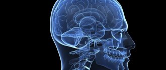
Human nervous system
Brief Definition
The spinal cord pathways or tracts are collections of nerve fibers located inside the spine that carry impulses from the brain to all parts of the body and back. The nerve endings, the combination of which form the pathways, are distinguished by a similar structure, development and general functions. They are divided among themselves according to the tasks assigned to them. The paths are classified as follows:
- Associative. Their main purpose is to unite gray matter cells from different segments to form their own anterior, lateral or posterior bundles.
- Commissural. These fibers connect the gray matter from the two hemispheres. With their help, the coordinated work of individual areas, nerve centers, and both hemispheres occurs.
- Projection. With the help of such pathways, the work of the overlying and underlying parts of the brain is combined. They are the ones who provide the projection of pictures of the surrounding world, as on a monitor screen.
Projection pathways, in turn, are efferent and afferent. They form the basis of the central nervous system, and are divided into ascending (centripetal or sensitive) and descending (centrifugal, motor).
Important! Nerve fibers provide a constant, unbreakable connection to the brain located in the skull and spine. It is thanks to them that the impulse is quickly transmitted, all body movements are coordinated with each other.
The pathways of the brain and spinal cord differ from each other, but they always act harmoniously, ensuring the passage of an incredibly large number of nerve signals from receptors to the central nervous system. Paths are formed from long axons, special fibers that are capable of creating connections among themselves, thus connecting individual segments of the spinal trunk, providing control of effector organs.
[node:field_similarlink]
The meaning of the spinal cord
There are only two main and most important functions:
- Reflex. To put it simply, a whole series of reflex arcs are closed on this organ. It is thanks to this that reflexes are carried out (the so-called spinal reflexes).
- Conductor. The organ in this case acts as a conductor. It conducts signals that come from various organs to the brain. It is through this organ that the brain receives all information and processes it. Everything works the same way in the opposite direction.
Meaning
To perform a basic movement, you need to use thousands of individual neurons, instantly turn on the connection between them and transmit the desired signal. This happens every second, so all departments must be coordinated as much as possible.
It is difficult to overestimate how important the spinal cord is to life. This anatomical structure is of utmost importance. Without it, life is absolutely impossible. This is the link that connects the brain and different parts of our body. It quickly transmits the necessary information encoded in bioelectric impulses.
Knowing the structural features of the departments of this amazing organ, their main functions, you can understand the principles of operation of the whole organism. It is the presence of segments of the spinal cord that allows us to understand where it hurts, aches, itches or feels cold. This information is also necessary for making a correct diagnosis and successful treatment of various diseases.
Spinal cord location
The organ is located in the spinal canal (located inside the human spine). This canal is quite long and practically reaches the lower vertebrae. In fact, this is a special canal, which is an oblong hole in which the spinal cord lies. From the sides it is protected by vertebrae, as well as intervertebral discs.
The organ is also located at the lower edge of the foramen magnum, where connections with the brain occur. It is in this place that there is a huge number of roots that directly connect to the human brain. This connection is called the left and right spinal nerves.
Spinal cord location
The bottom ends at the level of 1-11 vertebrae. Afterwards the organ turns into a thin terminal filament. In fact, it is still the spinal cord, because it contains nervous tissue.
What are the segments?
The sections of the spinal cord differ from those used to conventionally divide the areas of the spine. Their length varies; the fewest elements are found in the coccygeal part. The segments are connected to certain areas in the body by nerve conductors. In general, the brain is divided into the following segments:
Prognosis for spinal cord myelitis
- Neck – 8.
- Chest – 12.
- Sacral – 5.
- Lumbar – 5.
The main length falls on the thoracic segments of the spinal cord, after which 23.2% is allocated to the neck and only 7.3% to the lower back. They are posterior and anterior nerve roots that alternate regularly. They are located above the vertebrae with the same number. Their main task is to report movements and be responsible for muscle contractions. Therefore, the anterior nerve roots are also called motor, and the posterior - sensitive.
The intervertebral foramen contains the roots of each individual segment. Their direction is not the same, because the spinal column is filled with the brain. In the cervical region they lie horizontally, in the thoracic region they are directed diagonally, and in the lower part they are almost vertical.
The shortest segments are the neck segments, and the longest ones are in the lower back and sacrum. In the lower part they form the so-called “horse tail” - this is a bunch of roots located below the 2nd vertebra.
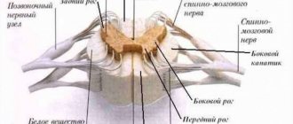
A visual representation of what a spinal cord segment looks like in cross section
No ads 1
Topography and shape of the spinal cord
Let's look at the features of location (topography) and shape.
To do this, consider a number of features:
- The average length is 42-43 centimeters. In men, the length is often several centimeters longer, while in women, on the contrary, it is shorter.
- Weight 33-39 grams.
- There is a middle gap on the front part, it is clearly visible. You can notice that it seems to be growing into the organ. In essence, it creates a kind of partition that divides the brain into two sections.
- In the cervical and lumbosacral regions it is possible to
- mark two fairly serious thickenings. This is due to the fact that innervation of the upper and lower extremities occurs here. In simple terms, this is where the nerve endings from the limbs “attach” to the spinal cord, which
- Allows them to transmit the necessary signals.
- The spinal cord is topographically practically not connected with the vertebrae. The various sections are not located depending on a specific vertebra or several vertebrae.
The increase in volume in these areas is due to the fact that this is where the largest number of nerve cells are located, as well as fibers through which signals are transmitted from the limbs and back.
Despite the fact that the spine is a kind of “storage place” for the organ, the location of the nerve endings, especially in the lower part of the spine, does not correspond to specific vertebrae. This is due to the fact that the length of the spinal cord is less than the length of the human spine.
That is why it is necessary for doctors to know the exact location of each of the segments, because they will not be able to navigate along the spine.
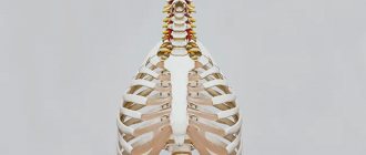
Location in the spine
Appearance
The spinal cord is shaped like a long thin cord that begins in the cervical region. The cervical medulla securely attaches it to the head in the area of the large foramen in the occipital part of the skull. It is important to remember that the neck is a very fragile area where the brain connects to the spinal cord. If it is damaged, the consequences can be extremely serious, including paralysis. By the way, the spinal cord and brain are not clearly separated; one smoothly passes into the other.
At the transition point, the so-called pyramidal paths intersect. These conductors carry the most important functional load - they ensure the movement of the limbs. The lower edge of the spinal cord is located at the upper edge of the 2nd lumbar vertebra. This means that the spinal canal is actually longer than the brain itself, its lower sections consisting only of nerve endings and membranes.
When a spinal tap is performed for analysis, it is important to know where the spinal cord ends. A puncture to analyze the cerebrospinal fluid is performed where there are no longer nerve fibers (between the 3rd and 4th lumbar vertebrae). This completely eliminates the possibility of damage to such an important part of the body.
The dimensions of the organ are as follows: length - 40-45 cm, diameter of the spinal cord - up to 1.5 cm, weight of the spinal cord - up to 35 g. The weight and length of the spinal cord in adults are approximately the same. We have indicated an upper limit. The brain itself is quite long; along its entire length there are several sections:
- cervical;
- chest;
- lumbar;
- sacral;
- coccygeal
The departments are not equal to each other. In the cervical and lumbosacral regions there may be significantly more nerve cells, since they provide motor functions of the limbs. Therefore, in these places the spinal cord is thicker than in others.
At the very bottom is the conus of the spinal cord. It consists of segments of the sacrum and geometrically corresponds to the cone. Then it smoothly passes into the final (terminal) filament, on which the organ ends. It already completely lacks nerves; it consists of connective tissue, which is covered with standard membranes. The terminal thread is attached to the 2nd coccygeal vertebra.
Characteristics of the spinal cord depending on age
Let's consider the features, depending on the age of the person:
- In a newly born child, the length of the organ is 13.5-14.5 centimeters.
- At 2 years, the length increases to 20 centimeters.
- At about 10 years old, the length can reach 29 centimeters.
- Growth ends in different ways, depending on the characteristics of a particular person’s body.
Let's look at the external features and changes depending on age:
- In infants, cervical and lumbar thickening is more noticeable than in adults. The same applies to the width of the central channel.
- The above features become almost invisible by the age of two.
- The volume of white matter grows many times faster than the volume of gray matter. This is due to the fact that the segmental apparatus is formed earlier than the pathways that connect the brain and spinal cord.
Otherwise, there are practically no age-related characteristics, because from birth the spinal cord performs almost all functions, as in an adult.
Reflex function
An equally significant task facing the spinal cord is the implementation of autonomic and motor reflexes. Impulses coming from the brain along descending pathways are responsible for the movements of the entire torso and limbs. It is thanks to the passage of impulses that motor, food and vasomotor reflexes are performed.
Basic reflex activity of the spinal cord:
- Regulation of muscle tone.
- Formation of normal walking.
- Contraction of the anterior and abdominal muscle wall.
- Reflex movement of the limbs: rhythmic, extension, flexion, posture.
The reflex function of the spinal cord is based on communication with the brain. When a signal is received, the flexion and extension reflexes of the spinal cord are activated. They themselves are quite simple in nature. With repeated stimulation, the strength and duration of the reflex increases significantly. The reflex and conduction function of the spinal cord is controlled by the overlying parts of the central nervous system.
The pathways of the brain and spinal cord are a single system that always works harmoniously. This is what ensures the consistency of all body actions and its normal reaction to a given situation. For example, the receipt of a signal along the ascending pathways from receptors that the street is slippery allows, during the sliding process, impulses to be transmitted along the ascending pathways to ensure balance is maintained.
Features of the structure of the spinal cord
Now let's look at the structural features, one by one examining each segment separately that makes up the organ.
Spinal cord membranes
The spinal cord is located in a kind of canal, but at the same time it has protection, which also performs a huge number of functions.
The spinal membranes of the spinal cord, of which there are three in total:
- hard shell;
- arachnoid;
- soft shell.
All shells are interconnected, and below they fuse with the terminal filament.
Spinal cord membranes
White and gray matter
The spinal cord contains white and gray matter.
Let's try to figure out what it is:
- White matter is a complex system of pulpal and non-pulphate nerve fibers, as well as supporting nervous tissue.
- Gray matter is made up of nerve cells and their processes.
White and gray matter of the spinal cord
Spinal cord sections
There are five main sections of the spine, let’s look at them starting from the top:
- cervical;
- chest;
- lumbar;
- sacral;
- coccygeal
Structure
The local location of the spinal cord along the entire back has its own characteristics. This physiology ensures that the organ performs its basic functions. The organ begins at the level of the 1st cervical vertebra, where it is gently rebuilt into the brain, but there is no clear division in them. At the junction there is a crossover of the pyramidal tracts responsible for the motor activity of the limbs. The spinal cord ends in the region of the 2nd lumbar vertebra, so it is shorter in length than the entire spine as a whole. This feature allows lumbar puncture to be performed at the level of the 3-4 lumbar vertebrae, without the risk of damaging the spinal cord.
What is special about the structure? The oblong tube has two grooves at the front and back. The brain is covered by three membranes:
- Solid. It is the tissue of the periosteum of the spinal canal, followed by the epidural space and the outer layer of the dura mater.
- Cobweb. A thin, colorless plate that fuses with the hard shell in the area of the intervertebral foramen. In the place where there is no fusion, the subdural space is located.
- Vascular. A soft membrane separated from the previous one by a subarachnoid space with cerebrospinal fluid. The membrane is adjacent to the spinal cord and consists mainly of choroid plexuses.
The space between them is filled with cerebrospinal fluid - cerebrospinal fluid. The gray matter is located in the center of the organ. It consists of intercalary and motor neurons. It also contains two types of horns: the anterior ones, which contain motor neurons, and the posterior ones, where interneurons are located.
No ads 1
External characteristics
The external structure of the spinal cord largely follows the contours of the spine, since the structures adapt to its physiological curves. Two thickenings are observed in the area of the neck and lower thoracic, the beginning of the lumbar calving. These places are characterized as exits of the roots of the spinal nerves responsible for the innervation of the arms and legs.
The external structure can be briefly described by the following characteristics:
- The shape is cylindrical, flattened on the front and back sides.
- Visually, the spinal cord looks like an elongated “cord” with processes.
- On average, the length of the organ is 42-44 cm, but directly depends on the person’s height.
- The mass is 34-38 g, which is 50 times less than the organ of the brain.
- There are two grooves in front and behind, which visually divide the organ into two symmetrical parts.
- In the middle there is a canal, which in the upper part communicates with one of the ventricles of the brain. Inferiorly, the central canal expands, forming the terminal ventricle.
The thickness of the spinal cord is uneven and depends on in which part the measurement is taken. The organ also has four surfaces: two rounded lateral, a convex posterior and a flattened anterior. The external structure is in many ways reminiscent of the internal part of the ridge, since the organ fills the entire canal. The organ is reliably protected by bone tissue.
[node:field_similarlink]
Internal structure
The spinal cord is made up of nerve tissue cells called neurons. They are concentrated closer to the center and form gray matter. According to rough estimates by scientists, the entire organ contains about 13 million cells, which is many times less than in the head section. The gray matter is located inside the white matter, and if you make a cross section, it will be shaped like a butterfly. This is especially clearly visible in the diagram.
Schematic cross-section of the spinal cord
This unique anatomy allows the spinal cord to be divided into several structures. It is arranged as follows:
- Front horns. They are distinguished by their rounded, wide shape and consist of neurons responsible for transmitting nerve impulses to the muscles. It is precisely because they perform such a task that they are called motor. The anterior roots of the spinal nerves begin in the anterior horns.
- Hind horns. They are distinguished by a long, narrow shape and consist of interneurons. They bear this name due to their ability to receive incoming signals from the sensory roots of the spinal nerves, otherwise they are called dorsal roots.
- Side horns. They are present only in the lower segments of the organ, and contain vegetative nuclei responsible for the dilation of the pupils, or the functioning of the sweat glands.
The physiological function of nerve endings is to transmit signals from the brain to the spinal cord, as well as deliver received impulses in the opposite direction. This ensures interconnection at all levels and areas of the nervous system. Nerve fibers are combined into bundles and are present along the entire length of the spinal cord.
Metamer and segmental structure
Each part of the spinal cord is a constituent element of a specific metamere of the body. Moreover, if there is a “piece” of the spinal cord that includes a section of gray matter with a pair of roots, then the metamer includes the spinal segment itself, muscle fiber (myotome), a section of the epidermis (dermatome), a bone component (scletorome), an internal organ (splanchiotome), controlled by this segment. In humans and higher representatives of the animal world, radicular metamerism is observed - the spinal cord is confined to certain parts of the body.
The skin areas of the body consisting of sensory fibers that approach the corresponding segment of the spinal cord are called dermatomes. They are strips of epidermis controlled by sensitive nerve endings. They are located throughout the body and sometimes overlap each other.
Visual representation of the connection between the skin and the spinal cord
Myotomes are muscle groups that receive motor fibers from certain areas of the brain. Thanks to the study and knowledge of their location, the process of damage and diagnosis of spinal cord lesions is greatly simplified. Damage to a certain segment of the spinal cord provokes sensory and motor disorders.
Connection between the spinal cord and muscle fiber
Segmental structure
The spinal cord is conventionally divided into five sections, although it is a single whole. The name of each directly depends on its location in the body. In total, a person may have 31-33 segments, which consist of:
- Cervical area - includes 8 segments.
- Thoracic region - 12 segments.
- Lumbar region – 5 segments.
- Sacral – 5 segments.
- Coccygeal – 1-3 segments.
This division allows us to examine the organ in more detail and simplify the process of diagnosing various pathologies.
No ads 2
White and gray matter
In cross-section, the symmetrical halves can be seen in detail and the anterior medial fissure and connective tissue septum can be seen. The part located inside is darker and is called gray matter (GM), it is located in a lighter substance - white matter (WM). Most of the SV is located in the lumbar region, the least is observed in the thoracic region. What are the main functions of gray matter:
- Transmission of pain impulses.
- Response to temperature changes.
- Closure of reflex arcs.
- Obtaining information from muscle tissue, tendons, ligaments.
- Formation of pathways.
What is the structure of white matter? It consists of myelinated, unmyelinated nerve fibers, blood vessels and a small amount of connective tissue. Its main task is to trigger simple reflexes and provide connections with skeletal muscles.
Functions of the spinal cord
Let's move on to looking at functions. For convenience, we will consider each separately.
Reflex and motor functions
This function is responsible for human reflexes. For example, if a person touches something very hot, he will reflexively withdraw his hand. This is a reflex or motor function. But let’s take a step-by-step look at how it’s all tripled and how it’s connected to the spinal cord.
It’s best to look at everything using an example, so let’s imagine a situation where a person touched a very hot object with his hand:
- When touched, the signal is primarily received by receptors that are located throughout the human body.
- The receptor transmits a signal to the nerve fiber.
- The signal is sent along the nerve fiber to the spinal cord.
- On the approach to the organ there is a spinal ganglion, where the body of the neuron is located. It was along its peripheral fiber that the impulse transmitted from the receptors was received.
- Now the impulse is transmitted along the central fiber to the posterior horns of the spinal cord. At this moment, a kind of switching of the impulse to another neuron occurs.
- The processes of the new neuron transmit the impulse to the anterior horns.
- Now the return journey begins, because the anterior horns transmit the impulse to the motor neurons. They are responsible for the movement of the upper limbs.
- Through these neurons, the impulse is transmitted directly to the hand, after which the person removes it (motor function).
Reflex arc in the circuit
As a result of this whole process, a person withdraws his hand from the hot object and a reflex arc closes. The whole process takes a split second, so when touching any object, a person immediately feels its temperature, consistency and other features.
Conductor function
In this situation, the organ acts as a conductor. The conductor in this case is between the receptors and the brain. The receptors receive an impulse that is transmitted to the spinal cord and then to the brain. The information is analyzed there and transmitted back.
Thanks to this function, a person receives sensitivity, as well as a sense of himself in space. This has been proven many times, and this becomes especially evident in cases of serious spinal injuries.
Integrative function
This function is often forgotten, but it is no less important for a person than others. The integrative function manifests itself in reactions that cannot be classified as simple reflexes. In order for the body to respond, it is necessary to involve other parts of the human nervous system. This is how the spinal cord can form a connection between organs.
These include reflexes of chewing, swallowing, regulation of digestion, breathing and much more. In fact, this is an invisible function that ensures normal functioning.
Functions
The spinal cord performs two main functions:
- reflex;
- conductor.
It allows you to get sensations and make movements. In addition, it is involved in the normal functioning of many internal organs.
This body can easily be called a control center. When we withdraw our hand from a hot pan, this is clear confirmation that the spinal cord is doing its job. It provided reflex activity. Surprisingly, the brain is not involved in unconditioned reflexes. It would take too long.
It is the spinal cord that provides reflexes designed to protect the body from injury or death.
Spinal cord dysfunction
Impaired function can lead to serious consequences and often even death. Disorders often occur either due to injury or due to various diseases.
For example, due to dysfunction of the spinal cord, a person may lose sensitivity, in which case he may, for example, stop feeling temperature. In the worst case, the disorder can lead to uncontrollable actions of the limbs (or paralysis), disruption of the functioning of internal organs and the nervous system as a whole.
Spinal cord puncture
Puncture of cerebrospinal fluid (CSF) is a procedure that pursues diagnostic, anesthetic and therapeutic purposes. The procedure itself consists of an injection of an angle under the arachnoid membrane between the 3rd and 4th vertebrae, and then a certain amount of cerebrospinal fluid is extracted for research.
During the procedure, the brain itself is not affected, so there is no need to worry about violations. However, this procedure is quite serious and painful.
Spinal cord puncture