Causes
Not a single theory of the origin of the disease has been reliably confirmed.
Presumably, the infectious factor plays an important role in its development. There is often a natural connection between the occurrence of arteritis and previous influenza and group B hepatitis. The genetic programming of pathology also has its supporters. Cases of this rather rare disease have been observed in close relatives and identical twins. The leading role in the formation of inflammation of the vascular wall belongs to immunological disorders, and this is recognized by adherents of all etiological concepts. With temporal arteritis, the immune system reacts inadequately to its own tissues - the process proceeds as an autoimmune one.
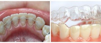
What is the temporomandibular joint - TMJ
We use this joint while talking, chewing, yawning, laughing - very often. Around it are muscles and tendons that allow the jaw to move in different directions for different purposes.
TMJ problems occur for many reasons:
- violation of the dental system, for example, congenital anomalies of teeth, gums or bone tissue, malocclusions, injuries, poor-quality prosthetics;
- congenital anatomical abnormalities of this joint;
- increased or decreased tone of muscle fibers around the TMJ;
- active conversational load, for example, among actors, singers, speakers;
- central nervous system problems;
- disruptions in the functioning of the endocrine system;
- excessive tension of the jaw muscles, for example, due to the bad habit of biting nails.
What is the temporomandibular joint and why do problems arise with it? Detailed and clear - in the video:
Actors and singers with active oratorical articulation are at risk for TMJ arthrosis
Symptoms
The disease has no characteristic onset. Several options are possible: acute, subacute, but more often with a long period of precursors, which can last several weeks or more than one month.
The set of symptoms preceding the height of the disease and united under the general name polymyalgia rheumatica includes the following manifestations:
• general malaise;
• slight increase in body temperature within 37.2–37.5°C;
• excessive sweating, especially at night;
• aching joints;
• pain in the muscles;
• sleep disorders;
• weight loss.
Later, vascular disorders come to the fore; their nature and severity depend on the location and degree of damage to the artery. More than half of patients suffer from vision impairment. Patients note:
• headache of varying intensity, often sudden, in various areas (temporal, frontal, parietal, less often - occipital);
• hyperesthesia (increased sensitivity) of the scalp, making it difficult to comb and wear a hat;
• transient pain and numbness in the tongue and lower jaw, which intensifies when talking and chewing;
• painful, stringy compaction along the inflamed artery;
• visual impairment (decreased visual acuity, diplopia (double image), blindness);
• neurological, mental disorders.
When the aorta, coronary, renal, and mesenteric arteries are involved in the pathological process, which happens in severe untreated variants of the disease, the development of aneurysm, angina pectoris and heart attack, impaired blood supply and kidney and intestinal function are possible.
Treatment: surgical removal of the tumor
Osteomas are treated in one way - only surgery. The operation is indicated for people with severe cosmetic defects or when there is compression of neurovascular structures caused by osteoma.
Such tumors are removed by an oncologist surgeon. If the tumor is small and does not compress nearby anatomical structures, you should not rush into surgery to remove the osteoma. It is advisable to consult a neurosurgeon. And be observed by a doctor, undergoing regular skull x-rays or tomography.
If osteoma manifests itself in eye symptoms, a feeling of fullness, neurological signs, intense and frequent headaches, and increased blood pressure, then in such cases the tumor must be removed.
The smaller the tumor, the fewer problems there will be during surgery. Small osteomas are usually removed endoscopically. They are performed under general anesthesia and are removed piece by piece. Large tumors are removed using craniotomy. And this means the mandatory removal of the plate located close to the osteoma, a healthy bone. This bone area is then replaced with a titanium plate.
Rehabilitation
As after any other operation, a patient who has had an osteoma removed needs time for rehabilitation. Rehabilitation begins already in the surgical hospital. There, doctors are taking measures aimed at preventing secondary infections and accelerating tissue regeneration processes. The second stage of rehabilitation is organizing the correct regimen for the patient and following a special calcium diet.
Important!
After surgery for osteoma of the frontal bone, at least in the first 6 months, take care and avoid ARVI.
Treatment at home, folk remedies
If osteoma is discovered and the patient is waiting for osteoma management, then it cannot be treated independently with any compresses, ointments, or heating. These actions will only speed up the process of tumor development.
Folk remedies for treating osteoma include celandine. But it needs to be collected during flowering, usually in May. At this time, celandine is most useful. It is ground in a meat grinder, then the juice is squeezed out, pouring it into a jar. For 2 weeks, this product, covered with a lid, should ferment. Then the juice is applied to the location of the neoplasm daily, while simultaneously consuming a few drops of it orally.
But still, you should not self-medicate to avoid complications. If you have symptoms of osteoma of the frontal bone, you should immediately consult a doctor. A specialist who has completed the diagnosis will recommend making a decision on the method of treatment.
Be healthy and take care of yourself!
Diagnostics
The diagnosis of arteritis can be established by performing a histological examination of a section of the superficial temporal artery obtained by biopsy. The sample collection is carried out under local anesthesia and is not difficult. The detection of granulomatous inflammation of the vascular wall with the presence of giant cells is indisputable evidence of this pathology.
But histologists are able to identify typical changes only in half of the cases. The fragmentary nature of the lesion does not always allow for successful selection of the segment for biopsy. However, a negative result does not mean the absence of the disease, since the main criterion for diagnosing Horton’s disease is the totality of clinical manifestations.
Criteria have been formulated and generally accepted to recognize temporal arteritis. The diagnosis is reliable if three or more of the following factors are present:
• age over 50 years;
• headaches with severe intensity;
• vision problems;
• presence of complaints characteristic of polymyalgia rheumatica;
• decrease in the number of red blood cells and hemoglobin level in the blood, increase in ESR.
Sphygmography, rheovasography, and Dopplerography of the affected arteries are of auxiliary value for differential diagnosis. For the same purpose, the presence of C-reactive protein and the level of sialic acid and fibrinogen in the blood are determined.
Treatment
At the moment, there are two directions in the treatment of temporal arteritis: therapeutic and surgical . Surgical methods are resorted to in the event of the development of complications such as aortic aneurysm and thrombosis of blood vessels, especially those supplying blood to the eyeball.
The basis of treatment for the disease, without which it is impossible to achieve positive results, is the prescription of steroid hormones (prednisolone). There is no alternative to glucocorticoids. They are prescribed as early as possible and taken for a long time. Doses and regimen are selected individually, under constant laboratory monitoring of clinical and biochemical blood parameters. A combination of hormones and drugs that suppress immune responses is possible. Symptomatic treatment is also carried out using anticoagulants, agents that improve microcirculation, and vitamins.
Forecast
Temporal arteritis is a serious disease. If not recognized in a timely manner and treated inadequately, the pathology poses a threat to the health and life of the patient. A timely diagnosis and compliance with doctors’ recommendations avoid complications and make the prognosis favorable.
In our department of vascular surgery, a full examination, qualified interpretation of the results obtained and professional implementation of all types of treatment measures are possible.
Causes and provoking factors
Bumps on the back of the head always form for pathological reasons. Large, small, hard or soft swellings indicate that at some stage a malfunction occurred in the body. Perhaps the sebaceous gland is simply clogged and a common pimple appears. But in case of unfavorable development of events, the formation of a benign or malignant tumor cannot be ruled out.
Often a person discovers a lump when it grows to a size of 1-2 cm. This is a signal of the progression of the disease, especially if the general state of health deteriorates. Seals in the scalp are dangerous and require careful examination of adults and children.
Injury
If a lump appears under the skin on the head after a bruise, then the relationship is easy to establish. This is the name of a closed traumatic injury, which occurs quite rarely in everyday life. Usually a person hits himself when he falls at home or on the street. Seals are formed due to sports and work injuries. New growths are especially dangerous after blows with a blunt object, traffic accidents and falls from a height. Indeed, in such cases they are often accompanied by skull injuries and (or) concussion.
Unlike other parts of the body, the scalp has almost no fatty tissue, tightly adjacent to the bone. Therefore, after an impact, the blood is not distributed evenly, but accumulates, forming a compaction. Symptoms characteristic of this type of injury:
- severe pain when pressed;
- the appearance of extensive hematoma or pinpoint hemorrhages;
- puffiness, swelling, redness of the skin.
As the lesion heals, the color of the dermis around the lump changes from dark purple to yellow-green. Its size decreases, and then it dissolves and disappears without a trace. If a person has a bump, then an ice pack becomes an excellent protection against hematoma and swelling.
You should consult a doctor immediately if, after the blow, in addition to the bump, you are bothered by attacks of nausea and vomiting, dizziness and headaches. These are specific signs of a concussion that do not go away on their own without consequences.
Allergy to insect bites
Allergies are another common reason why a lump appears on the back of the head. Only it does not develop through the consumption of food or certain medications, or the penetration of pollen or animal hair into the body. A lump in the scalp is the result of an insect attack. These could be mosquitoes, gadflies, horseflies, midges, human fleas living in basements. What indicates the development of an allergy:
- the seal does not hurt even with strong pressure. During a bite, many insects inject bioactive substances into the blood that have anesthetic properties;
- Itching occurs, which makes a person scratch constantly. At the same time, it involuntarily microtraumas the skin, provoking the development of a bacterial infection.
In people who are not predisposed to allergies, only a slight swelling forms after bites. It does not require treatment and disappears within a day. And allergy sufferers often develop large itchy bumps, which resolve after taking antihistamines.
Insects often attack an adult or child in their sleep. They prefer to bite through the thin, delicate skin behind the ears, in the temples and back of the head.
Atheroma
A lump on the head can be nothing more than atheroma. This is a sebaceous gland cyst, which is a cavity with a pasty secretion. Atheroma is a subcutaneous, round, raised, elastic formation. When touched, it is slightly pressed inward due to its soft consistency. Such a tumor easily moves to the sides and grows slowly. If left untreated, it reaches impressive sizes - up to 10 cm in diameter.
When the sebaceous gland is blocked, the secretion does not stop producing. It does not disappear anywhere, accumulates and gradually stretches the walls of the cavity. In this case, there is no unpleasant sensation, itching or pain, because compression of the nerve endings by compaction is excluded. Atheroma does not go away on its own; it requires surgical excision.
When trying to squeeze out the contents of the atheroma, there is a high probability of pathogenic microorganisms introducing into it. A person’s well-being will be aggravated by headaches, suppuration of the lump, and general intoxication of the body.
Lipoma
In everyday life, this benign tumor is called a wen. This is a soft lump on the head made up of mature fat cells. The lipoma is visualized as a round or oblong formation, soft, elastic, covered with unchanged skin. A wen forms on the forehead, in the back of the head, and less often on the top of the head. At first a tiny pea appears, but the swelling slowly increases in size, usually without causing discomfort.
Lipomas do not pose a serious health threat, especially small ones. A large seal can compress the nerve roots, causing pain. If treatment is not carried out, the person does not try to get rid of the wen, then tissue nutrition is disrupted until the formation of an ulcer and a limited area of skin necrosis.
Fibroma
Fibroma in the scalp is a benign tumor consisting of connective tissue. It looks like a small brownish growth, rising 0.5-1 cm above the skin. Fibroma is usually single, but in clinical practice there have been cases of the formation of multiple compactions. It has clear boundaries, is not painful when pressed, and is localized on the back surface of the head.
Like lipoma, fibroma does not pose a threat to human health. But only with its compact size. If the tumor formed several years ago and is constantly growing, then it compresses the surrounding tissues, disrupting their functioning, which is manifested by corresponding symptoms.
Fibroids must be treated. There is a risk of its degeneration and malignancy. It is minimal, but rapidly increases with constant microtrauma of the fibroma surface.
Hemangioma
This is the name of a benign tumor, which consists of cells on the inner surface of blood vessels and is usually detected in newborns. The seal is located on the crown, slightly behind the ears or on the crown. Hemangiomas are not symmetrical, they are localized randomly to the left and right of the longitudinal axis of the skull. Doctors can easily diagnose the following specific bumps:
- occur predominantly in female infants;
- painless when touched;
- rise above the surface of the skin only a few millimeters;
- have clear outlines;
- do not move when pressed.
Their formation does not have any impact on the child’s well-being. Even with the active growth of hemangioma in the first months of life, no discomfort occurs. In most cases, neoplasms in infants disappear on their own. If this does not happen, then treatment is required due to the risk of injury to the tumor.
Warts
In most cases, this is a benign neoplasm of predominantly viral origin, which has the appearance of a compact ball. If it pops up on the scalp, this indicates an increase in the activity of papillomaviruses. Warts are usually localized behind the ear, on the top of the head, in the temporal zone. They have several distinctive features:
- dense, rigid structure;
- the color of the skin is not changed, but a yellowish or pink tint may be present;
- are multiple in nature and tend to merge;
- the affected areas of the skin feel rough and uneven to the touch.
The wart is often injured and changes color, shape and size, can produce scars and scars, and lead to malignancy. When removing seals, there is a high risk of recurrence.