Article:
Dysarthria is a complex speech disorder, one of its most common forms is bulbar.
The word “dysarthria” itself comes from the Greek “dys” - “disorder” and “arthroo” - “pronounce clearly”. Bulbar dysarthria manifests itself as slurred, slurred speech with a dull nasal tone. A child with this disorder speaks as if he has porridge in his mouth. A similar disorder occurs due to damage to the speech apparatus of the CNS (central nervous system). The first detailed description of dysarthria appeared more than a century ago. It was compiled by A. Oppenheim and H. Gutzmann. This disorder was studied in more detail in 1888 by A. Kussmaul.
What is bulbar dysarthria
Bulbar dysarthria is a person’s loss of articulate expressive speech caused by a disorder of the articulation apparatus by damage to the cranial nerves (IX, X, XII pairs). The clinical picture of this disorder is as follows:
- slurred speech;
- slow pace (braking);
- monotone;
- paucity of speech;
- dysphonia;
- the swallowing reflex is impaired.
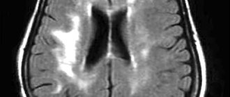
The bulbar type of dysarthria can be established after speech therapy and neurological examination. To identify it, a number of examinations must be carried out - MRI, CT, cerebrospinal fluid analysis and others. The clinical picture of bulbar dysarthria depends on the lesion.
Diagnosis of pseudobulbar dysarthria
The patient should be managed by a speech therapist, neurologist and other specialized medical specialists. For accurate diagnosis, the following research methods are used:
- speech therapy speech study;
- electroencephalography;
- electromyography;
- MRI of the head.
In some cases, a study of cerebrospinal fluid by lumbar puncture may be prescribed. This analysis identifies neuroinfectious causes. A neurologist should participate in the diagnosis. They must undergo an examination, special tests, and a study of the child’s motor skills and facial expressions. These methods make it possible to assess the degree, type, and causes of the disorder, and also, based on the data obtained, formulate an effective treatment program.
Reasons for the development of the disease
The main cause is damage to the bulbar nerves. If we consider from the point of view of anatomy, the glossopharyngeal nerve connects to the muscles of the pharynx, the vagus nerve to the palatine nerve, the hypoglossal nerve approaches the lingual muscle. Damage occurs precisely to these nerves. Experts identify the following causes of bulbar dysarthria:
| № | Factors of speech impairment | Manifestations |
| 1 | Perinatal factors | Complications during pregnancy, childbirth (improper position of the fetus, gestosis, premature birth, placental abruption, trauma during childbirth) |
| 2 | Head injuries | Bulbar dysarthria can develop due to traumatic brain injury of varying severity |
| 3 | Neoplasms in the brain | The growth of tumors in the brain disrupts the normal functioning of its parts. When localized in the speech department, compression of the bulbar structures occurs. Such tumors can be gliomas, medulloblastomas, benign formations |
| 4 | Neuroinfections | The most common of them are meningoencephalitis, viral encephalitis and others. The infection provokes inflammation and compression of the cranial nerve nuclei |
| 5 | Neurodegenerative processes | Destruction of the cranial nerve nuclei occurs |
| 6 | Encephalopathy and angiopathy | Occurs in somatic diseases (diabetes mellitus, abnormalities of cerebral vessels). As a result of such disorders, the blood circulation in the brain is destabilized. |
Before starting corrective work with bulbar dysarthria, it is necessary to accurately determine the cause of its occurrence. Without eliminating the cause, it is impossible to achieve complete restoration of speech and speech apparatus. Rehabilitation and reasons should be prescribed exclusively by specialized specialists in the field of speech therapy and neurology.
Causes
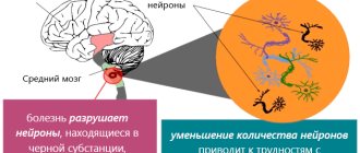
Based on the fact that motor neuron disease according to ICD-10 is classified as a systemic atrophy affecting predominantly the central nervous system, its main characteristics are degenerative processes in the central gyri of the frontal lobes. The destructive process spreads to the nuclei of motor neurons of the brain stem with changes in the corticospinal tracts. The result is slowly progressive muscle atrophy.
The causes of MND include heredity and environmental factors. In familial cases of the development of pathology, mutations of individual genes encoding DNA-binding proteins are noted. The hereditary form of the pathology occurs in 5-10% of cases. In other cases, the cause of the pathology remains unknown.
Provoking factors may be:
- shock and mechanical injuries of the brain and spinal cord;
- excessive physical activity;
- exposure to harmful substances.
Currently, motor nerve disease continues to be studied. Now theories are being developed about the connection of pathology with disruption of the chemical structures of RNA, failure of transport of metabolic products in cells, and disruption of energy metabolism. Research has shown that infectious diseases such as polio do not cause motor nerve disease, although they are characterized by progressive muscle atrophy. Although the pathologies have approximately the same end result, their mechanisms of action are different.
Symptoms
The main symptoms of bulbar dysarthria:
- The child's speech is slurred, inhibited, with poor articulation. The “R” sounds are replaced with fricative ones. Instead of the sound “B”, the sound “P” is pronounced “V”, the sound “P” changes to “F”.
- Consonant voiced sounds are completely or partially absent. Unstressed and stressed sounds are pronounced the same. The letter "A" sounds best.
- There is a noticeable rhythm disturbance. Speech sounds less emotional and monotonous. Expressive speech is not available to a child with dysarthria.
- The child often chokes and has problems swallowing when eating food and water.
- The child's voice is low and nasal.
- Facial expressions are poorly expressed, facial asymmetry is observed (with unilateral nerve damage).
A child suffering from this type of dysarthria has simple slurred articulation. In some cases, voiced sounds disappear from speech. The deaf begin to dominate them. Speech loses its expressiveness and emotionality; it is not melodic. Expressive speech causes great difficulty for the patient and quickly tires him.
The exact symptoms depend on which cranial nerves are affected. In some cases, all of these symptoms may occur.
Causes of syndromes
The main causes of bulbar syndrome include various diseases.
At the onset of the disorder, there is difficulty swallowing liquid food. However, as the disorder develops, along with weakness of the tongue, weakness of the facial and masticatory muscles appears, and the tongue becomes immobile.
Dystrophic myotonia most often affects men. The disease begins at 16 years of age.
Bulbar syndrome causes dysphagia, a nasal tone of voice, choking, and in some cases, breathing problems; attacks of muscle weakness.
The development of the disorder is provoked by increased salt consumption, carbohydrate-rich foods, stress and negative emotions, debilitating physical activity. Pseudobulbar syndrome involves two parts of the medulla oblongata.
Psychogenic disorders can be combined with psychotic disorders and conversion disorders.
This disorder manifests itself with central swallowing paralysis, changes in speech phonation and articulation.
Hypothalamic syndrome is a complex disorder of several body systems - you can learn how to fight it and how to prevent it from our material. Despite the apparent harmlessness, the consequences of a concussion can be extremely severe and lead to irreversible consequences.
Disorders manifest themselves in the following diseases:
- Alzheimer's disease;
- amaurotic idiocy;
- amyotrophy;
- Kennedy's disease;
- damage to the arteries of the brain;
- BASS;
- botulism;
- bulbar palsy;
- thrombosis of the venous sinuses;
- glycogenosis;
- Gaucher disease;
- diffuse sclerosis;
- cerebral palsy;
- encephalopathy;
- Creutzfeldt-Jakob disease;
- myasthenia gravis;
- olivopontocerebellar degeneration;
- brain formation;
- polyradiculoneuropathy;
- polyneuropathy;
- polio;
- myelinolysis;
- multiple sclerosis;
- syringobulbia;
- myopathy;
- encephalitis;
- encephalomyelitis;
- Binswanger encephalopathy.
Diagnosis of bulbar dysarthria
Diagnosis of bulbar dysarthria should be carried out only by specialists. To do this, it is necessary to conduct a number of specialized studies:
- Neurological examination. It allows you to determine the location of lesions of the cranial nerves. The doctor must assess the existing pathological process and its severity, as well as determine the causes of their occurrence.
- Speech therapy examination. The specialist evaluates the rhythm of speech and its speed, voice, mobility of the articulatory apparatus, and breathing rhythm during speech. Based on these data, the speech therapist chooses treatment tactics.
- CT studies, electroencephalography, ultrasound, MRI of the brain. It is necessary to obtain complete information about the pathology, the type of disorders, and on their basis decide on a correction program.
- Lumbar puncture. Necessary for identifying and identifying infectious pathogens.
The doctor may refer the child for additional tests and schedule consultations with other specialists. It may also be necessary to monitor the patient's health over time. Repeated studies may be required during the correction of dysarthria.
Also, the specialist must accurately distinguish the manifestations of bulbar dysarthria from pseudobulbar. The reason for the development of the second is damage to the corticobulbar nerves. To develop rehabilitation, it is necessary to exclude motor, dynamic and acoustic-mnestic aphasia from suspicion of diagnosis.
Diagnostics
There are several diagnostic methods and indicators that indicate the suffering of motor neurons and muscles.
Blood analysis
The main indicator that can indicate the suffering of motor neurons and muscles is the level of creatine phosphokinase. Levels of the enzyme increase as muscle tissue breaks down, and levels may be elevated in people with ALS. However, this indicator is not a specific sign of pathology, since it can also be an indicator of other diseases associated with muscle damage (myocardial infarction, myositis, trauma).
Electroneuromyography (ENMG)
For an accurate diagnosis of ALS, it is necessary to conduct both needle electromyography, using thin needles, with which the doctor assesses the state of electrical activity of damaged and undamaged muscles, and stimulation electroneuromyography to exclude disturbances in the conduction of sensory fibers, which in ALS almost always remain intact.
Transcranial magnetic stimulation (TMS)
This is a new method that can be performed simultaneously with ENMG. It is designed to assess the condition of motor neurons in the brain. TMS results can help make a diagnosis.
Magnetic resonance imaging (MRI)
MRI can detect damage in the brain and spinal cord caused by stroke, Alzheimer's disease, Parkinson's disease, multiple sclerosis, tumors and spinal cord and brain injuries. In some cases, MRI reveals signs that are characteristic only of MND/ALS - this is the luminescence of the pyramidal tracts, which are affected in this disease.
Other methods
To verify the diagnosis, it is also necessary to perform a lumbar puncture, assess the function of external respiration, and conduct cardiorespiratory monitoring. These studies make it possible to identify changes that in the early stages do not bother the patient, but make a detrimental contribution to the progression of the disease.
It should always be remembered that ALS is a disease of exclusion, and the final diagnosis is made only after a full, high-quality examination of the patient under the supervision of experienced neurologists who have extensive experience working with such patients.
Treatment of bulbar dysarthria
Treatment of bulbar dysarthria has better results with parallel rehabilitation measures. A variety of specialists should take part in this work, depending on the causes and severity of the speech disorder. Corrective work consists of several components:
- Etiopathogenetic treatment. It is carried out if dysarthria is caused by infectious diseases. If tumors are present, a special treatment plan is developed together with neurosurgeons.
- Neurometabolic correction. This treatment is aimed at restoring the functioning of cerebral neurons and fibers.
- Speech therapy classes. Aimed at establishing correct sound pronunciation and subsequent automation. Special speech therapy exercises and breathing exercises are used. At later stages, the rhythm of speech and its expressiveness are corrected.
- Rehabilitation therapy. Restores motor skills and psycho-emotionality of the child. This is necessary for proper social adaptation. This work is carried out jointly with a psychologist and psychotherapist.
Causes of speech impairment
Speech disorders occur when the pathways between the bulbar nerves and the cerebral cortex are damaged. This can occur for a number of reasons:
- Cerebrovascular disorders. These are strokes, cerebral ischemia, cerebral atherosclerosis. These diseases often lead to the development of pseudobulbar disorders, leading to disorders of the speech apparatus.
- Head injury. This reason is most common for children over one year of age.
- Perinatal pathology. This group includes fetal hypoxia, intrauterine infections, and birth trauma. As a result of these processes, functional corticobulbar disorders occur.
- Neuroinfections.
For all these reasons, with pseudobulbar dysarthria, the structure of the speech defect suffers and is disrupted.
Clinical picture
Bulbar and pseudobulbar syndromes have many similar symptoms and signs, since the common feature of these disorders is the effect on one muscle.
But if pseudobulbar deviation appears when there is a violation of the central motor neuron , then bulbar deviation when there is a violation of the peripheral neuron . That is, pseudobulbar palsy is central, and bulbar palsy is peripheral.
Accordingly, in the clinic of pseudobulbar disorder, the main thing is an increase in muscle tone, the occurrence of unhealthy reflexes and the absence of muscle atrophy.
In the bulbar clinic, the main factors are muscle weakness, the appearance of muscle twitching, the death of muscle tissue, and the absence of reflexes.
Manifestations of both types of lesions simultaneously occur in amyotrophic lateral sclerosis.
Treatment of the disease
In some cases, urgent assistance is necessary to save life with bulbar syndrome.
The main goal of this assistance is to neutralize the threat to the patient’s life before he is taken to a medical facility, where treatment will then be determined and prescribed.
The doctor, in accordance with the symptoms and nature of the disorder, can predict the outcome of the disease, as well as the effectiveness of the prescribed treatment for bulbar syndrome, which is carried out in stages, namely :
- resuscitation, support of body functions that were impaired due to the disorder - restoration of breathing, activation of the swallowing reflex, reduction of salivation;
- This is followed by treatment of manifestations aimed at alleviating the patient;
- treatment of the disease that caused the syndrome;
- Patients are fed using a feeding tube.
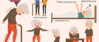
The photo shows special gymnastics for bulbar syndrome
Good results are achieved in the treatment of pseudobulbar syndrome using stem cells.
Their administration to a patient with pseudobulbar disorder leads to the fact that they replace functionally affected cells. As a result, the person begins to live normally.
In case of pseudobulbar and bulbar syndromes, it is quite important to carefully monitor the oral cavity , and also, if necessary, monitor the patient while eating so that he does not suffocate.
Prognosis and complications
Depending on the nature of the disorder, the clinical signs that provoked the syndrome, the doctor assumes the outcome and effectiveness of treatment, which is usually aimed at eliminating the causes of the main deviation.
It is also necessary to support and restore impaired body functions: breathing, swallowing, salivation, etc.
Thanks to the use of proven medications, it is often possible to achieve an improvement in the patient’s condition, but it is very rare to achieve a complete cure.