Today, there are more than 50 million people with epilepsy in the world. This disease is caused by non-infectious brain damage and is chronic. It causes seizures of varying levels of complexity, in which a certain clinical picture is observed: convulsions, fainting, salivation, etc. A single case of such symptoms is not a reason to visit a specialist; every person has experienced a similar condition at least once in their life. If the seizures are constant and prolonged, this becomes a good reason to visit a doctor. In order to find out about the presence of a disease and the extent of brain damage, it is necessary to undergo a lot of diagnostic measures that make it possible to establish the type of disease, as well as its effect on the body. One such method is MRI of epilepsy.
What does an MRI of the brain show in epilepsy?
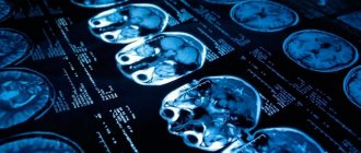
Magnetic resonance therapy for epilepsy shows the affected areas of the brain and is performed in almost all cases of the disease. This is an excellent way to determine the extent to which the disease has spread to healthy cells and the speed of its development. In terms of its information content and harmlessness, MRI is in many ways superior to even computed tomography. In addition, this technique has an obvious advantage: it does not contain X-ray radiation, and it does not affect the human body in any way. MRI of the brain for epilepsy is performed at any age, since this procedure does not provide for severe clinical consequences, it can be used by patients with a disease of any complexity and degree of development.
It is the deep conviction of international antiepileptic organizations that the process of undergoing MRI or CT scanning is mandatory and should be carried out at the request of a doctor.
Many modern clinics with the latest equipment provide the opportunity to diagnose this disease, so the procedure is mandatory. For a successful examination you need:
- Referral for research with a competent formulation of a possible diagnosis and the need for diagnostics. The referral must indicate possible suspicions of pathology and any of its manifestations. Based on previous examinations, including EEG, the neurologist can assume the possible location and nature of the pathological process.
- Modern MRI scanner with optimal power and amplitude. Such devices conduct research even with minimal sections.
- Radiation therapy specialist with in-depth knowledge of neurology, with a good understanding of the specifics of the nervous system. The doctor must know when to use additional data collection methods that are not provided for in the medical protocol.
Whether an MRI shows epilepsy depends largely on physicians and consultants, as well as MR imaging specialists, who are often unable to meaningfully analyze the collected data to establish a treatment modality and confirm a final diagnosis. However, according to statistics from the Ministry of Health, the number of cryptogenic epilepsies is significantly decreasing, which has a positive effect on further treatment.
Who is indicated for MRI to detect epilepsy?
Only an experienced doctor can recognize the symptoms of the early stages of epilepsy. But the patient himself or his relatives can help the neurologist by promptly seeking medical help.
The “risk group” is headed by relatives of a person who already has a confirmed diagnosis. And regular MRI examinations using high-quality equipment (MIBS centers in Moscow use modern tomographs with the main parameter - field strength, 1.5 Tesla, which are the “gold standard” in terms of the resolution of MRI images) - the key to identifying an early, asymptomatic stage epilepsy and timely initiation of treatment for the disease.
An important aspect is that if true epilepsy is suspected, it is recommended to undergo a special examination - MRI of the brain according to the epiprotocol, which determines characteristic changes in the cerebral cortex.
In a person with no family history of epilepsy, it is recommended to undergo an MRI of the brain if at least one of the symptoms of this disease occurs:
- severe convulsive seizures;
- short-term attacks without convulsions - a person freezes, sometimes throwing his head back, his gaze is unfocused, absent, his eyelids may twitch;
- a person regularly loses orientation or even consciousness, walks in his sleep;
- limbs become numb, increased/decreased muscle tone is noted;
- persistent headaches, hearing and vision impairment are observed.
In addition to epilepsy, these symptoms may signal other functional disorders or diseases (including brain tumors), which is an additional justification for the need to undergo an MRI of the brain. In fact, MRI of the brain shows most of the causes that contribute to the occurrence of epilepsy (tissue degradation, vascular abnormalities, the presence of neoplasms, traumatic injury, ischemic injury).
Is it possible to detect epilepsy using MRI?
Epilepsy as a disease has been known for more than 7 thousand years, but instrumental diagnostics began only in 1929. Magnetic resonance imaging in this case is a reliable instrumental method for diagnosing such a pathology, therefore complex procedures are practiced all over the world to determine the spread and development of the disease down to the last small detail. It makes it possible not only to identify the disease, but also to identify the causes that provoked it. A significant advantage of the method is high sensitivity, specificity, and absence of radiation. The Association of Radiologists has developed special protocols for identifying the disease. Thanks to this, it is possible to establish the causes of the pathology, of which there are many:
- defects and anomalies of the brain of various types;
- sclerotic changes;
- oncology and benign tumors;
- changes as a result of impacts and trauma experienced;
- infectious lesions;
- vascular pathologies;
- degenerative changes;
- metabolic disorders.
In other words, diagnostics using modern equipment allows us to examine the pathology from the inside and determine the sources and factors that provoked its development. This method undoubtedly influences the level of treatment, since the final conclusions regarding the patient’s diagnosis are based on MRI or CT readings.
Who prescribes MRI for epilepsy?
Any disease requires the attention of a specialist, and epilepsy is no exception. Especially considering the fact that the most important human organ is affected. After a detailed history, the therapist refers the patient to an appointment with a neurologist, who records the client for an EEG if his suspicions suggest the possibility of damage to tissue cells. This conclusion is required by the radiologist in order to:
- clarify the areas where epileptic activity was detected;
- if necessary, introduce a number of additional programs during the procedure;
- compare the indicators obtained from EEG and MRI, and determine structural changes in tissues.
The use of tomography has several goals pursued by a neurologist. To clarify suspicions and collected data, only those whose history has identified and described a seizure are sent for MRI, since the program allows for a detailed study of possible structural disorders of the areas that are most active during attacks. In addition, the procedure is recommended for patients with an already confirmed diagnosis who cannot get rid of attacks with the help of medications. Drug-resistant epilepsy is not one of the most common pathologies, but its development has certain risks, since drug treatment does not produce results, and the person may not meet the requirements for surgical interventions. MRI is important to clarify the need for surgical intervention for a specific diagnosis.
What is positron emission tomography (PET)?
PET is based on the phenomenon of recording two oppositely directed gamma rays of identical energies resulting from annihilation. The process of annihilation occurs in cases where, with the introduction of metabolites labeled with a short-lived radioisotope, a positron emitted by the nucleus of the radioisotope meets an electron in the patient’s tissues. To conduct research in the interictal period, 2-deoxy-2-(18F)fluoro-D-glucose is used. In case of temporal lobe epilepsy in the interictal period, identification of foci of hypometabolism in the area of epileptogenic foci is indicated. These foci are detected in approximately 80% of patients with temporal lobe epilepsy. However, foci of hypometabolism detected on PET are more extensive than structural changes on MRI and focal changes on EEG and may involve ipsilateral regions of the Sylvian fissure and ipsilateral regions of the parietal lobe.
CT or MRI - which is better?
Quite a lot depends on the choice of equipment, so random visits to such diagnostic centers are unacceptable. For more detailed diagnostics, the MRI unit is equipped with programs that were created to obtain additional information about the condition of the affected tissues. The absolute coverage of such tissues by the information field of the tomograph greatly simplifies the process of determining therapy and the level of risks for individual cases.
MRI according to the “Epilepsy 3 Tesla” program allows you to create optimal conditions for a patient who can succumb to an epileptic seizure at any moment, and collect the necessary information on the specifics and development of the disease in great detail. The use of modernized equipment in medical institutions has led to first-class diagnostic quality, but thereby increased its duration. However, this method is most in demand in cases where it is difficult to give a final conclusion or determine the damaged areas due to the presence of a tumor or vascular compactions. And whether epilepsy is visible on an MRI of the brain is a rhetorical question.
A CT scan is performed to obtain a detailed cross-sectional picture of the brain. Specialized equipment ensures the safety of the procedure and efficiency, and also guarantees the quality of the results obtained. This is a painless examination option, the only contraindication for which is pregnancy. Both tomography options have their advantages, but the need to use one or another device is determined by the attending physician.
Based on the characteristics, it should be noted that these options are suitable for many patients and do not carry great risks. Precautions and contraindications are always determined before the start of treatment and require general tests and a complete medical history. A detailed study of the pathology allows us to exclude possible complications and additional difficulties during therapy. The patient must provide complete information about the disease, personal intolerance to drugs and allergies, and indicate complications of illness or surgery.
What is computed tomography (CT)?
CT is a neuroimaging method based on layer-by-layer transverse scanning with a beam of X-rays (in step-by-step or spiral modes). Registration of radiation is carried out by special detectors, after which, from the received data, through computer processing, an image is obtained. Brain CT is a relatively inexpensive, uncomplicated neuroimaging method that allows for rapid examination of the bony structures of the skull and brain, providing a relatively reliable imaging modality for most patients. In addition, the latest generation of computed tomographs allows one to examine the brain within a few seconds. Although the use of CT in patients with epilepsy is less informative than MRI, CT is still the technique of choice for the evaluation of patients with epileptic syndromes and epilepsy in some pathological conditions. In newborns and infants, CT is often of secondary or additive value, but it serves as a significant addition to ultrasound examination of the child’s brain (neurosonography - NSG). CT allows for early accurate verification of intracerebral hemorrhage, cerebral infarctions, abnormalities of brain development, pathology of the ventricular system, as well as intracerebral calcifications. In older children and adults, CT scan of the brain is the technique of choice in the perioperative period, as it allows to identify recent hemorrhages, acute occlusive hydrocephalus, and space-occupying processes in the brain. CT examination has some advantages in the study of bone defects of the skull, identifying foci of ossification (calcification) of brain tissue in Farah disease, toxoplasmosis, tuberous sclerosis, Sturge-Weber syndrome, as well as in children under 3 years of age, since the processes are incomplete neuronal organization and myelination of conductors, manifested by the lack of a clear demarcation between the gray and white matter of the brain, sometimes does not allow the use of some advantages of MRI - identification of structural changes based on violations of the topography and integrity of neuronal conductor demarcation.
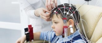
How is an MRI performed for epilepsy?
The main obstacle for many is the fact that during the tomography the patient must remain motionless for up to 50 minutes, which is extremely difficult for people with increased irritability or constant anxiety. The procedure itself is carried out according to the same standards as for any other disease that requires a similar diagnosis. Epilepsy is chronic, and it is quite difficult to predict the onset of the next attack. The main object of the search is focal cortical dysplasia, which is a specific sign of true epilepsy, and not symptomatic.
After anamnesis and mandatory tests with established results, the doctor sends you for diagnostics. The patient takes off all metal jewelry, belts, costume jewelry, precious metals and other paraphernalia and sits on the tomograph couch. After this, it enters the center of the ring magnet, and the diagnostic process itself starts. This manipulation does not cause any sensations or discomfort; any complications or inconveniences can arise exclusively in the technical part of the equipment.
The specialist performing the procedure is in another room behind glass and communicates with the patient via a microphone. There should be no foreign objects in the inspection area that could disrupt the operation of the unit. The patient should listen carefully to the doctor and follow his instructions, while remaining as still as possible. A long, but non-traumatic process extracts all the necessary information about the state of the cells and the extent of the spread of the disease.
How is epilepsy diagnosed?
Anamnesis
Diagnosis of epilepsy begins with a thorough interview of the patient and his relatives. Every detail is important: does the patient feel the approach of an attack, does a loss of consciousness occur, does the patient fall, do convulsions begin in all limbs at once or in one of them, in which direction does he turn his head during an attack, what does he feel after it, etc. .
Epilepsy is an insidious disease that sometimes remains unrecognized for a long time. An experienced epileptologist usually talks for a long time with the patient and his family to get the most detailed description of the disease. This makes it possible to determine the type of attack (generalized or focal, focal simple or focal complex) and suggest in which part of the brain the pathological electrical activity originates and where it spreads. It is very important to know how often attacks occur, at what age the disease began, and whether any immediate relatives had epilepsy.
Neurological examination
The presence of neurological symptoms in a patient such as headache, weakness on one side of the body (hemiparesis), unsteady gait and other signs indicate an organic disease of the brain, such as a brain tumor or multiple sclerosis.
MRI with contrast for epilepsy
It is known that epilepsy is a disease that has several factors that provoke characteristic seizures. This greatly expands the range of searches for affected tissue areas, which is why different research methods are used. Protocols for diagnosing chronic brain lesions involve the use of a contrast agent, which makes it possible to determine the extent of changes and the functioning of the naked brain during the study.
The procedure itself consists of administering a substance intravenously; it quickly spreads through the vessels and veins, ultimately reaching the brain. The contrast itself contains an acceptable amount of iodine and other compounds that, when released into the blood and under the influence of rays, display their own color. This is how the easily recognizable mesh of the venous system appears. On the computer image, clear contours of all channels and the circulatory system of the head are visible, the smallest changes in the structure of blood vessels are determined, and specialists see where inhibition or fluid accumulation occurs. This is another effective way to determine epilepsy by MRI, caused by vascular pathologies or degenerative changes.
Contraindications
There is no need to take any special measures before the diagnosis itself. The patient can continue medication treatment, adhere to the diet and all the doctor’s requirements. Usually the patient leads a full life, without restrictions and precautions. The main recommendation of doctors is a calm environment at work or at home. An isolated case is a patient who experiences a short interval between seizures. Such people are often kept under the supervision of specialists to establish the intervals between attacks, then acceptable options for examination are prescribed.
Despite all the harmlessness, the procedure still has a number of limitations. This is the presence of any metal implants in the body, implants of the inner and middle ear, as well as pacemakers. In addition, it is not recommended to undergo examination for people whose weight exceeds 130 kg. The first months of pregnancy in women are also contraindications. The procedure is contraindicated for people suffering from claustrophobia. Patients in this category undergo pre-treatment or a similar procedure using adapted devices.
MRI for epilepsy in children
Unfortunately, one of the causes of the disease is a congenital pathology of fetal development. Patients of neurologists and epileptologists often include children of all ages. MRI for epilepsy in children is performed in order to exclude the most dangerous causes that require surgical intervention:
- tumor;
- consequences of injury;
- congenital developmental anomalies;
- cortical dysplasia;
- manifestations of genetic diseases.
It is worth remembering that this examination requires standing still (in the case of examining children) for about 20–30 minutes, so this often becomes a problem. Children are afraid of noise, the absence of adults nearby and unfamiliar sensations. In order for the process itself to proceed quickly and efficiently, a qualified specialist is required who can identify the problem and is well acquainted with the changes in epilepsy. To diagnose all possible changes in brain tissue, the examination must be carried out on a high-field (not
Deciphering MRI for epilepsy
MRI images of the brain are interpreted exclusively by a qualified radiologist. Often this is a specialist who personally performed the diagnosis and works closely with the patient’s attending physician. Correctly interpreting and explaining images requires professional medical training and significant practical experience. At the clinic, the patient can receive tomography results a few hours after completion of the procedure or the next day.
Basic information is transmitted electronically to the attending physician. Specialists conduct a thorough examination of the scanograms, after which they report the results. The images are used to prescribe additional diagnostic measures if the disease causes controversial statements. None of the doctors with a narrow specialization in other areas has the right to interpret the results in any order. Any findings by doctors who are not radiation specialists or the patient's attending physician cannot be considered as a final diagnosis.
Where in Moscow can I get an MRI of the brain, which will exclude/confirm the diagnosis of epilepsy?
If you need a quick and accurate MRI of the brain according to the epiprotocol to confirm/refute the diagnosis of epilepsy, we invite you to one of the MIBS-Moscow centers. The epiprotocol for MRI of the brain at MIBS-Moscow has proven its effectiveness over the many years of operation of our MRI centers. Largely due to the accuracy and information content of our images and conclusions, leading neurologists in Moscow and the Moscow region prefer to work with the results of examinations obtained specifically at MIBS-Moscow.
Call now to give your doctor the most informative opinion about the condition of your body!
An MRI performed in accordance with the protocol is the only way to determine the presence of true epilepsy caused by specific disorders of the brain. Only processing of MRI images in a special mode can confirm or refute the diagnosis of epilepsy. This information is the only way to determine whether the cause of seizures is true epilepsy or whether another pathology should be looked for (trauma, swelling, tumors, etc.)
Prices for MRI for epilepsy in Moscow
The results of an MRI examination do not constitute a doctor's opinion. This is one of the diagnostic methods, which is largely educational in nature for the doctor, who uses several similar conclusions to determine the diagnosis and prescribe drug or surgical therapy. Therefore, the images and the information obtained appear in the protocols as the main and legal indications for starting and conducting treatment for the patient. This method of examination by specialists is one of the most popular for many diseases. A narrow range of contraindications allows the use of an objective examination of the whole body under any circumstances and in different patients.
The cost of diagnostics in each clinic is determined differently. This depends on the focus and status of the medical institution in which the person is being examined. In addition, the price is also affected by the specifics of the therapy. A patient who is an inpatient in a hospital receives the service free of charge. The cost varies depending on many factors that are often beyond the control of the organization itself. The emergence of modified equipment with the latest developments often affects the pricing of other units already in operation. But even with such price fluctuations, we can say that this is an affordable survey method. 55% of patients use MRI and CT services. Thus, these methods are recognized as one of the most effective, painless and informative methods in examining pathologies of various types and nature.
What is Single Photon Emission Computed Tomography (SPECT)?
The essence of SPECT is that a pharmacological drug connected to a radionuclide label is injected into a patient’s vein. Depending on the type of radiopharmaceutical (RP), it accumulates in one or another organ. A radioactive label, chemically bound to a pharmaceutical, emits gamma rays and is thus an indicator of its distribution. The features of this distribution make it possible to diagnose various diseases, determine the prevalence of the pathological process, and assess the functional state of brain tissue. SPECT has been used in the study and diagnosis of epileptic seizures for the past 10 years. SPECT is a more accessible diagnostic method compared to positron emission tomography (PET), and is much less expensive.
SPECT is not a mandatory diagnostic method for most patients with epilepsy, but is of great importance in evaluating candidates for epilepsy surgery. Numerous studies using dynamic and static SPECT methods to study the state of the brain in the interictal period have been published over the past 5-6 years. It has been shown that approximately 50% of patients with temporal lobe epilepsy have foci of hypoperfusion in the interictal period, which are detected in the region of epileptogenic regions of the brain. Unlike SPECT in the interictal period, SPECT examination during epileptic seizures (ictal period), for example in patients with temporal lobe epilepsy, is a more accurate method for localizing the epileptogenic focus. Interictal SPECT testing in other (extratemporal) forms of epilepsy is less relevant in providing useful information to the clinician. Studies in recent years have shown that bilateral or ill-defined decrease in perfusion is detected in 30% of patients. On the contrary, seizure (ictal) SPECT for extratemporal forms of epilepsy is highly informative. It has been shown that localization of the focus of seizure SPECT is reliable in 70-90% of cases in patients with frontal lobe epilepsy. With extratemporal seizures, especially if less than 20 seconds have passed since the attack, changes in the blood circulation of the brain are extremely rapid.
Delayed SPECT studies may not detect changes in brain perfusion, especially if they are performed late or early postictal. This is due to the peculiarities of connections and blood supply to the frontal cortex, leading to rapid dynamic focal changes in cerebral circulation during frontal epileptic seizures. Therefore, SPECT should be performed within the first 5-10 seconds from the onset of a frontal seizure to obtain accurate information about the localization of the focus of epileptic activity. Seizure (ictal) SPECT may also be useful for studying the pattern of spread of epileptic activity to other parts of the brain during a seizure. In temporal lobar epilepsy, the spread of epileptic activity to the basal ganglia from the ipsilateral side in the form of foci of hyperperfusion is often detected. This correlates with contralateral dystonic hand placement. On the contrary, in extratemporal forms of epilepsy the nature of the spread of epileptic activity to other parts of the brain is more complex. In epileptic seizures originating from the midfrontal regions of the brain, epileptic activity often involves the subcortical (basal) nuclei in the ipsilateral hemisphere or both hemispheres, as well as the contralateral hemisphere of the cerebellum. In dorsolateral frontal epileptic seizures, the spread of epileptic activity from the primary focus is most typical to the ipsilateral subcortical nuclei and the contralateral cerebellar hemisphere. The main limitation of the introduction of SPECT techniques into practical healthcare is the material and technical equipment of the neuroimaging departments of medical institutions and research institutes.