Scientists consider the frontal cortex as a set of formations that, from an early age, exhibit pronounced individuality in their anatomical structure. Among these formations there are those that are new, “ human ” fields that develop at a later age. These include field 46.
Field 46 is a “human field”, because it is an evolutionary neoplasm that differentiates late. Field 46 is the last to mature and reaches 630% of its original size. Because this field is inhibitory, you can notice that children do not control their movements and grab everything that is not lying well. This behavior is typical of monkeys.
General
It is impossible to specifically develop the frontal lobes of the brain in children. There is a misconception in society that physical activity promotes increased blood circulation in the brain, thereby developing all areas of the brain. Physical activity fills the motor centers of the brain, while the rest of the brain ' rests ', because When performing different tasks, the brain uses specific centers, rather than the entire brain.
Based on the above, in order to determine exercises for the development of the frontal lobes, we need to find out what functions the frontal lobes are responsible for, with which we can develop the frontal lobes.
The frontal lobe, like the others, consists of white and gray matter.
Assigned functions
Evolutionarily, it so happened that the active development of the frontal lobes is not associated with mental and intellectual activity. The frontal lobes arose in humans through evolution. The more a person could share food within his community, the more likely the community could survive. In women, the frontal lobes arose for a specific purpose: sharing food. The men got this area as a gift. Without those assigned tasks that lie on the shoulders of women, men began to use the frontal lobes in a variety of ways (thinking, building, etc.) to demonstrate Dominance.
Essentially, the frontal lobes are inhibitory centers . Also, many people ask what the left or right frontal lobe of the brain is responsible for. The question is not posed correctly, because... in the left and right frontal lobes there are corresponding fields that are responsible for specific functions. Roughly speaking, the frontal lobes are responsible for:
- thinking
- coordination of movements
- conscious control of behavior
- memory and speech centers
- display of emotions
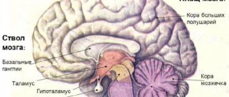
Limbic system
The limbic system (systema limbicum) consists of two large convolutions and several smaller parts of gray matter, as well as fascicles of white matter, which together form a ring surrounding the diencephalon. It is quite easy to remember the name of this system, because from Latin the word “limb” is translated as “ring”.
There are several other meanings of this word. According to one of them, “limb” means “edge”. This is due to the teaching of the Catholic Church, according to which the souls of unbaptized infants and virtuous people who died before the birth of Christ do not go directly to heaven, but end up in a place called limbo. If I'm not mistaken, in Catholic myths there is a separate limbus for infants (limbus puerorum) and a separate limbus for people who lived in ancient times (limbus patrum).
You can read about it in the Divine Comedy - when I first opened it, I was very surprised at the lack of jokes.
But, fortunately, today we are analyzing anatomy, so for us the limbic system is certain formations surrounding the diencephalon. Let's figure out what it includes.
Cingulate gyrus
The cingulate gyrus (gyrus cinguli) is what we see if we go down from the frontal and parietal lobes. On the superolateral surface we saw the lateral sulcus, below which the temporal lobe was located. Here we see a long gyrus, which has the shape of a semicircle and is called the cingulate gyrus.
The groove that separates it from above from the frontal and parietal lobes is called the cingulate groove (sulcus cinguli). From below, this gyrus is delimited by the groove of the corpus callosum (sulcus corporis callosi). This name is due to the fact that directly under the groove there is a large bundle of white matter, which is called the corpus callosum.
In the Wikipedia files I found an excellent illustration from the old-school Gray atlas, in which the cingulate gyrus is tinted yellow:
Unlike the image from the Sinelnikov atlas with which we are working, this shows the brain of a person looking to the right, not to the left.
Parahippocampal gyrus
As you remember, we must draw a limb, that is, a ring. We have half a ring, so we'll draw another half. This half will be the parahippocampal gyrus (gyrus parahyppocampalis), already familiar to us from the relief of the cortex of the lower surface. I am using a drawing with the cingulate gyrus already highlighted, on which I will add the parahippocampal gyrus in orange:
The anterior, posteriorly rounded part of the hippocampus is called the uncus. The posterior part of the hippocampal gyrus, without a sharp border, passes into the lingual gyrus (gyrus lingualis). The hippocampal gyrus is separated from the rest of the temporal lobe by the collateral sulcus (sulcus collateralis).
Components of the limbic system
In addition to the cingulate and parahippocampal gyri, the limbic system includes several other anatomical structures. To demonstrate some of them, it is necessary to dissect the temporal lobe to its deepest parts.
Amygdala
However, in the wonderful (already mentioned by me) Gray atlas there is a frontal section of the cerebral hemispheres, in which we see a rounded formation in the thickness of the temporal lobe. This is nothing more than the amygdala (corpus amygdaloideum), which is also called the amygdala.
Without selection, this area looks like this:
In classical textbooks and atlases, the amygdala is part of the basal ganglia of the brain. I will definitely talk about them in the next article.
Hippocampus
The hippocampus is a collection of gray matter, which is also located in the thickness of the temporal lobe medial to the parahippocompal gyrus and has approximately the same shape, only smaller in size. The hippocampus is located in close proximity to cavities called the lateral ventricles. I'll write an article about them, and then we can use them as a guide to find the hippocampus.
Gray's atlas comes to my aid again. Here we see the hippocampus, tinted turquoise:
I also managed to get this blurry illustration from the Wikipedia archives. Here we see both hippocampi, which are tinted in red:
Olfactory analyzer
All parts of the olfactory analyzer, except the olfactory organ itself and the thalamus, are part of the limbic system. These are the olfactory bulbs, olfactory tracts, olfactory triangles and anterior perforated substance, as well as the mastoid bodies. We studied all these formations in the previous lesson and in the lesson about the diencephalon.
In total, the components of the limbic system are:
- Cingulate gyrus (gyrus cinguli);
- Parahippocampal gyrus (gyrus parahpocampalis);
- Hippocampus;
- Amygdala (amygdala);
- Olfactory bulbs (bulbus olfactorius);
- Olfactory tracts (tractus olfactorius);
- Olfactory triangles (trigonum olfactorium);
- Anterior perforated substance (substantia perforata anterior);
- Mastoid bodies (corpus mamillare);
We looked at the boundaries of the frontal, parietal and occipital lobes, as well as the limbic system. Now we can study the individual grooves and convolutions of these anatomical formations.
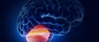
Occipital lobe
Yes, we will start with the occipital lobe, because, firstly, this is where the most noticeable changes are compared to the superolateral surface, and secondly, it will be a little easier for us to navigate.
As I already said, the occipital lobe is separated from the parietal lobe by the deep, prominent parieto-occipital sulcus (sulcus parietooccipitalis). It is this groove that is projected onto the superolateral surface, wanting to show the boundaries of the occipital lobe, although there are no landmarks for it on the superolateral surface.
In addition to the parieto-occipital groove, we can notice a large groove crossing the occipital lobe and dividing it into two halves. This groove is called the calcarine groove (sulcus calcarinus), and the upper of the two halves is called the wedge (cuneus).
Parietal lobe
The parietal lobe on the medial surface looks slightly different than on the superolateral surface. On the medial surface, the parietal lobe is limited by the parieto-occipital sulcus from below, and from above it does not have any landmark, therefore the projection of the central sulcus is used to separate the frontal and parietal lobes. You can see this in our illustration from Sinelnikov’s atlas.
However, another groove is located here - the cingulate. It, dividing the frontal lobe and the cingulate gyrus, continues posteriorly, towards the longitudinal fissure of the cerebrum. The posterior part of the cingulate gyrus crosses the parietal lobe and forms the precuneus. I highlighted the boundaries of the wedge in blue, and the boundaries of the precuneus in green. The arrow points to the posterior part of the cingulate sulcus.
The illustration I have already used from Gray’s atlas is very informative. Here we see both the wedge and the precuneus, clearly separated by the corresponding grooves:
Frontal and temporal lobes
On the medial surface we can see the same convolutions of the frontal lobe that we could see on the superolateral surface. So, in the figure above we see the superior and middle frontal gyri. The only difference is that from below the frontal lobe is limited not by the lateral groove, but by the cingulum (sulcus cinguli), below which part of the limbic system is located (see above).
The relief of the temporal lobe on the medial surface repeats the relief of the temporal lobe of the lower surface - remember the two extended, large temporo-occipital gyri, medial and lateral. The temporal lobe on the medial surface, as we have already said, is limited by the parahippocampal gyrus, which is part of the limbic system.
What fields are included?
Fields and subfields are responsible for specific functions that are generalized under the frontal lobes. Because The polymorphism of the brain is enormous; the combination of the sizes of different fields makes up a person’s individuality. Why do they say that over time a person changes. Throughout life, neurons die, and the remaining ones form new connections. This introduces an imbalance in the quantitative ratio of connections between different fields that are responsible for different functions.
Not only do different people have different margin sizes, but some people may not have these margins at all. Polymorphism was identified by Soviet researchers S.A. Sarkisov, I.N. Filimonov, Yu.G. Shevchenko. They showed that the individual ways in which the cerebral cortex is structured within one ethnic group are so great that no common features can be seen.
- Field 8 - located in the posterior parts of the middle and superior frontal gyri. Has a center for voluntary eye movements
- Area 9 - dorsolateral prefrontal cortex
- Area 10 - Anterior prefrontal cortex
- Area 11 - olfactory area
- Area 12 - control of the basal ganglia
- Field 32 - Receptor area of emotional experiences
- Area 44 - Broca's Center (processing information about the location of the body relative to other bodies)
- Field 45 - music and motor center
- Field 46 - motor analyzer of head and eye rotation
- Field 47 - nuclear zone of singing, speech motor component Subfield 47.1
- Subfield 47.2
- Subfield 47.3
- Subfield 47.4
- Subfield 47.5
Literature
- Collins A., Koechlin E. Reasoning, learning, and creativity: frontal lobe function and human decision-making //PLoS biology. – 2012. – T. 10. – No. 3. – P. e1001293.
- Chayer C., Freedman M. Frontal lobe functions // Current neurology and neuroscience reports. – 2001. – T. 1. – No. 6. – pp. 547-552.
- Kayser AS et al. Dopamine, corticostriatal connectivity, and intertemporal choice // Journal of Neuroscience. – 2012. – T. 32. – No. 27. – pp. 9402-9409.
- Panagiotaropoulos TI et al. Neuronal discharges and gamma oscillations explicitly reflect visual consciousness in the lateral prefrontal cortex // Neuron. – 2012. – T. 74. – No. 5. – pp. 924-935.
- Zelikowsky M. et al. Prefrontal microcircuit underlies contextual learning after hippocampal loss // Proceedings of the National Academy of Sciences. – 2013. – T. 110. – No. 24. – pp. 9938-9943.
- Flinker A. et al. Redefining the role of Broca's area in speech //Proceedings of the National Academy of Sciences. – 2015. – T. 112. – No. 9. – pp. 2871-2875.
Symptoms of the lesion
Symptoms of the lesion are revealed in such a way that the selected functions are no longer adequately performed. The main thing is not to confuse some symptoms with laziness or imposed thoughts on this matter, although this is part of frontal lobe diseases.
- Uncontrollable grasping reflexes (Schuster reflex)
- Uncontrolled grasping reflexes when the skin of the hand is irritated at the base of the fingers (Yanishevsky-Bekhterev Reflex)
- Extension of the toes due to irritation of the skin of the foot (Hermann's sign)
- Maintaining an awkward arm position (Barre's sign)
- Constantly rubbing your nose (Duff's sign)
- Speech Impairment
- Loss of motivation
- Inability to concentrate
- Memory impairment
The following injuries and illnesses may cause these symptoms:
- Alzheimer's disease
- Frontotemporal dementia
- Traumatic brain injuries
- Strokes
- Oncological diseases
With such diseases and symptoms, a person may not be recognizable. A person may lose motivation, and his sense of defining personal boundaries becomes blurred. Impulsive behavior associated with the satisfaction of biological needs is possible. Because disruption of the frontal lobes (inhibitory) opens the boundaries to biological behavior controlled by the limbic system.
Frontal, parietal and occipital lobes
The central sulcus, which divided the frontal and temporal lobes, is only partially present here, here it is:
Accordingly, the frontal lobe is located in front of it, and the parietal lobe is located behind it. On real preparations of the cerebral hemispheres, which are kept in the anatomical hall of my university, the central sulcus descends much lower and is much more distinguishable.
But here we can finally fully separate the parietal and occipital sulci of the deep, noticeable occipital-parietal sulcus (sulcus parietooccipitalis):
You can navigate by the shares thanks to this excellent illustration from Sinelnikov’s atlas. Here we see the boundaries of the frontal lobe (highlighted in yellow), parietal lobe (highlighted in green) and occipital lobe (highlighted in blue).
I will definitely look at all the structures within these lobes, but for now I encourage you to look at where the temporal lobe is located on the lateral surface. Here we see several strange formations in the center, which seem to be surrounded by a ring. This is the same limbic system that I briefly mentioned in the last article. It's time to get to know her in more detail.