- Home >
- Symptoms >
- Pain in the left side under the ribs
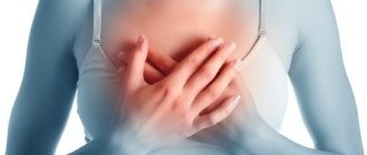
Most people associate pain under the ribs on the left side with heart problems.
However, a similar syndrome can be a sign of other diseases - from intercostal neuralgia to renal failure. There is no clear reason that will allow you to initially determine the diagnosis based on the localization of pain. That is why pain under the left rib requires consultation with a specialist. A visit to the clinic under such circumstances should not be postponed in order to avoid unforeseen health consequences. Timely diagnosis will help determine the cause, and treatment prescribed by a doctor will relieve you of your ailments in the shortest possible time.
Causes of pain on the left under the ribs
Localization of discomfort in the area of the left rib may indicate the development of an acute process or the presence of chronic diseases of the nervous, endocrine, respiratory, cardiovascular systems, gastrointestinal tract, injuries of the diaphragm, ribs, liver, kidneys, spleen, etc.
Periodicity, irradiation, accompanying syndromes (nausea, vomiting, headaches), factors that intensify the pain syndrome (physical activity, respiratory activity, change of position, eating or drinking water) - this information provides the basis for a detailed analysis of pain and understanding why it hurts on the left side. ribs.
To diagnose pain, a list of studies (laboratory, instrumental) is used: CBC, blood biochemistry, urine analysis, radiography, ultrasound examination, MRI, CT. Depending on the medical history, external examination and research results, a consultation with a specialized specialist (gastroenterologist, traumatologist, cardiologist, endocrinologist, surgeon, etc.) is scheduled.
In the structure of radiology diagnostics of the abdominal organs, visualization of the spleen occupies a special place due to the low incidence of pathological processes. However, we must not forget about the variations in the normal anatomy of the spleen and the possibility of regenerating its tissue after removal (reparative regeneration).
Clinical example
In an outpatient patient K., 70 years old, an ultrasound examination on January 28, 2013 revealed a mass formation in the left hypochondrium; there was a history of splenectomy (automobile trauma in 2002); multislice computed tomography (MSCT) with contrast enhancement was recommended as a follow-up examination.
An MSCT study with intravenous contrast enhancement on January 31, 2013 revealed an additional volumetric formation in the bed of the removed spleen with smooth, clear contours, dimensions 38×51×40 mm, the nature of the accumulation of the contrast agent was similar to the spleen tissue with the presence of a feeding vessel extending from the splenic arteries, most likely ectopic tissue of the spleen (splenosis; Fig. 1, a-c).
Figure 1. MSC tomogram of the abdominal organs with intravenous contrast enhancement, arterial phase in patient K. a - hypervascular formation in the bed of the removed spleen (arrow)
Figure 1. MSC tomogram of the abdominal organs with intravenous contrast enhancement, arterial phase in patient K. b - maximum intensity projection (MIP): a feeding vessel extending from the splenic artery to the formation in the bed of the removed spleen (arrow)
Figure 1. MSC tomogram of the abdominal organs with intravenous contrast enhancement, arterial phase in patient K. c - multiplanar reconstruction (MPR; coronal projection): hypervascular formation in the bed of the removed spleen (arrow).
A control CT study with intravenous contrast after 6 months did not reveal any growth dynamics of the previously detected spleen tissue.
Discussion
Normal anatomy and developmental variants of the spleen
Spleen
- an unpaired parenchymal organ of the hematopoietic and lymphatic systems, which is located in the upper floor of the abdominal cavity, deep in the posterior part of the left hypochondrium. The dimensions of the spleen are variable: length 12-14 cm, width 8-10 cm, thickness 3-4 cm, density characteristics of the parenchyma in MSCT studies 40-50HU. The main blood supply to the spleen is carried out by the largest branch of the celiac trunk - the splenic artery. It is important to be aware that during the arterial phase of contrast enhancement, a characteristic “marbled” (pallisading) pattern of enhancement of the splenic parenchyma is detected, which is likely due to differences in pulp perfusion and should not be confused with a focal lesion.
Normally, 10% of adults have, in addition to the normal spleen, additional spleens called accessory lobules of the spleen, or spleens. They are usually found in the hilum of the spleen or near the tail of the pancreas, but can also be found in the mesentery of the small intestine or greater omentum [1].
Anatomical asplenia (absence of the spleen) and polysplenia (several small vestigial bodies of the spleen) are associated with congenital heart disease and isomerism (bilateral right or left-sided anatomy of the heart, bronchi and abdominal organs). This is influenced by the fact that the development of the spleen and heart occurs during the same period of intrauterine development.
Splenosis
Splenosis is an autotransplantation of splenic tissue, usually as a result of traumatic rupture of the spleen or splenectomy. The average interval between injury and the onset of abdominal or pelvic splenosis is 10 years (range 5 months to 32 years). On average, the period of onset of thoracic splenosis is 21 years (3-45 years) [2]. Thoracic splenosis usually occurs with splenic rupture accompanied by a simultaneous diaphragmatic rupture and is therefore less common [3]. Subcutaneous splenosis is a rare condition, the pathogenesis of which may be explained by mechanical implantation. In all cases where he was observed, there was surgery or post-traumatic scarring.
It is important to distinguish splenosis from accessory lobules of the spleen, since both conditions are manifestations of ectopic splenic tissue. In the first case we are dealing with an acquired process, and in the second with a congenital condition. The accessory lobule of the spleen is histologically represented by unchanged splenic tissue, in contrast to splenosis, in which the structure is changed due to poorly formed white pulp, normal content of red pulp and the absence of trabeculae. Also, splenic tissue with splenosis has fewer elastic fibers, the absence of a hilum, and a poorly formed capsule [4]. In addition, accessory spleens are located near the gastrosplenic ligament, left hypochondrium, while splenosis can be in any floor of the abdominal cavity or even have an extraperitoneal localization. Autografts of splenic tissue after splenic trauma are often numerous, have variable size and shape, and are located along the peritoneum and pleura, retroperitoneum, pericardium, lungs, and even subcutaneously.
Pathogenesis
Splenosis begins at the time of splenic rupture or splenectomy, when the splenic pulp enters the abdominal cavity [5]. The number of ectopic splenic tissue nodules that develop in the peritoneal cavity is hypothesized to correlate with the severity of splenic injury.
Another mechanism of splenic tissue transplantation is splenic vein embolism or hematogenous spread of splenic pulp, which occurs in intrahepatic and intracranial splenosis.
Splenosis has rare clinical manifestations. Occasionally, patients experience nonspecific abdominal pain (likely due to tissue infarction), intestinal obstruction due to external mechanical compression by ectopic splenic tissue, gastrointestinal bleeding, or hydronephrosis. Pleurisy and hemoptysis may be symptoms of thoracic splenosis.
A particular difficulty in diagnosis is splenosis, which simulates a primary disease of other organs (liver, pancreas).
Patient A., 48 years old, was routinely admitted to the surgical department of the Federal State Budgetary Institution Central Clinical Hospital with a polyclinic for additional examination and surgical treatment. On an outpatient basis, ultrasound and MSCT with intravenous contrast were used to diagnose her as a “formation of the tail of the pancreas with focal liver damage of a secondary nature.”
However, during a preoperative examination during ultrasound of the abdominal organs and retroperitoneal space, a volumetric formation was revealed in the bed of the removed spleen (splenectomy in 1998, torsion of the elongated pedicle of the spleen) measuring 31×33 mm, comparable in echogenicity and echo structure to spleen tissue (Fig. .2).
Figure 2. Ultrasound data of the abdominal organs. a — isolated scanning mode: in the bed of the removed spleen a formation is visualized, comparable in echo structure and echogenicity to spleen tissue (arrows)
Figure 2. Ultrasound data of the abdominal organs.
b — energy mapping mode, longitudinal scanning: single vessels are visualized in the formation. No focal liver damage was detected by ultrasound. A routine MSCT examination of the abdominal cavity with intravenous contrast enhancement revealed an additional structure with smooth, clear contours in the bed of the removed spleen measuring 30×33×32 mm, closely adjacent with its lateral surface to the tail of the pancreas, the nature of the accumulation of the contrast agent is similar to the spleen tissue, without signs of hypervascularization, the liver parenchyma is homogeneous (Fig. 3).
Figure 3. MSC tomogram of the abdominal organs with intravenous contrast enhancement, arterial phase in patient A. a - hypervascular formation at the level of the tail of the pancreas, according to the density characteristics and type of contrast enhancement corresponding to the spleen tissue (arrow)
Figure 3. MSC tomogram of the abdominal organs with intravenous contrast enhancement, arterial phase in patient A. b - maximum intensity projection (MIP; arterial phase): a feeding vessel extending from the splenic artery to the ectopic tissue of the spleen, simulating the formation of the tail of the pancreas ( arrow)
Figure 3. MSC tomogram of the abdominal organs with intravenous contrast enhancement, arterial phase in patient A. c - multiplanar reconstruction (MPR; coronal projection): a formation in the bed of the removed spleen at the level of the tail of the pancreas (arrow).
Thus, taking into account the anamnesis, the absence of a clinical picture, and the results of radiation imaging methods, the incoming diagnosis was changed to “splenosis of the left subdiaphragmatic region.”
Diagnostics
Since most patients do not complain, the presence of ectopic splenic tissue is often an incidental finding on ultrasound, CT, or MRI. If a mass similar to normal splenic tissue is found during diagnosis in a patient with a history of splenic trauma or splenectomy, the diagnosis of splenosis should be considered.
According to ultrasound data
a well-demarcated (for example, with perihepatic splenosis), hypoechoic formation with clear contours and single arterial and venous vessels is determined.
With MSCT
A hypodense round formation with smooth, clear contours is determined, possibly multiple. The density and features of contrast enhancement of the formation are similar to those of the spleen tissue: ~50HU, hyperdense in the arterial phase, isodense with the liver parenchyma in the portal phase (with the perihepatic variant of splenosis), and hypodense in the parenchymal phase.
With MRI
Normal splenic tissue is hypointense on T1-weighted images (WI) and hyperintense on T2-weighted images. Occasionally, the spleen may be hypointense on T2-weighted images due to excess iron deposition. Typically, splenic tissue implants for splenosis have the same signal intensity and contrast enhancement characteristics on MRI. Typically, heterogeneous enhancement of splenic tissue occurs in the arterial phase of MR studies, while in the delayed phase this ectopic tissue becomes homogeneous.
In splenosis simulating a liver mass, a hypointense rim around the mass may be noted on T1-weighted images. The presence of a peripheral rim of the lesion is characterized by low signal intensity on T1-weighted images and T2-weighted images and represents a thin layer of fat or fibrous capsule around the lesion and is described as a characteristic feature of splenosis. This sign would be nonspecific for primary liver disease.
Scintigraphy
A widely used radionuclide diagnostic method for identifying additional splenic tissue after ineffective splenectomy is scanning with the introduction of colloidal radiopharmaceuticals labeled with 99mTc and 111In. Their spectrum is quite wide, but colloidal sulfur labeled with 99mTs has the greatest sensitivity. Scintigraphic isotopes, such as 99mTc, are detected in reticuloendothelial tissue (liver, spleen, bone marrow) [6]. Using this method, even small areas of spleen tissue can be visualized.
However, a more specific method is scanning with the introduction of 99mTc-labeled red blood cells that have been heat-treated. The physiology of this method lies in the fact that damaged red blood cells will selectively accumulate in the spleen tissue. Few radioisotope laboratories are equipped for this technique, so its use is limited.
MRI with intravenous iron oxide
Another specific method for diagnosing splenosis is MRI with the introduction of nanoparticles as an alternative to traditional contrast agents.
For this purpose, intravenous administration of superparamagnetic iron oxide nanoparticles is used, which are subject to nonspecific capture by cells of the reticuloendothelial system.
In this regard, iron oxide nanoparticles are used for imaging the liver, spleen and lymph nodes. Superparamagnetic iron oxide nanoparticles are highly detectable
even at very low concentrations. There is evidence of imaging of individual cells containing these nanoparticles and even individual nanoparticles. Therefore, this type of nanoparticles
actively used for tagging individual
cells and tracing their migration routes in vivo [7].
Differential diagnosis
- metastatic lesion;
- enlarged lymph node (abdominal lymphadenopathy);
- depending on the location, it can imitate malignant formations of other organs and tissues;
- endometriosis.
Conclusion
Splenosis is a benign condition that is often misdiagnosed as a neoplastic process, and therefore splenosis should be included in the differential diagnosis for neoplasms, single or multiple, in the abdominal cavity or even in atypical locations (intrahepatic, pleura, lungs, pericardium, retroperitoneum , or subcutaneously) in patients with a history of splenic trauma or after surgical removal of the spleen.
It is necessary to know the normal anatomy of the spleen, variants of the anatomical structure and be able to interpret data from various imaging methods for the correct diagnosis of splenosis.
Diseases associated with pain on the left under the ribs
The reasons why it hurts under the left rib may be:
- cardiomyopathy, myocardial infarction;
- enlarged spleen, splenic rupture;
- intercostal neuralgia;
- gastrointestinal dysfunction (ulcer, gastritis, gastroduodenitis, stomach cancer, pancreatitis);
- kidney dysfunction;
- pulmonary diseases (pneumonia, pneumonia, cancer, pleurisy);
- endocrine disorders;
- vegetative-vascular dystonia;
- injuries to the ribs, diaphragm, internal organs;
- musculoskeletal system (osteochondrosis, protrusion, etc.).
The nature of the pain (sharp, dull, aching, pulling, shooting) also provides information about the possible source of the problem. Pain in the left side under the ribs often accompanies injuries and ruptures of internal organs. Dull, diffuse, long-lasting pain distinguishes gastrointestinal diseases; they are also indicated by pain in the morning in the epigastric region. Morning pain after a long sleep of a shooting nature with sudden movements is a characteristic of osteochondrosis. Permanent aching pain is a sign of disturbances in the functioning of the heart, and aggravated by nausea and vomiting is a sign of stomach ulcers.
If you have any questions, ask our specialist! Ask a Question
Why does my left side hurt under my ribs?
Pain in the left side is characteristic of diseases of the abdominal organs. Sometimes there are cases when the cause of pain can be pathologies of the lungs, heart and neuralgia. To identify the exact cause, instrumental diagnostics are used using ultrasound, x-rays and radio wave studies. Depending on the cause of the pain in the left side, appropriate treatment is prescribed. Below, we will talk in detail about the most common pathologies that cause pain. But you should understand that self-diagnosis and self-medication will most likely only worsen the condition, so you should contact a qualified specialist for help as soon as possible.
- Diseases of the spleen.
The spleen is a hematopoietic organ. Inflammation is characterized by a rapid increase in size of the spleen (splenomegaly), and with injuries there is a high risk of rupture of the organ capsule with heavy bleeding. Diseases of the organ are characterized by bursting, pressing pain in the left hypochondrium, which does not depend on physical activity and food intake. - Intestinal diseases.
Pathology of the small intestine leads to visceral or referred pain in the left side. A tumor of the left colon in the later stages of the disease causes aching pain that intensifies 2-3 hours after eating. The pain syndrome occurs against a background of weakness, weight loss, and aversion to food. - Stomach diseases.
Gastritis and gastric ulcer cause pain in the epigastric region and in the left hypochondrium. In the case of gastritis, the pain is stabbing, drilling, shooting, and intensifies after eating spicy and fatty foods. Characterized by a feeling of heaviness in the stomach, belching rotten or sour, heartburn, nausea. - Diseases of the pancreas.
Inflammation of the pancreas is called pancreatitis. Chronic pancreatitis causes indigestion and periodic pain in the left side. Characterized by diarrhea, greasy stools with lumps of undigested food, and flatulence. Pain of a stabbing, bursting, shooting nature occurs 1-1.5 hours after eating. Often the pain syndrome becomes girdling - spasmodic pain compresses the left and right hypochondrium in a “ring”. - Muscle disease.
Muscle inflammation - myositis can be accompanied by pain in the left side. The pain syndrome is shooting and cramping, intensifies when muscle fibers are stretched while bending in the opposite direction. Dull or burning pain develops when food passes through the esophagus and bends the body after eating. - Intercostal neuralgia.
Inflammation of the intercostal nerves is commonly called intercostal neuralgia. In most clinical cases, the disease develops with herpes zoster. An acute burning pain occurs, which is localized in the left hypochondrium and can spread to the back in the area of the shoulder blades and spine. Discomfort increases when bending towards the affected side. - Disease of the left lung.
Pain in the left side may appear with left-sided lower lobe pneumonia involving the pleura in the pathological process. There is a cutting, stabbing, boring pain in the left hypochondrium, which intensifies at the height of inspiration. Pneumonia is characterized by an increase in temperature up to 40 degrees, shortness of breath, weakness, dry or productive cough, wheezing when listening to breathing. - Heart diseases.
During myocardial infarction, pain occurs in the left half of the chest, between the shoulder blades, and radiates to the arm on the affected side. The atypical course of the disease leads to referred pain in the left hypochondrium. If pain occurs in the left hypochondrium, you should consult a doctor for examination and treatment. Timely consultation with a specialist reduces the risk of disease progression and complications.
Elimination of pain on the left under the rib
Localization of pain makes it possible to focus on dysfunction of specific organs. Pain in the left side under the ribs in front, retrosternal pain is associated with functional disorders of cardiac activity - clinical forms of coronary heart disease (myocardial infarction). The same localization of pain is observed in gastrointestinal ulcers, stomach cancer and gastritis with high/low acidity. Damage to the diaphragm is characterized by pain on the left under the rib in front, radiating to the supraclavicular region or under the scapula, aggravated by respiratory activity, coughing, and sneezing.
Painful sensations accompanied by headaches, migraines, and manifestations of convulsions are a sign of disorders of the nervous system. The pain is localized on the left under the ribs on the side and is paroxysmal in nature. The same symptoms are characteristic of a viral disease – herpes zoster. The pain is aching at first, eventually turning into a sharp one. Pain syndrome precedes the appearance of herpetic eruptions.
Kidney disease and vertebral osteochondrosis are distinguished by pain in the back left under the ribs. Acute severe pain indicates renal colic. Tolerable pain of a constant nature indicates an enlargement of the organ and the development of inflammation. With osteochondrosis, pain changes the level of intensity with physical activity. The answer to the question of what hurts in the left side below the ribs is in most cases the same - the spleen. Disturbances in its work entail the appearance of aching pain.
The patient’s understandable desire is to relieve acute pain with the help of antispasmodics and analgesics. However, this is a situational temporary solution that does not eliminate the cause and is very dangerous to health. If you have pain on your left side under the ribs, especially in combination with pallor/blueness of the skin, nausea, vomiting, increased pain when lying down, or increased temperature, contact a medical facility immediately.
At the MART clinic on Vasilyevsky Island
- Experienced doctors (including those practicing in the USA and Europe)
- Prices affordable for everyone
- Expert level diagnostics (MRI, ultrasound, tests)
- Daily 8:00 — 22:00
Make an appointment
Treatment of pain in the left side
Based on the causes and nature of pain in the left side near the rib, various treatment methods can be used. To eliminate pain, our clinic specialists use:
- manual therapy : designed to reduce inflammation, relieve pain, improve tissue regeneration;
- reflexology : the use of various therapeutic techniques (acupuncture, stimulation with applicators, superficial acupuncture, heat therapy, etc.) eliminates pain and the cause of its occurrence;
- physiotherapy : used as an addition to the main treatment, blocks pain, increases blood flow, has anti-inflammatory and anti-edematous effects, helps eliminate movement disorders;
- therapeutic massage : recommended for relieving muscle spasms and improving blood circulation in the problem area.