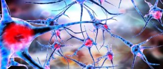
Localization
Color agnosia occurs in most cases with lesions in the left occipital lobe that extend to the temporal or parietal lobe. There are known cases of color agnosia with bilateral occipital lesions. Functional and localization studies indicate that areas V4, V8 of the visual cortex and the lingual gyrus are involved in color processing. Thus, studies of single neurons have shown a possible role in the development of this syndrome of the V4 region, which is responsible for color perception. This area contains many cells that are sensitive to the shape and color of visual stimuli. It is assumed that the color properties of the stimulus can be integrated with the system that describes contours and shapes, and their connection with the development of color agnosia can be destroyed. However, it is unclear under what conditions lesions of area V4 lead to the development of visual object agnosia and under what conditions to color agnosia. Probably, color agnosia occurs when a lesion spreads from the medial occipitotemporal region to various parts of the temporal and parietal lobes involved in the process of recognizing a visual stimulus based on the properties of shape, contour and spatial relationships, which in certain specific areas are integrated with the results of color processing information. This assumption requires additional research. Modern studies report cases of covert processing of information about colors, when subjects with color agnosia successfully coped with implicit color recognition tasks. This led the authors to propose that color agnosia arises from a failure to access and retrieve chromatic information. This study supports the involvement of multiple neural mechanisms and different neuroanatomical regions in color processing.
Agnosia - what is it, causes, types, symptoms, treatment, prognosis
Agnosia is a consequence of certain disorders that are often observed in the brain. Perception is distorted, which becomes a symptom of agnosia, which comes in several types. Its causes lie in diseases that require treatment.
A person perceives the world through the senses. The eyes see, the ears hear, the tongue tastes, the nose smells, etc. These analyzers can work fully. However, perceptual error can occur at the level of information processing in the brain. A person perceives everything that surrounds him distortedly. This may be agnosia, which will be discussed in the online magazine psytheater.com.
Symptoms of agnosia
Symptoms of agnosia are recognized by the manifestations that occur. Depending on the area of brain damage, certain types of agnosia arise, which are considered.
For example, damage to the occipital-parietal lobe leads to the fact that a person is able to see an object, but cannot name it.
And damage to the temporal region leads to the inability to perceive familiar speech (it is heard as a set of sounds).
- A person is not able to navigate in space and on a map.
- A person cannot recognize objects by touch.
- A person does not perceive the presence of defects in himself, although they undoubtedly exist.
- A person is indifferent to the fact that he has defects.
- A person cannot recognize sounds, their nature, source of origin, etc.
- A person incorrectly perceives his own body, which seems to him to be of a different shape or with a different quantity.
- A person does not recognize familiar faces, but remembers information about these people.
- A person does not see the picture as a whole, although he recognizes individual objects in it.
- A person sees only half of space.
go to top
Causes of agnosia
The question naturally arises as to what causes agnosia. No person would want to face such a disease. Avoiding factors can help achieve this desire.
The main cause of agnosia is damage to the parts of the brain that are responsible for processing information received through analyzers. Factors that lead to damage are:
- Chronic circulatory disorder in the brain (stroke), which leads to dementia.
- Alzheimer's disease, which is accompanied by the accumulation of amyloid, a protein that should normally break down.
- Inflammatory processes in the brain: encephalitis.
- Parkinson's disease.
- Tumors in the brain.
- Consequence of traumatic brain injury.
go to top
Treatment of agnosia
Agnosia occurs quite rarely, but it significantly prevents an ordinary person, who has all his senses and sensitivity, from living fully.
Efficiency and quality of life decrease because a person cannot adequately and fully perceive the world around him.
Treatment is prescribed after diagnosing agnosia, where the cause and location of the pathology are first identified.
Complaints are collected (when the disease appeared, how quickly symptoms develop, what preceded agnosia), after which the following measures are prescribed:
- Neurological examination to identify changes and pathologies in tissues. The sharpness of the analyzer is checked.
- Examination by a neuropsychologist who conducts tests and surveys.
- CT and MRI to identify damaged areas in the brain.
- Consultation with a psychiatrist, otolaryngologist, cardiologist, ophthalmologist as necessary.
There are no specific treatments for agnosia. Doctors focus all their efforts on eliminating the cause that provoked damage to the brain, after which the individual is sent for additional corrective work as rehabilitation:
- Speech therapy sessions, especially for auditory agnosia.
- Occupational therapy.
- Psychotherapeutic sessions.
- Working with teachers.
Treatment of the underlying disease is prescribed individually. Here, tumors can be removed from the brain, blood pressure can be monitored, and medications can be prescribed to improve blood circulation in the brain and medications to eliminate neuropsychological pathologies.
The rehabilitation period can last about 3 months. In case of serious damage to a part of the brain, treatment may take 10 months or more.
go to top
Causes of agnosia
Agnostic deviations develop against the background of various pathological changes in the cerebral cortex during the process of ontogenesis, the existence of the organism from the embryonic state to death. In medicine, there are different causes of the disease; depending on the nature of agnosia, a diagnosis is made and treatment is prescribed.
Disorders of the circulatory system in the brain
The death of neurons containing information collected by the senses leads to its loss and reduction in cognitive function. This disease affects people who have experienced an ischemic or hemorrhagic stroke - spontaneous hemorrhage as a result of the destruction of blood vessels.
This form of the disease appears suddenly; depending on the location of the effusion, the occipital, temporal or frontal region is damaged. Timely localization of the site will increase the likelihood of future cure.
Hemorrhagic stroke, which led to an accelerated course of agnosia, is a consequence of the voluntary destruction of blood vessels due to high blood pressure. Symptoms increase quickly; when they are detected, it is necessary to accurately determine the location and cause of the pathology, and carry out treatment with possible surgical intervention.
Chronic cerebral ischemia
Progressive degradation of the cerebral circulatory system leads to the appearance of various types of dementia, including serious disorders. A secondary consequence of the disease can be an almost complete cessation of higher mental activity, in which the patient lives like a plant, unable to satisfy minimal needs on his own.
Description of symptoms:
- Disorders of various types of memory - the patient becomes forgetful, cannot remember the simplest things. To ensure that important information is always at hand, the patient is forced to create tips similar to school cheat sheets.
- Decreased stamina. A person always looks tired, he loses interest in the world around him and becomes apathetic, avoiding unnecessary movements.
- Pain and noise in the head, this manifestation intensifies over time.
- Poor orientation in the surrounding space.
- Sleep disturbance.
- Tearfulness and hot temper, replacing each other.
- Loss of ability to walk.
- Disorder of gnostic functions.
- Inability to focus on one object or express emotions.
And also against the background of loss of normal perception of reality, periodic dizziness and impaired swallowing function appear.
Brain tumors
With the development of cancer, loss of various cognitive functions is possible. Tumors disrupt the normal functioning of the organ, and depending on the location of the tumor, various consequences can occur. Neoplasms destroy neurons; when they grow rapidly, agnosia can progress rapidly in a short period of time.
Disturbances in the processes of recognition and touch during oncogenesis and the development of cancer pathologies are accompanied by:
- Left-sided or right-sided (depending on location) headaches, appearing most often in the morning or evening. The sensations may intensify from rotating the head and tensing the abdominal muscles. Painkillers do not work, but taking an upright position can relieve pain.
- Slowing brain activity. The auditory-speech function is impaired if the tumor has affected the lower parietal part of the brain, apraktoagnosia is observed - loss of the ability to perform habitual, long-learned actions.
- Involuntary muscle contractions in the legs or arms with dullness of tactile function. The next stage in the development of cancer is periodic loss of consciousness.
- Hallucinations, impaired coordination, unreasonable sensation of temperature - the patient thinks that the room is cold or hot.
Further damage to the brain by the tumor leads to irritability, hormonal imbalances and agnostic disorders - the patient suffers from the inability to write anything or remember the purpose of objects.
Traumatic brain injury
In pathopsychology, several disorders are identified that are a direct consequence of physical damage to the brain or cortical lobes. Neuronal destruction is observed immediately at the time of injury or as a consequence of post-traumatic processes.
The likelihood of restoration to a normal state depends on the severity of the injury and the presence/absence of secondary pathologies. If the damage is irreversible, the symptoms do not worsen. The patient, depending on the location of the injury, may lose certain cognitive abilities.
Encephalitis
A dangerous disease characterized by acute inflammation of brain tissue. It can be a consequence of a tick or other insect bite, acute flu, or complications after vaccination. There are several signs that, together with gnostic disorders, indicate the development of encephalitis:
- increase in temperature, sudden or gradual;
- characteristic fever starting at temperatures above 39 degrees;
- severe headache accompanied by speech impairment - it is difficult for a person to understand the semantic structure of sentences and put his wishes and thoughts into words;
- nausea and vomiting, after which there is no relief;
- seizures and development of epilepsy;
- disorders of consciousness in the form of drowsiness, lethargy; if unilateral inflammation spreads to the second hemisphere, the patient may fall into a coma;
- paresis of the limbs - gait disturbance, feet drooping when lifting the leg, a person cannot get up from a sitting position or keeps his head constantly tilted forward.
If such signs are detected, accompanied by a violation of verbal or taste function, you should contact a neurologist.
Degenerative diseases of the central nervous system
Pick's disease, Huntington's chorea and Alzheimer's disease - these diseases lead to serious damage to brain function.
More than 9,000 people have gotten rid of their psychological problems using this technique.
Each of them can be recognized by its characteristic features:
- Pick's disease. Progressive brain damage leading to complete cessation of higher mental activity.
- Alzheimer's disease. The first symptom is a tremor or tic of the left or right hand, accompanied by a gradual breakdown of short-term memory and dysfunction of the musculoskeletal system.
For these and other diseases, agnosia is a secondary manifestation that occurs due to irreversible destruction of nerve cells and brain tissue. Orientation in space and recognition of objects are preserved, but sometimes there is a loss of amodal meaning - the world gradually ceases to be recognizable to humans.
Treatment of agnosia
There is no specific therapy for this pathological condition. Basic treatment of agnosia is aimed at treating conditions that have led to damage to the cortical and subcortical structures of the brain. In each individual case, the therapeutic tactics are determined by the severity of the manifestations, the severity of the condition and the location of the pathological changes, the course of the disease and the presence of complications.
To develop a plan for adequate therapeutic care, it is necessary to conduct a full diagnostic examination:
— conducting a complete examination of the patient, collecting anamnesis, determining the presence of hereditary diseases;
— diagnostic manipulations aimed at identifying the tumor process, the presence of trauma, the presence of vascular accidents;
— consultations with specialists of a narrow profile (ophthalmologist, otolaryngologist, cardiologist, psychiatrist) to exclude other possible causes of this symptomatology;
— conducting diagnostic tests that reveal the degree of change in perception;
— carrying out diagnostic procedures aimed at identifying areas of damage to the brain cortex (CT, MRI).
To directly correct the manifestations of agnosia, it is necessary to work with a neuropsychologist, speech therapist, and the use of occupational therapy.
The recovery period is about three months. The severe course of the disease leading to agnosia and its complications can increase the duration of therapeutic procedures to up to a year. If necessary, treatment is repeated, but when the cause is eliminated and agnosia is completely corrected, relapses often do not occur.
According to the latest statistics, with proper and timely diagnosis of the underlying disease and its manifestations, adequate and complete therapy and corrective measures, carried out fully, will lead to a complete restoration of the analyzers’ functioning.
If you do not consult a doctor in a timely manner, ignore prescribed recommendations or do not implement them in full, or use self-medication, the prognosis may be unfavorable, and the risk of developing irreversible processes in the structure of the brain cortex may increase. The unfavorable outcome of the disease may be influenced by the patient’s age, nature and severity of the disease.
The impact of agnosia on the patient’s quality of life depends on the type of this pathology, for example, simultaneous agnosia or spatial perception disorder significantly worsens the patient’s quality of life, reduces work activity, and impairs communication skills. Whereas, tonal or finger agnosia occurs almost unnoticeably.
Primary prevention of agnosia comes down to the prevention of major diseases, the manifestations of which may be agnosia - maintaining a healthy lifestyle, a nutritious, healthy diet, prevention of stressful conditions. If the first signs of pathology occur, you should immediately contact a specialist.
Correction of agnosia
Agnosia is rarely independent and is more often accompanied by serious illnesses or brain damage. A complete examination and thorough diagnosis help to identify the causes of this or that type of agnosia, only after this individual symptomatic drug therapy is selected.
- complete recovery is possible if the underlying disease is cured;
- long-term remission supported by prophylactic doses of drugs;
- adaptation of a person with lifelong agnosia: various activities aimed at socialization and assimilation of lost skills in partial form through the development of new neural connections.
Symptoms
Symptoms of the disease depend on the damage to one or another analyzer and can serve as a diagnostic criterion for differentiating damage to parts of the brain. Depending on their manifestation, I distinguish the following types of agnosia:
- Prosopagnosia
is a violation of memorization and subsequent recognition of faces with preservation of mnemonic functions relating to objects. In severe cases, patients cannot recognize their own reflection in the mirror; - Lissauer object agnosia
is a violation of the recognition of individual objects; - Color agnosia
- the inability to determine the color belonging to certain objects, as well as to select identical colors or their shades; - Simultaneous agnosia
- limitation of the visual field to only one visible object or the ability to perceive only one object, regardless of its shape, color and size; - The weakness of optical concepts is the inability to create a description of any characteristics of a visible object;
- Balint's syndrome
is the inability to direct gaze towards an object and then fixate it, while maintaining the ability to move the eyeballs; - One-sided
spatial agnosia - loss of one side of space from the visual field; - Violation of stereoscopic vision
- the impossibility of comparing a single whole of visual images entering each eye; - Deep agnosia
is the inability to correctly determine the location of objects located in front of the patient; - Violation of topographic orientation
- loss of orientation in places familiar to the patient, while maintaining other cognitive functions; - Auditory speech agnosia
- recognition of speech as a set of unrelated sounds; - Simple auditory agnosia
- the inability to recognize individual sounds; - Tonal agnosia
- the inability to recognize timbre, as well as the emotional coloring of speech, while maintaining other functions of the auditory analyzer; - Autotopagnosia hemicarpus
- complete ignorance or denial of the presence of the left half of the body with complete or partial preservation of its functioning; - Somatic aloesthesia
is a feeling of the presence of static or dynamic extra limbs; - Autotopagnosia of posture
is the inability to determine the position of the body or its individual parts in space; - Pretman syndrome
(finger agnosia) - the inability to correctly point to the finger shown by the interlocutor; - Violation of “right-left” orientation - the inability to point to the right or left parts of the body.
Kinds
The disease has three main types: visual agnosia, auditory agnosia and tactile agnosia. In addition, there are several less common types of the disease (spatial agnosia and other perceptual disorders).
In visual agnosia, the lesions are localized in the occipital lobe of the brain. This type is characterized by the patient's inability to recognize objects and images, despite the fact that he retains sufficient visual acuity for this. Visual agnosia can be expressed in different ways and manifest itself in the form of the following disorders:
- object agnosia (damage to the convexital surface of the left part of the occipital region): inability to recognize various objects, in which the patient can only describe individual features of an object, but cannot say what kind of object is in front of him;
- color agnosia (damage to the occipital region of the left dominant hemisphere): inability to classify colors, recognize identical colors and shades, correlate a specific color with a specific object;
- visual agnosia, manifested in weakness of optical representations (bilateral damage to the occipital-parietal region): inability to imagine any object and characterize it (name size, color, shape, etc.);
- agnosia for faces, or prosopagnosia (damage to the lower occipital region of the right hemisphere): a violation of the process of recognizing faces while maintaining the ability to distinguish between objects and images, which in especially severe cases can be characterized by the patient’s inability to recognize his own face in the mirror;
- simultaneous agnosia (damage to the anterior part of the dominant occipital lobe): a sharp decrease in the number of simultaneously perceived objects, in which the patient is often able to see only one object;
- Balint's syndrome, or visual agnosia caused by optomotor disorders (bilateral damage to the occipital-parietal region): inability to direct the gaze in the right direction, focus it on a specific object, which can be especially pronounced when reading - the patient cannot read normally, since It is very difficult for him to switch from one word to another.
Auditory agnosia occurs when the temporal cortex of the right hemisphere is damaged. This type is characterized by the patient’s inability to recognize sounds and speech, while the function of the auditory analyzer is not impaired. The following disorders are distinguished in the category of auditory agnosia:
- simple auditory agnosia, in which the patient cannot recognize simple, familiar sounds (the sound of rain, rustling paper, knocking, creaking doors, etc.);
- auditory-verbal agnosia - the inability to distinguish speech (for a person suffering from this type of auditory agnosia, native speech is presented as a set of unfamiliar sounds);
- tonal auditory agnosia - the patient cannot perceive the tone, timbre, or emotional coloring of speech, but at the same time he retains the ability to normally perceive words and correctly recognize grammatical structures.
With tactile agnosia, the patient is unable to identify objects by touch. One type of tactile agnosia is the patient’s inability to recognize parts of his own body and evaluate their location relative to each other. This type of tactile agnosia is called somatoagnosia. Tactile agnosia, in which the process of recognizing objects through touch is disrupted, is called astereognosia.
There are also spatial agnosias, which are expressed as a violation of the identification of various parameters of space. With lesions of the left hemisphere, it manifests itself in the form of impaired stereoscopic vision; with lesions of the middle parts of the parieto-occipital region, the disease can be expressed as the patient’s inability to correctly localize objects in three spatial coordinates, especially in depth, as well as to recognize parameters further or closer.
There are also types of agnosia such as unilateral spatial agnosia - the inability to recognize one of the halves of space (usually the left), and spatial agnosia, expressed in a violation of topographic orientation, in which the patient may not recognize familiar places, but he does not have any memory impairment .
One of the rarest types of agnosia is impaired perception of time and motion - a condition in which a person is unable to judge the speed of time and perceive the movement of objects. The last disorder (inability to perceive moving objects) is called akinetopsia.
Gnostic visual disorders. Classification of visual agnosias.
Higher gnostic visual functions are provided primarily by the work of the secondary fields of the visual system (18th and 19th) and the adjacent tertiary fields of the cerebral cortex. Secondary fields 18 and 19 are located on both the outer convexital and inner medial surfaces of the cerebral hemispheres. They are characterized by a well-developed layer III, in which impulses are switched from one area of the cortex to another.
With electrical stimulation of the 18th and 19th fields, not local, point excitation occurs, as with stimulation of the 17th field, but activation of a wide zone, which indicates broad associative connections of these areas of the cortex.
From studies conducted on humans by W. Penfield, G. Jaspero and a number of other authors, it is known that with electrical stimulation of the 18th and 19th fields, complex visual images appear. These are no longer isolated flashes of light, but familiar faces, pictures, and sometimes some vague images. Basic information about the role of these areas of the cerebral cortex in visual functions was obtained from the clinic of local brain lesions. Clinical observations show that damage to these areas of the cortex and the adjacent subcortical zones leads to various disorders of visual gnosis. These disorders are called visual agnosia.
This term refers to disorders of visual perception that occur when the cortical structures of the posterior parts of the cerebral hemispheres are damaged and occur with the relative preservation of elementary visual functions (visual acuity, visual fields, color perception). In all forms of agnostic visual disorders, elementary sensory visual functions remain relatively intact, that is, patients see quite well, they have normal color perception, and their visual fields are often preserved; in other words, they seem to have all the prerequisites to perceive objects correctly. However, it is the gnostic level of the visual system that is disrupted in them. In some cases, patients, in addition to gnostic ones, have sensory dysfunctions. But these are, as a rule, relatively subtle defects that cannot explain the severity and nature of disorders of higher visual functions.
The first description of visual agnosia belongs to G. Munch (1881), who, working with dogs with lesions in the occipital lobes of the brain, discovered that “the dog sees, but does not understand” what it sees; the dog seems to see objects (since it does not bump into them), but “does not understand” their meaning.
Naturally, visual impairment in humans is much more complicated. In the clinic of local brain lesions, various forms of disorders of higher visual functions, or various forms of visual agnosia, have been described. The term “agnosia” was first used by Z. Freud (1891), who was not only the founder of psychoanalysis, but also the largest neurologist who studied the functions of the nervous system. The cases of violations of higher visual functions described by him were designated as “visual agnosia.” After Z. Freud, many authors studied visual agnosia; we can say that of all the disorders of mental processes that are observed with local brain lesions, it is visual agnosia that has been best studied at the phenomenological level.
Contributions to the problem of studying visual agnosia included D. Nielsen, G. Teuber, A. P. Luria, O. Zangwill, E. P. Kok, G. Ekaen, D. Brown, I. M. Tonkonogiy, J. A. Meerson and pl. etc. It should be noted that both domestic and foreign publications are devoted mainly to the description of what happens to patients when certain areas of the “wide visual sphere” are damaged - the occipital-parietal areas of the cortex, i.e., the primary study of visual dysfunctions on a phenomenological basis level.
The nature and structure of mental disorders in visual agnosia and their brain mechanisms are much less studied. There is still no general theory to explain the occurrence of various forms of disorders of higher visual functions, which directly affects the classifications of visual agnosia existing in neuropsychology and clinical neurology. All of them are based on phenomenological differentiation of types of visual impairment. Thus, at present there is no unified classification of visual agnosia, since there is no single explanation for the nature of these disorders. Some authors explain visual agnosia by intellectual defects, a decrease in the “abstract attitude”, others consider them as a consequence of speech disorders, etc.
Most authors, based on clinical phenomenology, identify six main forms of
visual gnosis disorders:
Attention!
If you need help writing a paper, we recommend turning to professionals. More than 70,000 authors are ready to help you right now. Free adjustments and improvements. Find out the cost of your work.
Cost calculationGuaranteesReviews
1) if the patient, correctly assessing individual elements of an object (or its image), cannot understand its meaning as a whole - this is called object agnosia;
2) if he does not distinguish human faces (or photographs) - facial agnosia;
3) if he is poorly oriented in the spatial features of the image - optical-spatial
agnosia;
4) if he, while copying letters correctly, cannot read them - letter agnosia;
5) if he distinguishes colors, but does not know which objects are painted in a given color, that is, he cannot remember the color of familiar objects - color agnosia;
6) simultaneous agnosia
- a violation of visual gnosis when the patient can perceive only individual fragments of the image, and this defect is observed even when the visual fields are preserved.
Let's take a closer look at the different forms of visual agnosia.
Object agnosia
- one of the most common forms of visual gnosis disorders, which, to one degree or another, occurs in most patients with damage to the occipito-parietal parts of the brain. In its rough form, object agnosia is observed only with bilateral damage to the occipital-parietal parts of the brain, i.e., with bilateral damage to the 18th and 19th fields.
Object visual agnosia is associated with damage to the lower part of the “wide visual sphere.” It is characterized by the fact that the patient sees as if everything, he can describe individual signs of an object, but cannot say what it is. A particularly severe violation of the ability to correctly evaluate an object occurs when there is bilateral damage to the lower parts of the wide visual sphere: the patient, looking at an object, cannot identify it, but when palpating it, he often solves this problem correctly. In their daily lives, such patients behave almost like they are blind, and although they do not bump into objects, they constantly feel them or navigate by sounds. However, in such a rough form, object agnosia is relatively rare; more often it manifests itself in a latent form when performing special visual tasks: for example, when recognizing contour, crossed out, superimposed on each other, inverted images, etc.
With object agnosia, difficulties in recognizing the shape of objects are primary, and in their most “purest” form they appear when recognizing the contours of objects; at the same time, copying their drawings can be safe.
In patients with object agnosia (as well as with other forms of visual gnosis disorders), the temporal characteristics of visual perception are grossly altered. Tachistoscopic studies have established that in such patients the thresholds for image recognition sharply increase; and, as a rule, they increase by several orders of magnitude. If a healthy person perceives simple images in 5-10 ms (without a background erasing image), then in patients the time of recognition of simple images increases to 1 s or more. Thus, with visual agnosia, a completely different mode of operation of the visual system is observed, which causes great difficulties in processing visual information.
Optical-spatial agnosia
associated primarily with damage to the upper part of the “wide visual sphere”. In a particularly severe form, it is observed with bilateral damage to the occipital-parietal parts of the brain. However, even with unilateral lesions, these disturbances are also quite clearly expressed. With optical-spatial agnosia, patients lose the ability to orient themselves in spatial features of the environment and images of objects. Their left-right orientation is disturbed; they cease to understand the symbolism of the drawing, which reflects the spatial characteristics of objects. Such patients do not understand the geographical map, their orientation in the countries of the world is impaired. The book “The Lost and Returned World” (1971) by A. R. Luria is devoted to a description of such a violation, which tells about a patient, a former topographer, who was injured in the occipital-parietal region of the left hemisphere of the brain.
In severe cases, patients experience disorientation not only in left-right, but also in superior-inferior coordinates. In patients with optical-spatial (as well as with object) agnosia, as a rule, the ability to draw is impaired (with relative preservation of the ability to copy an image). They do not know how to convey spatial characteristics of objects in a drawing (further-closer, more-less, left-right, top-bottom). In some cases, even the overall pattern of the drawing falls apart. Thus, patients, when drawing a person, separately depict parts of his body (arms, legs, eyes, nose, etc.) and do not know how to connect them. The pattern is more often disrupted when the posterior parts of the right hemisphere are damaged .
In a number of cases (usually with right-hemisphere lesions),
unilateral optical-spatial agnosia is observed,
when patients, even when copying a drawing, depict only one side of the object or grossly distort the image of one (usually the left) side. At the same time, their ability to visually afferentate spatially organized movements, i.e., “postural praxis,” is often impaired. Such patients cannot copy the posture shown to them by the experimenter; they don’t know how to position their hand in relation to their body; they lack the immediate ease of perception of spatial relationships that is inherent in healthy people, and this makes it difficult to copy poses from a visual model (performed with one or two hands).
This is associated with various difficulties in everyday motor acts in which spatial orientation of movements is required. These patients poorly perform movements that require basic visual-spatial orientation, for example, they cannot put a blanket on the bed, put on a jacket, trousers, etc. Such disorders are called “apraxia of dressing.” Combinations of visuospatial and motor-spatial disorders are called “apractoagnosia.”
Optical-spatial disorders sometimes affect reading skills. In these cases, difficulties arise in reading letters that have “left-right” characteristics. Patients cannot distinguish between correctly and incorrectly written letters, and this task can be one of the tests for determining visual orientation in the spatial characteristics of objects. In such cases, impairments in the recognition of letters with mirror spatial features, as a rule, reflect a general defect in spatial orientation in objects.
A special form of visual agnostic disorder is letter agnosia.
In its pure form, letter agnosia manifests itself in the fact that patients, although copying letters completely correctly, cannot name them.
Their reading skills disintegrate (primary alexia).
Such a reading disorder occurs in isolation from other disorders of higher visual functions, which gives grounds to classify this defect as an independent form of agnosia. Such patients correctly perceive objects, correctly evaluate their images, and even correctly navigate complex spatial images and real objects, but they “do not understand” letters and cannot read.
This form of agnosia, as a rule, occurs when the left hemisphere of the brain is damaged - the lower part of the “wide visual sphere” (in right-handed people).
Color agnosia
also represents an independent type of visual gnostic disorders. A distinction is made between color agnosia itself and impaired recognition of colors as such (color blindness or defects in color perception). Color blindness and impaired color perception can have both peripheral and central origin, i.e., be associated with damage to both the retina and the subcortical and cortical parts of the visual system. It is known that color perception occurs through the action of three different types of cones (retinal detectors), which are sensitive to different colors: blue-green, red-green and yellow. This ability of cones to be reactive to certain color stimuli is the basis of color perception, and a defect in this ability can be caused by various types of retinal lesions (degeneration, etc.).
There are known disturbances in color discrimination associated with damage to the NKT and occipital cortex (field 17), which indicates the existence in the visual system of a special channel (or channels) designed to carry information about the color of an object.
Color agnosia, in contrast to color discrimination disorders, is a violation of higher visual functions. The clinic describes violations of color gnosis, which are observed against the background of preserved color perception. Such patients correctly distinguish individual colors and name them correctly. However, it is difficult for them, for example, to correlate a color with a certain object and vice versa; they cannot remember what color an orange, a carrot, a Christmas tree, etc. are. Patients cannot name objects of a certain specific color. They lack a generalized idea of color and therefore are unable to perform the color classification procedure, which is not due to difficulties in distinguishing colors, but due to difficulties in categorizing them. It is known that a person perceives a huge number of shades of colors, but there are relatively few color names (categories). Therefore, in ordinary life, a healthy person constantly solves the problem of color categorization. It is this categorization of color sensations that is difficult in patients with color agnosia.
A special form of visual agnosia is simultaneous agnosia.
For a long time it was known as
Balint syndrome.
This form of visual gnosis impairment manifests itself in the fact that the patient cannot simultaneously perceive two images, since his scope of visual perception is sharply narrowed.
The patient cannot perceive the whole, he sees only its part (or parts). Balint's syndrome is always accompanied by complex eye movement disorders called gaze ataxia. The patient's gaze becomes uncontrollable, the eyes make involuntary jumps, constantly being in motion. This creates difficulties in organized visual search, as a result of which the patient cannot consider the object consistently. It is assumed that the cause of simultaneous agnosia is the weakness of cortical visual cells, which are capable of only narrowly localized foci of excitation. The connection between Balint's syndrome and the side of the lesion and the localization of the lesion within the “wide visual sphere” has not yet been established.
Facial agnosia
- a special form of visual gnosis disorders, which manifests itself in the fact that the patient loses the ability to recognize real faces or their images (in photographs, drawings, etc.). In the severe form of facial agnosia, patients cannot distinguish between female and male faces, as well as the faces of children and adults; do not recognize the faces of their relatives and friends. Such patients recognize people (including those closest to them) only by their voice. Facial agnosia is clearly associated with damage to the posterior parts of the right hemisphere (in right-handed people), to a greater extent to the lower parts of the “wide visual sphere.”
In general, the question of the connection between various forms of visual agnostic disorders and the side and area of damage to the occipital-parietal parts of the brain has not been completely resolved. Many authors indicate that various forms of visual agnosia appear especially clearly when the commissural fibers of the splenium of the corpus callosum, connecting the 18th and 19th fields of the left and right hemispheres of the brain, are damaged.
In general, as clinical observations show, disorders of visual gnosis are heterogeneous. The nature of agnosia depends, apparently, on the side of the brain lesion, and on the location of the lesion within the “wide visual sphere,” and on the degree of involvement in the pathological process of commissural fibers that unite the posterior sections of the left and right hemispheres. It is important to note that different forms of visual gnosis disorders occur in isolation. This indicates
the existence of separately, autonomously functioning channels that process different types of visual information.
However, it should always be remembered that different forms of visual perception are not realized only with the help of special visual channels; in all cases, the entire brain as a whole, all its three main blocks, takes part in the implementation of higher visual functions (or visual gnostic activity), as follows from the theory of systemic dynamic localization of higher mental functions. Therefore, disturbances in visual gnosis can occur, for example, when the frontal lobes of the brain are damaged; then they are secondary in nature and are designated as pseudoagnosia.
Thus, neuropsychological data confirm the general concept that the visual system is organized as a multichannel apparatus that simultaneously processes a variety of visual information, various “blocks” (channels) of which can be affected in isolation while other “blocks” (channels) operate intact. As a result, disturbances in the perception of only objects, or faces, or colors, or letters, or spatially oriented objects may appear.
We will help you write any paper on a similar topic.
- Essay
Gnostic visual disorders. Classification of visual agnosias.
From 250 rub.
- Test
Gnostic visual disorders. Classification of visual agnosias.
From 250 rub.
- Course work
Gnostic visual disorders. Classification of visual agnosias.
From 700 rub.
Receive completed work or specialist advice on your educational project
Find out the cost
Treatment
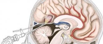
Initially, treatment is aimed at eliminating the underlying disease, for example, antibiotic therapy for brain abscesses, surgery, radiation therapy for brain tumors, etc.
Rehabilitation through speech therapy, correction of disorders by speech pathologists, psychological support, consultations with a neuropsychologist who can help adapt to the characteristics and manifestations of the disease can be effective.
The doctors
specialization: Neuropsychologist / Neurosurgeon / Speech therapist / Psychologist
Chesnova Tatyana Andreevna
10 reviewsSign up
Find a doctor and make an appointment
Medicines
Cavinton Picamilon Fezam Lutsetam
Conservative drug treatment is carried out using drugs that improve metabolism and cerebral blood flow. These include:
- Cavinton is a psychostimulant nootropic drug that helps reduce the severity of neurological and mental disorders. Intended for intravenous infusion at a rate of 80 drops/min, 20 mg per day.
- Picamilon is a nootropic drug that dilates blood vessels in the brain with additional tranquilizing, psychostimulating, antiplatelet and antioxidant effects. The standard daily dose of 60-150 mg can be divided into 2-3 doses, the duration of treatment is 1-2 months, it is recommended to repeat the course six months later.
- Trental is a vasodilating agent that can reduce viscosity and improve the rheological properties of blood, as well as promote microcirculation in areas of impaired blood flow. Usually doctors prescribe taking 1-2 tablets 3 times a day, maximum 15 tablets.
- Phezam is a drug that improves metabolic and microcirculatory processes in the brain. Recommended daily dose: take 1-2 caps. 3 times a day for 1-3 months, repeat the course of treatment 2-3 times a year.
- Lucetam is a nootropic drug that can increase concentration and improve cognitive function. Typically, the daily dose is 2.4-4.8 g/day, divided into 2-3 doses, but it is best that the treatment regimen is selected by the attending physician, depending on the characteristics and severity of the disorders.
Patients are most often recommended individual psychotherapy sessions, sessions with speech pathologists and speech therapists.
Types of agnosia
The described disorder is characterized by three main types: tactile, visual and auditory perceptual disturbances. In addition, we can distinguish a number of less common forms of the disease in question (for example, spatial agnosia).
Visual agnosia is characterized by the presence of a lesion in the occipital region of the brain. This form of the disease manifests itself in the inability of patients to recognize images and objects while maintaining visual acuity. The type of pathology in question can manifest itself in different ways. The following forms of visual agnosia are distinguished: object, color, visual, simultaneous agnosia, prosopagnosia and Balint's syndrome.
Auditory perceptual dysfunctions arise due to damage to the temporal cortex of the right hemisphere. This type of agnosia is represented by the inability of individuals to recognize speech and sounds against the background of normal functioning of the auditory analyzer. Auditory agnosia, in turn, is divided into simple auditory perception disorder, auditory speech and tonal auditory agnosia.
A simple disorder of auditory perception is characterized by the inability of people to recognize simple, previously familiar sounds, such as the sound of rain, the rustling of the sea, knocking, a doorbell, creaking, etc.
Auditory speech agnosia is the inability to recognize speech. To a person suffering from the described form of agnosia, native speech seems to be a set of unfamiliar sounds.
Tonal hearing disorder is characterized by the inability to perceive the emotional coloring, tone, and timbre of speech while maintaining the ability to adequately perceive words and correctly distinguish grammatical structures.
Tactile agnosia is the inability to identify objects or things by touch. The following types of agnosia are distinguished: somatoagnosia, astereognosia and disturbance of spatial perception. The patient's inability to recognize parts of his own body and evaluate their location relative to each other is called somatoagnosia. A disorder of tactile perception, in which the process of recognizing objects and things through touch is called astereognosia.
There are also disturbances in spatial perception, expressed in the form of incorrect identification of space parameters. Damages to the middle areas of the occipital-parietal region are revealed in the inability to measure quantities closer or further away, as well as to correctly place objects in three-dimensional space, especially in depth; damage to the left hemisphere entails spatial agnosia, manifested by impaired stereoscopic vision. In addition, there are such types of agnosia as a unilateral violation of spatial perception and a perceptual disorder consisting in the inability to topographically navigate the terrain. Unilateral spatial agnosia is the inability to recognize one half of space. Violation of topographic orientation is expressed in the inability to recognize familiar places against the background of intact memory function.
One of the rarest types of agnosia is dysfunction of the perception of movement and time. This disease manifests itself in a violation of the correct understanding of the movement of objects and an adequate assessment of the speed of the passage of time. The inability to perceive objects in motion is called akinetopsia.
Diagnosis of agnosia
The diagnosis of agnosia is made based on medical history (trauma, stroke, tumor) and the clinical picture of the disease. Special tests are also performed to determine the type of agnosia.
The patient is asked to identify simple objects using various senses. If the doctor suspects denial of half of space, then he asks the patient to identify paralyzed parts of his body, or objects in different parts of space.
Conducting a neuropsychological examination helps determine the presence of more complex types of agnosia.
Brain imaging methods (MRI or CT) are also used to establish the nature of central lesions (hemorrhage, infarction, massive intracranial process) and to identify areas of cortical atrophy.
To identify primary disorders of certain types of sensitivity, a physical examination is performed.
Treatment of the disease
In medicine, there is no single protocol for the treatment of agnosia, since it depends on the root cause of the disease, its type and degree of neglect. In parallel with the treatment of the main illness, work is carried out with a psychiatrist, speech therapist, and neuropsychologist. This is necessary in order to help a person adapt to life in the presence of such pathologies. There are cases when treatment brought instant results and when it lasted for many years. The effect depends on how quickly the patient sought professional help. The most commonly used drugs are the following groups: vascular drugs, neuroprotectors and nootropics, B vitamins.
Symptoms of agnosia
Damage to the cerebral cortex, which is responsible for the analysis and synthesis of information, gives rise to agnosia. Therefore, the symptoms will depend on the location of the affected area of the brain. For example, as a result of damage to the left zone of the occipital region, object agnosia arises, which consists of the patient losing data about the object and its purpose. In other words, an individual suffering from this perceptual disorder sees an object and can describe it, but is unable to name it or talk about its purpose. When the temporal region is damaged, an auditory-verbal perception disorder occurs: the patient perceives the speaker’s speech as if it were an ordinary set of sounds; he is unable to perceive the meaning of phrases and distinguish individual words. Statistics confirm that the disorder in question is quite rare.
The causes of agnosia are as follows: dysfunctions of the temporal and parietal areas of the brain, where data on the use of familiar objects are stored (more often it occurs suddenly after a stroke, heart attack or head injury, when the cortex and nearby subcortical formations of the brain are affected, and damage to the cortex can cause a tumor process ). In addition, the pathology in question may arise as a result of degeneration of areas of the brain that are responsible for the integration of perception, memory and identification processes.
Thus, the main causes of agnosia are damage to the parietal and occipital areas of the cerebral cortex, which occur, in addition to the above pathologies, with the following ailments:
- chronic circulatory disorder in the brain, which later develops into dementia;
- inflammatory processes of the brain (for example, encephalitis);
- Alzheimer's disease, which is associated with the accumulation of amyloid in the brain (a specific protein that normally breaks down quickly in the brain);
- Parkinson's disease, characterized by the occurrence of progressive muscle stiffness, tremors and a number of neuropsychological disorders, including apraxia.
There are different types of perceptual dysfunction depending on the location of the affected area in the brain. For example, if the parieto-occipital zone is damaged, a violation of topographic orientation occurs; if the right subdominant part of the parietal lobe is damaged, anosognosia occurs, which is the absence in patients of a critical assessment of their own illness or defect. For example, people suffering from this form of dysfunction consider themselves completely healthy even against the background of immobility on one side of the body (state of paralysis).
Many people far from medicine wonder about agnosia, what is it, what are the symptoms of this disease, how do they manifest themselves?
The following manifestations and symptoms of agnosia can be distinguished:
- violation of spatial orientation and the ability to “read” on the map, that is, to understand the location of cities, regions and other places on the map;
- a disorder in the ability to recognize objects by touch (sick people find it difficult to determine the texture, configuration and shape of an object;
- denial of the fact of having a physical defect or illness (for example, blindness, deafness), despite the indisputability of the existing defects;
- indifference to the existing defect (a person may be little worried about sudden deafness, blindness or other defects;
- impairment of the ability to recognize sounds (the patient is not able to distinguish the nature of the sound, understand where it is coming from, for example, when he hears a bell in his own house or the voice of a relative;
— dysfunction of perception of one’s own body (people are not able to correctly determine the number of their limbs or their length);
- a disorder in the ability to recognize the faces of friends, along with this, patients are able to name their approximate age or gender;
- impaired recognition of complex visual images, while patients retain the ability to recognize individual components of these images, for example, an individual, looking at an image, recognizes a jug on the table, but is not able to understand that the presence of a jug, glasses, plates, food on the table, shows that the picture shows a feast;
- ignoring part of the visible space (for example, a patient, while eating food, eats food only from the right side of the plate).
Object agnosia
Among the factors that contribute to the development of cognitive disorders, a distinction is made between those that can be corrected with medication and those that cannot be changed. Most vascular and degenerative diseases of the brain cannot be corrected and are irreversible. Uncontrollable risk factors also include the patient’s age (usually people over 60-70 years old), female gender, genetic predisposition, and previous traumatic brain injuries.
Among those that affect the development of memory impairment, but can be corrected, there are significantly more conditions:
- Existing arterial hypertension;
- Increased lipid levels in the blood;
- Diabetes mellitus, hypothyroidism, thyrotoxicosis;
- B12 folate deficiency anemia;
- Increased level of homocysteine in blood plasma;
- Vascular encephalopathy;
- Alzheimer's disease;
- Dementia;
- Huntington's chorea;
- Infectious diseases of the brain
- Cerebral infarction and stroke;
- Insufficient physical and intellectual activity.
Object agnosia occurs when a “wide zone” of the visual analyzer is damaged and can be characterized as the absence of a recognition process or as a violation of the integrity of the perception of an object with the possible recognition of its individual features or parts. It can have varying degrees of severity - from maximum (agnosia of real objects) to minimum (difficulty recognizing contour images in noisy conditions or when superimposed on each other). As a rule, the presence of extensive object agnosia indicates bilateral damage to the occipital regions.
With unilateral lesions, the structure of visual object agnosia differs. Damage to the left hemisphere is manifested to a greater extent by a violation of the perception of objects by the type of enumeration of individual details, while the pathological process in the right hemisphere leads to the actual absence of the act of identification.
Classification of impaired perception
Depending on the location, the types of agnosia differ.
Agnosia comes in three main types, each of which in turn is divided into subspecies.
Visual agnosia
With this type of disorder, brain damage is localized in the occipital region. With visual agnosia, the patient cannot recognize an object or picture. The patient does not have visual impairment.
This type of agnosia manifests itself in different ways:
- Subject. With damage to the surface of the brain, which is adjacent to the left side of the occipital bone: objects are unrecognizable (the patient describes it in general terms).
- Colored. With lesions in the occipital region of the left hemisphere, which is considered dominant: the patient has difficulty distinguishing colors and shades, cannot match an object and a color.
- Visual, manifested in optical weakness (optical spatial). If the occipital and parietal region is affected on both sides: the patient cannot imagine the object and describe it.
- Face perception disorder. With lesions of the lower region of the back of the head of the right hemisphere: the patient cannot recognize faces, even his own reflection, but distinguishes between objects and pictures.
- Simultaneous. With lesions of the anterior part of the dominant occipital lobe: the patient cannot simultaneously perceive several objects. Autotopagnosia is characterized by the inability to recognize one's body.
- Balint's syndrome. It is noted with bilateral damage to the occipital-parietal region: the patient cannot look in the right direction, focus his gaze, and cannot read normally.
Auditory agnosia
With this type of disorder, damage occurs to the cortex in the temporal region of the right hemisphere.
A patient with auditory agnosia cannot recognize speech or sounds, but a disorder of the auditory analyzer is not diagnosed.
This type of pathology has such disorders that characterize each subtype of auditory agnosia, such as:
- Simple. The patient does not recognize simple sounds.
- Hearing-speech. The patient is unable to distinguish speech.
- Tonal. The patient does not grasp tone, timbre, or emotionality, but at the same time perceives phrases and words normally.
Disorders of tactile and spatial sensations
Tactile agnosia is characterized by the fact that the patient cannot recognize an object by touch. It happens that the patient cannot even recognize a part of his own body and evaluate its location (somatoagnosia). With astereognosia, the patient cannot recognize an object using touch.
Spatial agnosia:
- damage to the left cerebral hemisphere: impairment of spatial vision (optical-spatial), in this case there is often an intersection with visual agnosia;
- damage to the middle part of the parietal and occipital region: the patient cannot localize an object in space.
There is also a spatial neurological disorder:
- which is expressed in the violation of one of the halves of the space;
- which is expressed in a violation of topographic orientation.
A rare type of disorder is considered when the patient does not perceive time frames and movements (akinetopsia).
Classification
Existing types of agnosia can be divided into nosological groups according to the similarity of the nature of the manifestation
Visual agnosia
Damage to the cortical parts in the posterior parts of the brain leads to deviations in the perception of objects and objects visible to the eye.
These include the following groups:
- Subject. The meaning of visible images is lost, although brain activity remains active. A.R. Luria and E.D. Chomskaya indicate that for the patient, attempts to identify a visible object become a complex process of deciphering perceived images.
- Optical-spatial. A person is unable to perceive three-dimensional signs of the environment, loses orientation in space, and cannot perform simple movements. Accompanied by the inability to read and recognize letters.
- Colored. The patient can distinguish shades, but does not know how to associate them with specific objects.
These and other pathologies most often occur in adults.
Auditory agnosia
Speech is formed on the basis of the auditory system; it allows you to navigate noise, music, and speech. The focus of pathology development is the middle part of the temporal lobe.
The following varieties are distinguished:
- Auditory agnosia itself. The patient does not recognize what exactly sounds, although he is able to indicate pitch, timbre and frequency.
- Auditory arrhythmia. Recognition of rhythmic structures becomes impossible; he cannot determine how many beats are contained in the rhythm pattern.
- Amusia. The ability to play the melody or learn by ear is lost. Musical themes become one of the causes of severe headaches.
- Dysarthria. Articulation of speech disappears, which is caused by paralysis of the muscles of the speech apparatus due to the development of pathology of the medulla oblongata. In mild cases, speech therapy can help relieve symptoms.
All types are accompanied by increased irritability, since the inability to recognize sounds causes anxiety in the patient.
In childhood, it is possible to develop acquired or congenital alalia - absent or underdeveloped speech function.
Olfactory agnosia
As the name suggests, the disease is associated with an impairment of the ability to detect odors. A person cannot give an exact description of an audible smell or make a comparison, but he is able to recognize or distinguish them.
Tactile agnosia
It is difficult for a person to understand what shape an object has while maintaining its sensory basis and tactile perception.
Existing types:
- Astereognosis. When taking the test, the patient cannot describe or identify certain features of the object.
- Lissauer's Agnosia. There is a holistic perception, but the patient cannot recognize or name the objects he saw.
- Agnosia of object structure. The fingers feel the texture of an object, but due to disturbances in the functioning of the brain, the patient cannot describe the basic property of the surface - whether it is rough, smooth or sticky.
- Tactile alexia. A rare condition in which a person cannot read the characters written on the skin.
With the development of Somatoagnosia, orientation in one's own body is lost - the transmission of impulses that occur when an irritant is exposed to the skin is disrupted.
Pseudoamnesia
A disruption of the memorization process that appears as a result of extensive pathologies of the frontal lobes of the brain. Involuntary memory is preserved for the most part, but active memorization of data becomes impossible, and passive retention remains instead.
They develop as a result of the breakdown of programming and control of the brain over voluntary activity. It is difficult for a person to organize the collected information and reproduce it. It is impossible to impose a requirement on the patient to remember certain data.
Cauda equina syndrome
Pathology leading to gait disturbances, pain in the lower back, fecal and urinary incontinence and a number of other symptoms. Unlike the above-mentioned types of agnosia, its cause is damage to the spinal cord roots in the lumbar and sacral areas. It becomes a direct consequence of previous injuries, hernia, spinal stenosis or congenital defects.
Causes and symptoms
According to statistics, agnosia is a fairly rare disease. The main causes are head injuries, stroke, brain tumors. The type of pathology depends on the area of damage to the cerebral cortex: with lesions of the posterior parietal and anterior occipital cortex, visual agnosia develops, auditory agnosia develops in the temporal lobe of the left hemisphere, and spatial agnosia develops in the parieto-occipital lobe.
Visual agnosia is a disorder of object recognition with completely normal vision. The following varieties are distinguished:
- Simultaneous – a disorder in the ability to perceive a group of images, limiting perception to one object.
- Color vision is a violation of the ability to distinguish colors, although color vision is not impaired; this pathology should not be confused with color blindness.
- Letter - letter recognition disorder. In this case, speech is practically not affected, but the person does not distinguish between letters and is not able to read and write. Occurs when the dominant hemisphere of the occipital cortex is damaged.
- Facial – loss of the ability to distinguish the faces of familiar people; in severe cases, a person is unable to recognize himself when looking in the mirror. Occurs with lesions in the lower occipital region of the right hemisphere.
The essence of tactile agnosia is a violation of the recognition of objects by touch. There are the following types:
- Object - a violation of the ability to determine by touch the shape of an object, its size and the material from which the object is made, although a person can visually describe the object.
- Finger - a disorder due to which a person with his eyes closed is unable to independently determine which finger the doctor (or any other person) is touching.
- Somatoagnosia is a disorder of recognition of body parts and their location. Occurs when various parts of the right hemisphere are damaged.
Auditory agnosia is a disturbance in the perception of sounds and speech, sometimes a complete loss of the ability to distinguish extraneous noise from useful sound information. Often accompanied by an inability to write text from dictation or read aloud. There are the following types:
- Simple auditory agnosia is the loss of the ability to recognize the simplest sounds, for example, creaking, knocking, rustling leaves, clicking, etc.
- Hearing speech is the inability to recognize human speech, which is perceived as a set of unfamiliar sounds or noise.
- Tonal - the patient does not grasp the emotional nuance of speech, timbre of voice, tone, although he clearly perceives the meaning of what he heard.
Spatial agnosia is characterized by loss of the ability to orientate oneself in the area; the patient cannot distinguish between right and left, confuses the hands on a watch dial, confuses letters and numbers when reading. In some cases, the patient sees only part of the spatial image, for example, only the left half of the road, the other part of the road is not recognized.
Identification of agnosias
Despite the fact that agnosia is not a common pathology, its diagnosis should be carried out comprehensively. Most often, gnostic dysfunction is found in adults. However, cases of identifying symptoms of agnosia in a child are not uncommon (at a younger age, we are talking about a delay in the formation of gnosis centers; in puberty, true agnostic disorders can be diagnosed).
A patient with suspected cognitive impairment should be evaluated by a neurologist to identify focal neurological deficits. The presence of additional symptoms can help carry out topical diagnosis and identify the area of brain damage. The symptoms of true cognitive impairment and pseudoagnosia are similar.
To clarify the type of agnosia, a number of neuropsychological tests are carried out. It includes specially developed materials that allow the assessment of higher cortical functions in general and their individual manifestations in particular.
To assess the state of visual gnosis, the patient is asked to look at images of objects, people, animals, plants, and color schemes. Some pictures may be shaded or covered with a curved line (so-called noisy pictures). Additionally, the patient is asked to look at images of parts of an object; with their help, simultaneous agnosia can be identified.
When checking for the presence of pathology of acoustic gnostic functions, the patient is asked to close his eyes and the most common sounds are played (most often they clap their hands, let them listen to a ticking alarm clock, rattle keys).
To identify astereognosis, the doctor gives the patient an object, which he must feel with his eyes closed, and then determine what it is. Violations of the body diagram are established by interviewing the patient.
To clarify the degree of formation of gnostic functions in children, there are similar neuropsychological materials adapted for a child of a certain age.