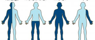
Causes of benign rolandic epilepsy
The exact causes of the disease are unknown. However, there is a hypothesis that confirms the hereditary nature of the pathology. The disease may occur due to a pathological disorder in the maturation of the cerebral cortex in the central temporal region.
As for benign epilepsy, its dependence on the patient’s age has been proven, since it is often diagnosed in children. The pathology may be caused by increased excitability of the children's brain. This can happen for a variety of reasons:
- predominance of excitatory neurotransmitters;
- structural and functional features of epileptogenic areas (hippocampus, neocortex, amygdala);
- low GABA content;
- increased number of excitatory reentrant synapses;
- immaturity of GABA receptors.
As the child's brain matures, its excitability decreases, and foci of pathology gradually disappear. Usually, after children reach puberty, the number of seizures decreases until they disappear completely.
Diagnostics
Electroencephalography (EEG) is the most basic diagnostic method. However, for some, no changes are observed during the daytime, and they are noticeable only during sleep, so for such patients additional studies using polysomnography or monitoring are necessary.
A characteristic feature of the disease is the presence of sharp waves and high amplitude peaks in the central and temporal parts. They exist against a background of normal and normal brain activity. In such a situation, the “Rolandic complex” often occurs. It lies in the fact that after the peak slow waves follow. Its duration is usually about 30 ms.
Such complexes can be compared to the QRST complexes on an electrocardiogram. Almost always, “Rolandic complexes” occur on the side that is not subject to convulsions, but cases of bilateral manifestations cannot be excluded. Another distinctive sign of pathology during diagnosis is the ability of EEG patterns to change from one recording to another.
Symptoms of benign rolandic epilepsy
With rolandic epilepsy, patients periodically experience seizures that are moderate in nature. In this case, the attack occurs without the patient losing consciousness. Often, the patient experiences a sensory aura in front of him, which is characterized by sensations such as tingling, tingling, numbness in the gums, face, throat, tongue and lips. Then strong tonic and muscle contractions occur.
If a patient experiences a hemifacial attack, they experience muscle spasms on one side of the face. During a pharyngeal attack, unilateral convulsions of the tongue, lips, larynx and pharynx occur, which are inevitably accompanied by speech impairment. Due to convulsive contractions of the muscles of the larynx, guttural sounds appear that resemble gurgling or gargling.
Patients' attacks occur mainly at night. They are often observed during the period of falling asleep or waking up. In rare cases, both daytime and nighttime attacks are observed. In 20% of all cases of the disease, brachiofacial seizures occur, which are characterized by the spread of facial spasms to the homolateral arm. Patients may also experience more severe forms of seizures, in which spasms involve all the muscles of the body. Typically, such extensive convulsions cause the patient to lose consciousness.
How does the disease progress?
The peaks of Rolandic epilepsy pass without loss of consciousness and take the form of attacks with tingling, numbness and tingling of half the face (the affected sides may change with each new case. Sometimes benign epilepsy in children ends with convulsions at night. Experts associate this with the sleep process.
At the onset of a seizure, observant parents often notice changes in the child’s behavior - children complain of pain in the tongue and gums, as well as “goosebumps crawling across the face.” After the onset of an attack, the following symptoms of benign epilepsy are noted:
- spasms affecting the face and throat (usually half the muscles contract);
- spasms affecting the lips and tongue;
- increased salivation;
- speech impairment (due to numbness of the tissues of the oral cavity - primarily the tongue);
- strange throat sounds, reminiscent of wheezing when breathing.
Often attacks are sporadic and occur mainly at night, so some parents are not aware of their child’s illness.
Course of benign rolandic epilepsy
Benign rolandic epilepsy is characterized by short-term attacks, the duration of which is about 2-3 minutes. Typically, such attacks are isolated and occur several times a year. At the beginning of the disease, epileptic seizures may occur frequently, but over time they occur much less frequently. It is important to note that this type of epilepsy will not be dangerous for children, since it does not provoke a delay in their psychomotor development and mental retardation. However, in rare cases, neurological damage occurs.
Course of the disease
A huge advantage of the pathology is certainly the fact that attacks are very rare, and their duration does not exceed 2-3 minutes. There are many known cases when they were isolated and were repeated no more than several times a year. Of course, attacks may occur more frequently, but as time passes and the child gets older, they are guaranteed to happen less and less often.
If we compare benign Rolandic epilepsy with Jacksonian epilepsy, then the first has absolutely no effect on the child’s development, including psychomotor and physical, and will not cause mental retardation.
However, this disease cannot be called completely safe. In a small number of carriers of the disease, namely 3%, epilepsy leads to incomplete paralysis of half the face, which is called transient hemiparesis.
Diagnosis of benign rolandic epilepsy
The disease can be most accurately diagnosed using electroencephalography (EEG). This technique is used to study the functional activity of the brain and identify pathological disorders in it. However, it is worth noting that in some patients, changes in the EEG may only be noticeable during sleep. That is why the doctor may additionally prescribe an overnight EEG or polysomnography.
A neurologist can make a diagnosis if the following pathology is detected: identification of high-amplitude sharp waves or peaks that are localized in the central temporal leads. At the same time, the functional activity of the brain is normal. The EEG results in this case show that the peaks or sharp waves are immediately followed by slow waves, which together constitute the “Rolandic complex”. This symptom can be either bilateral or unilateral.
International Neurological Journal 5(21) 2008
Idiopathic epilepsies in children represent more than half of all cases of epilepsy and suggest the absence of an detectable morphological substrate, as well as a neurological deficit in the interictal period. They are most likely genetically determined syndromes, exhibit an age-dependence and a benign course in most cases. The list of these epileptic syndromes is gradually expanding [3, 10].
The classic representative of this group of epilepsies is Rolandic epilepsy (RE), or benign childhood epilepsy with centrotemporal adhesions. It is one of the most common forms of childhood epilepsy and accounts for up to a quarter of cases of epilepsy between the ages of 5 and 14 years. Its prevalence ranges from 7.1 to 21 per 100,000 children under 15 years of age. The true incidence of this disease may be higher because not all cases are diagnosed.
Simple focal seizures make up the bulk of seizures in EC and are observed in 70–80% of patients. The most typical is the onset of an attack with a somatosensory aura: a sensation of tingling, numbness, “passage of electric current” in the pharynx, tongue, and gums on one side. At this point, either the paroxysm ends, or after the aura a focal motor attack develops.
The following possible attacks are possible:
- hemifacial - unilateral tonic, clonic or tonic-clonic spasms of the facial muscles;
- pharyngooral - unilateral convulsions of the lip, tongue, pharynx, larynx, which are often associated with anarthria and hypersalivation.
In 20% of patients, seizures can spread from the face to the homolateral arm (brachiofacial seizures); in approximately 8% of cases, the leg is also involved in the process (hemiconvulsive seizures). Seizures can change their direction. Secondary generalized seizures are observed in 20–25% of patients with EC. They are most typical for younger children and are often the debut symptom.
The duration of attacks is usually short - from a few seconds to 2-3 minutes. In 11–22% of patients, the duration of paroxysms exceeds 10–15 minutes. In isolated cases, severe prolonged attacks are observed, which end in transient post-attack hemiparesis (Todd's palsy). The frequency of attacks is also usually low - on average 2-4 times a year.
A characteristic interictal electrographic correlate is distinct high-amplitude peaks followed by a pronounced slow wave, appearing singly or in series in the central-middle temporal (Rolandic) region (T3 and C3 or T4 and C4) with the formation of a dipole in the frontal leads (Fig. 1–2). The number of Rolandic peaks increases during slow-wave sleep. The specificity of this EEG pattern is, of course, not absolute, but very high and can be compared with the specificity of generalized 3 Hz peak-wave complexes for typical absence seizures.
Diagnosis of Rolandic epilepsy is based on a combination of clinical manifestations, normal psychoneurological status, normal brain image according to neuroimaging data and a characteristic EEG pattern.
Criteria for the diagnosis of Rolandic epilepsy (Loiseau, Duche, 1989):
- debut at the age of 3 to 13 years;
— normal mental and neurological status before the onset of the disease;
- partial motor seizures, often with somatosensory aura, provoked by sleep;
— focus of peaks in the centrotemporal (rolandic) region with normal background activity;
- spontaneous recovery in adolescence.
The above criteria are relevant for classical Rolandic epilepsy. However, not all patients with a rolandic EEG pattern or hemifacial motor seizures correspond to them. Atypical clinical manifestations (exclusively or predominantly daytime attacks, severe Todd's paresis, prolonged attacks, status course, early or late onset, interictal neurological deficit and atypical EEG pattern) are not exceptionally rare and can account for up to half of all cases [1–3, 7]. Atypicality of EEG manifestations of RE (unusual morphology and localization of the maximum of epileptic phenomena, combination with other EEG patterns) also occurs quite often [1, 3, 10].
Over the past 10 years, 743 children with an electrographic picture of rolandic epilepsy have been under observation in our clinic. Various variants of the above clinical deviations were identified in 150 children (20.2%). 2 children with a typical Rolandic EEG pattern had completely atypical attacks: 1 child experienced nocturnal hemidysesthesias lasting up to 5 minutes, and the other experienced rare short paroxysms of abdominal pain during sleep. In both cases, the disease ended in remission and the disappearance of interictal discharges on the EEG.
Atypical variants of EC include cases when this syndrome develops against the background of a known damaged brain. In our practice, there are 13 cases of a typical electroclinical picture of RE in children with cerebral palsy.
EEG changes in our patients with the clinical picture of EC were typical only in 63% of cases. The rest had certain variations in the EEG pattern - the maximum amplitude and electrical negativity was not in the middle temporal lead; combination with peaks of a different localization (most often occipital and centrosagital); peaks exclusively in the centrosagital lead; combination with generalized discharges of peak-wave complexes or polyps; photosensitivity; recordings with very abundant epileptic activity that would meet the criteria for electrical status epilepticus (Figs. 3–6) [8–10].
The characteristics of the EEG in each individual patient can change significantly over time - peaks can disappear completely, reappear, change localization, increase in frequency to continuous and disappear again, to resume at the next examination, and then disappear forever. We consider such instability of electrographic manifestations as one of the signs of the idiopathic nature of epilepsy. Less commonly, relative stability of interictal activity is possible throughout the entire period of the disease.
The traditional idea of the benignity and excellent prognosis of idiopathic focal epilepsies of childhood, of which Rolandic epilepsy is a representative, has recently been significantly shaken. The concept of the famous epileptologist Panayiotopoulos of a benign childhood predisposition to seizures [4] cannot be applied to all cases of EC. With RE, not only frequent, severe and drug-resistant attacks are possible, but also a combination with permanent or transient behavioral, cognitive disorders, speech impairments and other types of psychoneurological deficits that are not typical for classical RE.
Such forms in modern literature are combined into the group “Rolandic epilepsy plus” [1–3, 5–8]:
— Rolandic epilepsy with transient cognitive impairment in the active phase;
— status of Rolandic epilepsy;
— acquired epileptic opercular syndrome (status opercular);
- Rolandic epilepsy with negative myoclonus.
The first category of these options includes Rolandic epilepsy, which occurs with transient cognitive disorders that occur during the period of greatest activity of the disease and directly correlate with the severity of epileptic activity on the EEG. Typically, clinically detectable abnormalities occur when the discharge rate is greater than 10 per minute, and normalization occurs when the discharge frequency is less than 5 per minute. Neuropsychological testing or cognitive evoked potential testing may be needed to identify such disorders.
The status of Rolandic epilepsy is represented by frequent Rolandic motor seizures with dys- or anarthria, drooling, oromotor dyspraxia, hemifacial seizures and atonic head nodding as part of negative myoclonus. Episodes of negative myoclonus may involve the arms and occur so frequently, literally with every shock, that it becomes difficult to manipulate the arms and may result in the child deliberately avoiding using the arm involved in the shocks. Interictal EEG is represented in such children by frequent bilateral (often synchronous) discharges of “peak-wave” or “sharp-slow wave” complexes with a maximum in the rolandic region and with a predominance over one of the hemispheres (Fig. 7–8). We observed two patients with RE with periods of pronounced negative myoclonus, which involved the half of the face and arm contralateral to the discharges. One of these cases also had congenital hypoplasia of both kidneys, the patient was on dialysis and no anticonvulsant therapy was administered. In the second case, pharmacoresistance was noted, and only corticosteroids and intravenous infusions of immunoglobulin led to short-term remissions.
Acquired epileptic opercular syndrome (or status opercularis) is clinically characterized by prolonged but clearly fluctuating episodes of pseudobulbar palsy with hypersalivation and drooling, and oromotor dyspraxia. The duration of each such episode can range from several hours to several months. Typical rolandic attacks do not occur during this period, but mild positive or negative perioral or facial myoclonus can be detected. An EEG recording while awake reveals abundant bilateral rolandic epileptic activity, which, however, does not reach the level of electrical status epilepticus. During periods of deterioration, electrical sleep status epilepticus may also develop. We observed three children with typical orofacial seizures and profuse rolandic discharges at onset, followed by long periods of severe drooling, moderate dysarthria, swallowing disorders and cognitive impairment. In all cases, anticonvulsant therapy led to relief of this condition.
At the onset of the disease, the clinical picture and EEG patterns may look completely harmless. However, its course can change, and after a few months or years the child’s condition worsens - seizures become more frequent, other types join in, and epileptic activity sharply increases; ordinary RE can transform into one of the variants from a large group of “disorders associated with rolandic epilepsy” [2, 6–9].
This group is represented by various syndromes of epileptic encephalopathy with a rolandic EEG pattern, electrical status epilepticus of sleep and/or wakefulness, variable combinations of seizures, including rolandic, - atypical benign focal epilepsy (pseudo-Lennox syndrome), Patry syndrome, acquired epileptic frontal syndrome, syndrome Landau-Kleffner and variants of autistic epileptiform disorders (disintegrative disorder, regressive autistic disorder) [1, 2, 6–9]. Among our patients with a rolandic EEG pattern, such variants were recorded in 57 cases (7.8%). The course of the disease in these children was characterized by the pharmacoresistance of attacks and the predominance of cognitive and behavioral disorders over the ictal manifestations themselves.
Thus, it can be argued that classical Rolandic epilepsy is not a monolithic nosological form, but represents only a small part of the spectrum of Rolandic epilepsies, which in turn are an integral part of a wide range of idiopathic focal epilepsies, within which combined variants or transformation of one syndrome into another are possible.
In this continuum, at the marginal position on one side are patients with a rolandic EEG pattern without any clinical manifestations, and at the opposite extreme are represented by severe disabling epileptic encephalopathies. Epileptic encephalopathies arising from focal epilepsies, in their clinical manifestations, approach generalized epileptic encephalopathies represented by Lennox-Gastaut, Dravet, Douse syndromes (Fig. 9). Common features of most epileptic encephalopathies are cognitive impairment, atypical absence seizures, negative myoclonus phenomenon, and electrical status epilepticus.
In this regard, we can talk about the benignity and good prognosis of Rolandic epilepsy only in relation to patients who exactly fit the criteria for classical Rolandic epilepsy. Unfortunately, full compliance with these criteria can often be established only retrospectively, when the disease has already ended or the tendency of its course has become obvious.
Considering the possibility of an unfavorable or at least atypical course of rolandic epilepsy, in our opinion, it is advisable to abandon the widespread belief in its benignity, not to use the term “benign” in formulating the diagnosis and to be cautious in the prognosis of the disease.
Differential diagnosis of benign rolandic epilepsy
Symptoms of benign rolandic epilepsy are similar to those of other neurological pathologies. First of all, this disease should be differentiated from other types of epilepsy provoked by traumatic brain injuries, neoplasms, inflammatory lesions of the brain due to encephalitis, meningitis, and abscess. The main distinguishing feature of the pathology is the absence of behavioral disorders and serious changes in neurological status in the clinical picture, as well as normal EEG activity and complete preservation of intelligence. A more accurate differential diagnosis can be made using MRI of the brain.
Neurologists note that it is most difficult to differentiate benign rolandic epilepsy from nocturnal epilepsy. In this case, speech disorders that are quite typical for pathology are often interpreted as disorders of consciousness. Therefore, to make a correct and accurate diagnosis, it is extremely important to conduct an EEG. If the patient has temporal lobe epilepsy or frontal lobe epilepsy, the results will show the presence of focal changes in brain activity.
Neurologists often have difficulty differentiating benign rolandic epilepsy and pseudolennox syndrome. The most pronounced signs in which a patient is diagnosed with pseudolennox syndrome will be: a complex of atypical absence seizures with typical rolandic epileptic seizures, the presence of intellectual-mnestic disorders, detection of slow complexes or diffuse peak activity during EEG.
Innovative medicine Treatment of epilepsy
Russian Medical Server / Treatment of epilepsy / Benign childhood epilepsy (rolandic epilepsy)
Benign childhood epilepsy is a form of epilepsy. A characteristic feature of this form of the disease is that the spasms affect the face and sometimes the body. Another name for this type of epilepsy is benign epilepsy with central temporal peaks. The term “benign” in the name of the disease indicates a favorable prognosis for the course of this form of epilepsy, since in most patients the disease resolves during adolescence.
The term “Rolandic” in the name of this disease indicates that the epileptic focus occurs in the Rolandic region of the brain. Such seizures are classified as partial, since their nature is associated only with a given area of the brain.
Who suffers from benign rolandic epilepsy?
Benign rolandic epilepsy occurs in 15% of children with epilepsy. On average, the first attacks of rolandic epilepsy occur between the ages of 6 and 8 years. This form of epilepsy does not occur in adults.
What is the cause of benign rolandic epilepsy?
The causes of rolandic epilepsy are unknown. Children whose close relatives have epilepsy have a slightly higher risk of developing rolandic epilepsy.
Manifestations of benign rolandic epilepsy
Like all forms of epilepsy, benign rolandic epilepsy is characterized by seizures. Usually they are moderate in nature. Most often the face is affected:
- Twitching of the face or cheek
- Tingling or unusual sensations in the face or tongue
- Difficulty speaking
- Drooling due to poor oral muscle control
In approximately every second child with benign rolandic epilepsy, the pathological focus from the rolandic region spreads to other areas of the brain. In this case, they speak of secondary generalized seizures. They are also called tonic-clonic. The manifestation of these seizures is as follows:
- The patient freezes and does not answer questions
- Clenching the muscles of the whole body for a short period
- Rhythmic spasms of the whole body
- Confusion and disorientation after consciousness returns
Typically, in benign rolandic epilepsy, seizures begin during sleep. Therefore, they may go unnoticed by others. Parents often find out about an epileptic attack due to noise in the child’s room at night.
Some children with benign rolandic epilepsy may also have learning difficulties and behavioral problems. These children require additional attention and treatment. However, in most cases, children cope well with schoolwork.
Diagnosis of benign rolandic epilepsy
When seizures are mild and occur only at night, the diagnosis of benign rolandic epilepsy may be easily missed. Often, parents bring their child to the doctor after tonic-clonic seizures during sleep.
Diagnosis of benign rolandic epilepsy is based on the type of seizures. Instrumental research methods are also used: EEG, MRI, neurological tests.
Treatment of benign rolandic epilepsy
Benign rolandic epilepsy often does not require any treatment. Attacks with this form of the disease are usually mild and infrequent. In fact, the child can grow out of the disease himself.
Treatment may be prescribed if benign rolandic epilepsy is accompanied by difficulty studying at school, problems with thinking or attention, behavioral problems, daytime seizures, or frequent seizures. Treatment is usually medication.
Most often, attacks of this form of epilepsy are controlled by antiepileptic drugs such as carbamazepine, lamotrigine or sodium valproate. They are taken daily, and the course of treatment lasts an average of two years.
+7 (925) 50 254 50 – Treatment of epilepsy
REQUEST TO THE CLINIC
Treatment of benign rolandic epilepsy
In neurology, the issue of treatment of benign rolandic epilepsy remains controversial. This is due to the fact that in children with age, all symptoms of the disease disappear without additional medical intervention. Therefore, some doctors defend the opinion that it is inappropriate to prescribe antiepileptic therapy. The opposite opinion is based on the complexity of differential diagnosis of the disease. It may happen that the diagnosis is made incorrectly or the pathology progresses to the pseudolennox stage. To prevent possible complications, doctors may still prescribe antiepileptic therapy to the patient.
When treating children with benign rolandic epilepsy, the principle of monotherapy is used - they are prescribed only one drug. Typically, treatment of children begins with the administration of valproic acid (Depakine, Convulsofin, Convulex). If treatment is ineffective or the child is intolerant to this drug, it is replaced with Topamax or levetiracetam. Children over seven years of age may be prescribed carbamazepine.
Parents often do not immediately notice single or rare epileptic seizures in children, since in most cases they occur at night. However, immediately after detecting epileptic seizures, the child must be shown to a neurologist. Diagnosis and treatment of pathology are selected for each case individually. The most careful monitoring is required for patients under three years of age, who may experience an increase in seizures due to the immaturity of nerve cells.
Parents of a sick child who has had an attack also need to know what they can do on their own before the ambulance arrives. If the baby has a complex seizure, which is accompanied by serious motor disturbances (tonic convulsions or muscle twitching of the face, tongue, larynx and pharynx), he needs to be slightly raised and calmed. It is extremely important to do this if the attack occurs while drinking, eating or sleeping. To prevent the tongue from sticking, you need to put the child's head on one side, and then put a handkerchief or some soft object in his mouth. If a child has a prolonged seizure, it is necessary to urgently call an ambulance.
Treatment
To treat or not to treat is a complex issue that remains a field of debate between representatives of different directions. On the one hand, even without therapeutic measures, the outcome is complete recovery. Seizures occur rarely and do not have a significant impact on the quality of life. All of the above raises doubts about the need to use antiepileptic drugs.
On the other hand, the possibility of error in determining the type of disease cannot be completely excluded. In addition, sometimes the Rolandic form transforms into pseudolennox syndrome. Given these circumstances, the use of anticonvulsants seems justified. Repeated episodes become the reason for prescribing drug therapy.
The standard is monotherapy (taking one drug with antiepileptic effect). At the initial stage of treatment, the drug valproic acid is usually prescribed. If there is no effect or intolerance to the medication, levetiracetam or topiramate is considered as an alternative. Carbamazepine is used with caution in children of the older age group, given that sometimes it causes not a cessation, but an increase in epileptic seizures.
The outcome is favorable. After adolescence ends, all disorders disappear, no residual effects are observed. Problems are rare and can arise when an incorrect diagnosis is made or when rolandic epilepsy changes to another form. To prevent such an outcome, a detailed examination and regular monitoring by specialists with possible correction of therapy are necessary.
Qualified neurologists have extensive experience in the diagnosis and treatment of benign epilepsy, which allows them to choose the optimal treatment option and prevent complications. Appointments can be made by phone..
Recommendations for epileptic seizures
There are a number of recommendations from doctors, adherence to which will help prevent an epileptic attack. First of all, you need to strictly adhere to your sleep and wakefulness schedule. The child must get a good night's sleep, as sleep problems can trigger a severe attack. In most cases, epileptic seizures occur in a child during sleep. At the same time, it is not always possible to notice an attack (usually this becomes clear only if the patient bites his tongue during an attack). Therefore, if the baby sleeps in a separate room, it is advisable to use a baby monitor - a device that allows you to hear sounds in the next room.
Patients with Rolandic epilepsy often experience insomnia at night and drowsiness during the day. As for drowsiness, it occurs mainly due to the use of medications. Insomnia can be caused by poor sleep hygiene: the patient should not sleep in a stuffy room, with the TV on, and should not drink or overeat before bed. The child should go to bed at a set time. If all these recommendations do not help and the attacks recur regularly, you should consult a neurologist.
Features of epilepsy in childhood
4. Features of epilepsy in childhood
The main features of childhood epilepsy are as follows:
- there is a large number of treatment-resistant forms of epilepsy;
- characteristic is a large polymorphism of epileptic paroxysms;
- the frequency of masked manifestations of the disease is high: behind many unclear pain attacks, umbilical colic, fainting, acetonemic vomiting, epileptic paroxysms of an organic nature may be hidden;
- Various non-epileptic phenomena, such as sleepwalking, night terrors, enuresis, migraines, syncope, and hysterical (conversion) seizures, are often mistaken for manifestations of epilepsy. At the same time, there is a tendency to regard similar and some other symptoms as epileptic in blood relatives of patients, using the term “diseases of the epileptic circle” to designate them. It should be taken into account that with any multifactorial disease, which includes epilepsy, many paroxysmal disorders of a non-epileptic nature can be found in the patient himself and his blood relatives. Therefore, at present, the terms “pre-epilepsy” and “diseases of the epileptic circle” are considered obsolete;
- in childhood epilepsy, a malignant course is often observed with the development of psychopathological symptoms and mental retardation;
- at the same time, in childhood, absolutely benign forms of epilepsy occur, ending with complete recovery, restoration of all body functions and successful socialization;
- In children there are also delayed forms of epilepsy, when seizures begin during the neonatal period, then they stop and then resume years later.
As for epileptic seizures, they have some common features in children:
- high frequency of undeveloped, incomplete, rudimentary forms of epiparoxysms;
- the presence of forms of epilepsy and types of seizures that do not occur in adult patients;
- high proportion of all absence forms of paroxysms;
- transformation of seizures as one gets older;
- frequent development of post-ictal focal neurological symptoms.
4.1. Epileptic syndromes in neonatal and infancy:
- benign idiopathic neonatal seizures. Appear around the 5th day. Have a favorable prognosis;
- benign idiopathic neonatal familial seizures. Occurs on the 2nd–3rd day of life. Someone in the family sometimes experiences the same cramps. The prognosis is favorable;
- early (neonatal) myoclonic encephalopathy: massive partial or fragmented myoclonus and partial motor seizures, short-term tonic seizures, spike fast-wave and slow-wave activity on the EEG. These signs indicate severe brain damage. Early death is common;
- early epileptic encephalopathy with the EEG phenomenon “suppression - discharges” - Ohtahara syndrome. Onset in the 1st month of life. Seizures are in the form of tonic ones, often grouped in a series of spasms. The EEG records alternating short periods of flattening of the curve and generalized epileptic activity. The prognosis is severe, and early death often occurs.
4.2. Epileptic syndromes of early childhood. These include the following.
1) Febrile convulsions (see paragraph 2.6).
2) Benign myoclonic epilepsy of early childhood. The earliest form of idiopathic epilepsy. Onset in the 1st–2nd year of life in children with normal development. Seizures in the form of generalized myoclonus. EEG shows spike-wave activity during slow-wave sleep. The prognosis is good. The treatment of choice is valproate.
3) Severe myoclonic epilepsy. One of the most malignant forms, absolutely resistant to therapy. Onset in the 1st year of life in healthy children with a family history of epilepsy and febrile seizures. Characterized by febrile unilateral clonic seizures, myoclonus, and often partial seizures. Generalized spike waves and polyspike waves are recorded on the EEG. There is a pronounced delay in mental development and severe neurological symptoms.
4) Myoclonic epilepsy (myoclonic status) in combination with non-progressive encephalopathy. Encephalopathy is detected from the first months of life in the form of an atonic variant of cerebral palsy, dyskinetic syndrome and severe mental retardation. Myoclonus varies from rhythmic bilateral to asynchronous. Polysomnographic studies during slow-wave sleep show an EEG picture of status epilepticus. Encephalopathy does not progress. Over time, the condition stabilizes, although myoclonus persists and absence seizures occur.
4.3. Childhood and adolescent forms of epilepsy.
1) Childhood absence epilepsy, or Friedman's syndrome (1906), pycnolepsy, pycnoepilepsy (Greek pyknos - frequent; lepsis - attack, grasping). Usually begins in children aged 6–7 years (according to other sources, at 4–10 or 2–8 years). It is more common in girls and is characterized by a high degree of hereditary predisposition (penetrance of the epilepsy gene). The main symptom of the disease is the presence of typical absence seizures, which in most cases occur with a tonic component and are retropulsive (the head falls back, the eyes rise up). It is noted that retropulsive seizures are often accompanied by a mild clonic component - twitching of the muscles of the face and eyelids. The frequency of seizures can reach dozens per day, absence seizure status is also observed. Focal postictal neurological symptoms, mental retardation and neurodestructive changes in the brain are not detected. The disease responds well to therapy. The treatment of choice is valproate, if necessary in combination with succinimides.
2) Juvenile absence epilepsy is an age-dependent form of idiopathic epilepsy. Begins at puberty, there are no differences in sexual preference. It manifests itself as a combination of simple absence seizures and primarily generalized tonic-clonic seizures. Seizures appear much less frequently, and their duration is longer than with pycnolepsy. The EEG shows paroxysms of generalized, synchronous, symmetrical spike waves with a frequency of 3.5–4 Hz. Responds well to therapy. The treatment of choice is valproate.
3) Juvenile myoclonic epilepsy, or impulsive Janz epilepsy. Age-dependent form of idiopathic epilepsy. It begins in adolescents aged 12–16 years, and is more common in girls. A hereditary predisposition is noted, the disease gene is mapped to the 6p21.3 locus. It manifests itself as generalized myoclonic seizures, mainly after sleep, and occurs in the form of sudden symmetrical or asymmetrical twitching in the muscles of the shoulder girdle - flinching of one or both arms (“wing flapping”). At the same time, the arms involuntarily fall to the side, objects fall out of the hands, the head and torso turn sharply. Often, the muscles of the pelvic girdle and legs are involved in a myoclonic attack - the patient may squat at this time, fall on his knees or buttocks (myoclonic-astatic attack). Seizures occur in series or in bursts of 5–20 times in a row over several hours. On the EEG during an attack, in response to photostimulation, generalized spike and polyspike waves are recorded, often with gigantic amplitude, both episodic and spontaneous. The most important factor in provoking seizures is sleep deprivation. Consciousness is not impaired during seizures. Characterized by the absence of pronounced neurological symptoms and intellectual impairment. Treatment requires the elimination of provoking factors and the use of valproate, sometimes in combination with clonazepam.
4) Epilepsy with generalized tonic-clonic seizures on awakening. The main role in the development of the disease is attributed to genetic factors, but it is believed that there may also be symptomatic forms. It begins at the age of 10–20 years, sometimes earlier and later. Seizures occur within 2 hours of waking up from sleep, regardless of whether it is at night or during the day. Seizures are usually preceded by a series of bilateral myoclonus. There may also be a second peak of attacks before bedtime, especially with late onset of the disease. The EEG is characterized by an increased number of slow waves and generalized spike-wave and polyspike-wave activity at a frequency of 2.5–4 Hz. The prognosis is favorable. The main treatment is valproate and phenobarbital.
5) West syndrome (West), or infantile spasm. These are symptomatic and cryptogenic forms of epilepsy. One of the most common age-dependent epileptic syndromes. May begin in infancy, with peak incidence occurring between 2–4 and 7 months of age, sometimes later. More common in boys.
In typical cases, West syndrome is characterized by a triad of symptoms: a) generalized myoclonic or myoclonic-tonic seizures; b) delayed mental, speech and motor development and c) hypsarrhythmia.
Seizures manifest themselves as:
- sudden sharp bending of the head (nodding);
- sudden sharp bending of the head and shoulders (peck);
- slow tonic bends with extension and abduction of the arms according to the type of eastern greeting (“Salaam convulsions”);
- sudden propulsive movements with falling forward. Seizures are accompanied by a scream, an expression of fear on the face, a grimace of a violent smile. Injuries due to falls during an attack are common.
The duration of attacks ranges from fractions to several seconds. Seizures can be single or occur in series, numbering several hundred attacks per day. Most often, seizures appear before bedtime, when falling asleep, but especially often in the morning - during awakening or immediately after waking up from sleep. There are “good” and “bad” days – with fewer and more frequent attacks.
On the EEG, a special type of disturbance is recorded in patients - hypsarrhythmia: the absence of main activity and the presence of asynchronous slow activity, which alternates with sharp waves or spikes. The prognosis is poor: as people get older, myoclonic and myoclonic-tonic seizures transform into grand mal seizures.
Therapy: ACTH or glucocorticoids in combination with valproate and benzodiazepines, large doses of pyridoxine (up to 100 mg/kg/day), in resistant cases immunoglobulin G IV.
6) Lennox-Gastaut syndrome. This is an age-related encephalopathy syndrome of unknown origin. The onset of the disease occurs between the ages of 1 and 8 years (according to other sources, from 3 to 6 years). Often this syndrome is transformed from a previous infantile spasm. In typical cases the following are observed:
- epileptic seizures mainly of the type of generalized tonic, atonic seizures and atypical absence seizures;
- severe delay in mental, speech and motor development;
- specific changes on the EEG.
Seizures most often occur in the morning, and there are “good” and “bad” days. Falls during seizures are often accompanied by injury. Tonic seizures are detected in 100% of patients. They last from 5 to 20 seconds and proceed according to the tonic-axial type (with rotation of the body around its axis). The EEG shows complexes of bilateral, synchronous spike waves of 1–2.5 Hz, irregular and unstable, transforming under the influence of sleep into rhythmic activity with a frequency of 10 Hz.
Subsequently, the seizures are replaced by generalized and complex partial seizures. In treatment, a combination of carbamazepine, valproate and benzodiazepines is used, in recent years - lamotrigine, vigabatrin, which are more effective in treating attacks of falling. The effectiveness of intravenous administration of immunoglobulin G has been established.
7) Duse syndrome, or myoclonic-astatic epilepsy. Symptomatic and cryptogenic form of the disease with age-dependent seizures. Begins in the 1st–2nd year of life with febrile and nonfebrile generalized tonic-clonic seizures. The main manifestation is myoclonic and astatic seizures. The EEG shows multifocal changes with generalized spike waves, spontaneous and in response to photostimulation. Spontaneous remissions are observed, but a malignant course is also possible. Treatment: Valproate is used, alone or in combination with ethosuximide, and sometimes with benzodiazepines. In the presence of generalized tonic-clonic seizures - ACTH, glucocorticoids. Carbamazepine is contraindicated.
 Epilepsy with myoclonic absence seizures. Age-dependent form of symptomatic and cryptogenic epilepsy with onset at 6–8 years. It manifests itself as myoclonic absences lasting 10–60 seconds with massive bilateral rhythmic myoclonus. The EEG shows the rhythm of typical absence seizures with high photosensitivity. Most patients experience infrequent generalized tonic-clonic seizures. Subsequently, mental retardation is revealed. Treatment resistance is common. Therapy: use valproate, phenobarbital, clonazepam and other benzodiazepines.
In children, especially in prepubertal age, there are rare forms of epilepsy with myoclonus of the eyelids and “self-evoking epilepsy” with seizures sensitive to the closing and opening of the eyelids and light flickers.
9) Rolandic epilepsy, or benign epilepsy with centrotemporal adhesions. Hereditary complications play a major role in the etiology of the disease. Onset between ages 3 and 13 years. Convulsions or paresthesias during a seizure involve the face, lips, tongue, and pharynx. Possible speech arrest or anarthria. Drooling is common. Consciousness remains clear. Seizures can spread, becoming brachiofacial or even unilateral in nature. There may be, less frequently, attacks with throat sounds and loss of consciousness, with secondary generalization of seizures. EEG - biphasic high-voltage spikes, following at short intervals in the central and temporal regions, unilateral or bilateral with a predominance on one side. Adhesions sharply intensify in a state of drowsiness or sleep. Therapy: phenobarbital and carbamazepine. After the cessation of attacks and normalization of the EEG, even with short-term treatment (1–2 years), it can be stopped. For rare seizures, anticonvulsants may not be prescribed.
10) Benign partial epilepsy with affective symptoms. There is a high degree of hereditary burden. The onset of the disease is at the age of 2–10 years in children with normal development. Seizures are characterized by sudden fear (to the point of horror) in combination with chewing, swallowing, and vegetative symptoms (pallor, hyperhidrosis, abdominal pain, etc.). Consciousness is usually altered. On the EEG there are complexes of a sharp wave - a slow wave in the frontal or parietal region on one or both sides. Therapy: carbamazepine and phenytoin. The prognosis is favorable.
(Landau-Kleffner syndrome - see Chapter 2.)
Return to Contents
Rolandic epilepsy
or
Benign focal epilepsy of childhood with centrotemporal spikes.
CLINICAL CASE DESCRIPTION
06.2013.
Appointment with an epileptologist in the outpatient department. Patient K.A., born in 2003 (age 9 years), visited an epileptologist for the first time.
We complained of attacks upon awakening: swallowing movements, drooling, cannot speak, clonic convulsions in the right half of the face and right arm, the child reacts to others, comes to his mother at night, “moos”, the attack lasts about 1 minute. After the attack comes sleep.
Onset of seizures 8 months ago. To date, no doctors have been contacted. Such attacks occur with a frequency of 1 time per month for 3 months, then 2 times per month, then 4 attacks per month.
From the Internet, parents learned that they should conduct an examination (EEG, MRI of the brain) and contact an epileptologist.
In addition to seizures, the child has other complaints. Hyperactive behavior. Difficulties in learning at school. He studies at school as a 3-4 student.
Peaceful sleep. Denies traumatic brain injuries. Headaches do not bother me.
From the life history: A child of young, healthy parents, from 1st pregnancy, which occurred with mild gestosis, chronic subcompensated fetoplacental insufficiency.
Childbirth at 39 weeks, spontaneous, uncomplicated.
Birth weight 3.6 kg, body length 52 cm, head/chest circumference 34/34 cm, Apgar score 8/9.
Maternity hospital diagnosis: Full term, maturity. Risk of CNS PP.
He grew and developed according to age: he began to hold his head from 1 month, to sit from 6 months, to walk from 11 months; phrasal speech from 1 year 10 months.
A neurologist performed preventive examinations. Received 2 courses of massage before 1 year.
Previous illnesses: infrequent acute respiratory viral infections.
Attended kindergarten and attends football classes.
There is no hereditary history of epilepsy.
Neurological status:
Hyperactive. Intelligence is normal.
The skull is round in shape, painless on percussion, head circumference 53 cm.
Cranial nerves: without pathology.
Motor sphere: no paresis, sufficient muscle tone in the limbs, tendon reflexes are lively and equal.
Performs coordination tests confidently.
Poor posture.
Results of the additional examination:
rolandic epilepsy
EEG (background dated 06.2013) – mild diffuse changes in the bioelectrical activity of the brain with irritation. Regional epileptiform activity in the form of acute-slow wave complexes in the central-parietal-temporal regions with distribution, high index of epiactivity .
MRI of the brain (06.2013) - no pathology was detected.
Taking into account the nature of the attacks, children's age, the absence of focal neurological symptoms, intact intelligence, the nature of epiactivity according to the EEG, the absence of pathology according to MRI, the type of course can be diagnosed:
Benign focal epilepsy of childhood with centrotemporal spikes (or Rolandic epilepsy).


