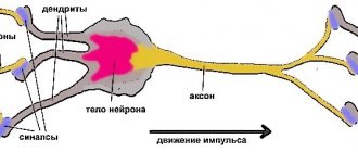
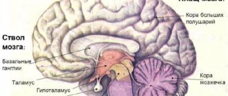
Structure and functions of the brain in the table
| Part | Structure | Function |
| Oblong (stem section) | Extension of the spinal cord located on the trunk. It has a white substance on the outside and a gray substance on the inside. Gray matter is contained in the form of nuclei. | Conductive, nutritional, protective, control of respiratory rate, control of heart rate, control of vital reflexes responsible for sneezing, swallowing, hunger. |
| Average | Connects the forebrain and hindbrain. Contains parts called quadrigeminal tuberosities. | Primary or subcortical centers of hearing and vision. Thanks to this, a person in the field of vision touches new objects or sound sources that appear. There are also centers responsible for muscle tone. |
| Intermediate | It consists of: thalamus, epithalamus, hypothalamus. The thalamus contains the centers of almost all sensory senses. The hypothalamus is the part of the intermedia that connects to and controls the pituitary gland. | Vision, tactile and taste sensations, sensations of body temperature and the environment, memory, sleep. |
| Cerebellum (hindbrain) | The subcortical part of the brain that has grooves. It consists of two hemispheres, which are held together by a worm. | Regulates coordination of movement, the ability to maintain the body in free space. |
| Cerebral hemispheres ( telencephalon) | It is formed from two parts (right and left), divided into grooves and convolutions, due to which the surface increases. They consist of a large amount of gray matter, which is located on the outside, and white on the inside. | Vision (occipital lobe), skin-articular sensitivity and muscle tone (parietal lobe). Memory, thinking, consciousness, speech (frontal lobe) and hearing (temporal lobe). |
Temporal lobe
Many researchers have found a decrease in neuronal density or a decrease in the volume of nerve cells in the temporal lobe in schizophrenia.
The results of a number of studies indicate an asymmetry in the size of the right and left temporal lobes in schizophrenia, with the former being larger than the latter.
As the disease progresses, the temporal lobe of the brain gradually shrinks in size. If in the early 90s only 50% of researchers adhered to this point of view, then already in 2001, with the expansion of the capabilities of neuroimaging methods for studying the brain, data on a reduction in the temporal lobe by an average of 3% in schizophrenia were confirmed in the works of more than 60% of scientists .
A decrease in the volume of the temporal lobes is less frequently recorded in patients with the first episode of schizophrenia compared to patients who have suffered from this disease for a long time. It was found that the volumes of the medial parts of the temporal lobes in patients with schizophrenia are always reduced compared to the volumes of these brain regions of healthy people.
Temporal lobe changes in schizophrenia
- Increased volume of the temporal lobe in childhood schizophrenia
- Reduction in the amount of gray matter in the superior temporal gyrus of both hemispheres
- Reduction in the volume of the left anterior superior temporal gyrus
- Reduction in the volume of the medial temporal lobes (amygdala)
- Reduction in the size of the fusiform gyrus
- Increased activity in the right temporal lobe and decreased activity in the left temporal lobe
- Increased activity in Wernicke's and Broca's areas
- Disruption of connections between neurons of the temporal and frontal cortex
- Disruption of connections between neurons of the temporal cortex
In schizophrenia, the activity of certain parts of the temporal lobe of the brain is increased, for example, Wernicke's and Broca's areas , and this activity is usually observed in those patients who experience auditory hallucinations and mistakenly perceive their thoughts as voices addressed to them.
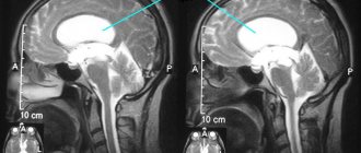
| | MRI allows you to diagnose changes in the size of the temporal lobe, and you can track brain activity in these areas using EEG |
Clinically, damage to the temporal lobe of the brain is relatively often manifested by auditory hallucinations and various disorders of thinking and speech. It is known that the superior temporal gyrus of the left hemisphere of the brain is the anatomical substrate for speech and communication. In Heschl's transverse gyrus there are 41 primary auditory cortexes associated with Brodmann's area. This area of the cortex is responsible for the non-interpretive awareness of auditory impulses that come from the inner ear and through the lemnicus lateralis, corpus geniculate mediale of the thalamus, reach the anterior transverse gyrus in the gyrus transversi. Interpretation of perceived sounds, previously distinguished only as sounds of different frequencies, occurs in the secondary auditory area of the cerebral cortex, which also serves as the sensory center for speech. Localized in Brodmann's areas 42 and 22 and anatomically corresponding to the planum temporale, it borders on the primary auditory cortex, located as if along its periphery. As a result of integration processes occurring in this area, sounds begin to be recognized as noises, words and melodies.
The auditory zone of the cortex is finely differentiated, each frequency has its own ending point, high frequencies are localized to a greater extent in the posteromedial area of the cortex, low frequencies, on the contrary, show anterolateral localization.
In patients with schizophrenia, the volume of the superior temporal gyrus is reduced compared to the volume of this region of the brain in healthy people. More noticeable is the change in the size of the left anterior superior and posterior superior parts of the temporal lobe. The fusiform gyrus in schizophrenia is also somewhat reduced in size.
Data were obtained indicating a correlation between thought disorder and the volume of the left anterior superior temporal gyrus. Moreover, the severity of thinking disorders correlated with the degree of reduction in the volume of this area of the brain (Shenton M. et al., 1992).
According to some authors, enlargement of the right superior temporal gyrus in childhood may be a biological risk marker (phenotypic risk indicator) for the onset of schizophrenia.
The most pronounced changes and, in particular, a decrease in size in schizophrenia are found in the gray matter of the superior temporal gyrus. However, when assessing the total size of the gray and white matter of the temporal lobe of the brain, the differences become less distinct.
A number of authors suggest that the pathological process in schizophrenia sequentially spreads from the deep structures of the brain, the parietal lobe to the frontal and then the temporal lobes, noticeably manifesting itself in the area of the superior gyrus of the right temporal lobe (gyrus temporalis superior).
The loss of cortical gray matter in schizophrenia is characterized by its dynamics and occurs in a specific order: starting from the parietal lobe, through the frontal lobe and the dorsolateral prefrontal cortex to the temporal lobe. At the same time, the temporal lobe of the brain and, especially, the superior temporal gyrus undergo structural changes much later than other brain structures. Note that the superior temporal gyrus is distinguished by the expression of highly differentiated dopamine receptors (D2), however, the density of these receptors here is still less than in the striatum.
Some studies have demonstrated a relationship between the severity of positive symptoms of schizophrenia and the degree of reduction in the gray matter of the superior temporal gyrus, and the intensity of auditory hallucinations correlated with a decrease in its anterior part, which includes Heschle's gyrus, and the size of the posterior part of the superior temporal gyrus (planum temporale) correlated with the severity thinking disorders. At the same time, according to some authors, noticeable cortical atrophy was not observed in patients with paranoid syndrome.
Some authors explained the decrease in the volume of gray matter in the temporal lobe and the increase in the volume of the basal ganglia with antipsychotic therapy. This process typically began following an increase in cortico-subcortical glutamate expression. In those patients with schizophrenia who did not receive antipsychotics, on the contrary, there was a decrease in the volume of the basal ganglia.
In schizotypal disorder, changes in the superior temporal gyrus have also been noted.
In childhood schizophrenia, in contrast to the decrease in the size of the temporal lobe recorded in adult patients with schizophrenia, on the contrary, an increase in the volume of this lobe of the brain was noted. The increase in the volume of the temporal lobe in childhood schizophrenia is explained as a secondary reaction to primary hypoplasia of adjacent brain structures. It can also be assumed that the temporal lobe of the brain is initially excluded from the destructive processes associated with the development of schizophrenia .
As childhood schizophrenia progresses, the right temporal lobe begins to shrink in size at a faster rate than the left. Thus, schizophrenia manifests itself in a progressive disorder of the structures of the temporal lobe of the brain (“neuronal developmental disorder”), in which early and late periods of change can be distinguished.
In schizophrenia in childhood and adolescence, some researchers have found an increase in the medial structures of the temporal lobe of the brain, in particular, the size of the amygdala.
For childhood schizophrenia, most researchers consider the presence of a defect in the genes responsible for the synthesis of factors involved in the development of certain brain structures to be significant. This defect also explains the disruption of neuronal connections, manifested in the form of speech deficits.
An MRI study of patients with schizophrenia revealed an association between changes in the superior temporal gyrus with cognitive deficits and hallucinations. Speech deficits in the premorbid period suggest a neuromorphological deficiency in this area of the brain long before the manifestation of clear clinical symptoms of schizophrenia. It is likely that a significant change in the structure of the temporal lobe of the brain in adolescence is a consequence not only of genetic predisposition, but also the result of developed psychosis, since clear correlations of structural changes in the temporal lobe of the brain with the severity of symptoms of psychosis have been shown in a number of studies.
In schizophrenia, the activity of the right temporal region, responsible for processing sound information, increases, and the activity of the left temporal region, associated with one's own speech, decreases. It is known that there is a certain connection between the left parietal-temporal region and the ability to understand mental states (Ferrari P., 2006).
Other studies have demonstrated abnormalities in the activity of brain regions associated with the perception of faces (fusiform gyrus), recognition of emotions (amygdala), and perception of human voices (superior temporal region).
At the neurophysiological level , with mental automatism syndrome, one can register increased activity in the temporal and parietal areas of the cerebral cortex , as well as in those structures that are responsible for the perception of one’s own actions and their consequences.
Experimental studies have shown that the more pronounced the hallucinations, the worse the connection between the temporal cortex and its frontal regions . Note that the auditory association cortex projects widely to other brain structures, including the amygdala and hippocampus.
It is a fair assumption that hallucinations can be a consequence of a “break” in any part of the “neural network” connecting these structures to each other.
Magnetic resonance spectroscopy using phosphorus (31P) revealed a significant decrease in the indicator characterizing the NAA/Cre ratio (N-acetylaspartate/creatinine) in the superior temporal gyrus in patients with schizophrenia who had suffered a first psychotic episode.
All multivoxel studies of patients with schizophrenia and their siblings found a decrease in the NAA/Cre index in the hippocampus and medial temporal regions. This fact indicates a genetic predisposition to schizophrenia, initially expressed in the inferiority of these parts of the brain. However, in the literature one can find studies whose results refute such conclusions.
Return to Contents
Other GM departments and their functions
The structure of the human brain is divided into several sections.
The formation of brain activity occurs during intrauterine development thanks to the rhomboid, midbrain, and forebrain.
The parts of our brain are responsible for various tasks, characterized by the telencephalon and medulla oblongata, intermediate and middle brain, as well as the hindbrain, pons and cerebellum.
Their functions are shown in the table:
| Medulla | Another name for this zone is the bulbus, located in the back of the skull, between the cerebellar region, the pons and the dorsal segment. The bulbus is a continuation of the spinal cord. The white matter of the brain in this area is represented by neurons, and the gray matter by nuclei:
If the functioning of this department is disrupted, heart problems will arise and the transmission of impulses to the brain centers will be disrupted. |
| Diencephalon | This brain region “filters” the impulses of neurons. It will accept all incoming data and decide where and how it will go next. It consists of a lower zone and a posterior zone, consisting of the epithalamus and thalamus. This department is responsible for the functioning of the endocrine system. The hypothalamus is part of the inferior region. This dense neuronal bundle regulates body temperature and the cycle of wakefulness and sleep. It synthesizes hormonal compounds that “tell” a person when to drink or eat. This is the pleasure zone, responsible for interest in the opposite sex. The medullary zone is connected to the pituitary gland, which regulates all glands. Impulses come from the hypothalamic zone to the pituitary gland, and the order to synthesize or stop the release of hormones is “executed.” The thalamus processes impulses from receptors responsible for vision, taste, hearing, and tactile sensitivity. The signals are distributed to the corresponding brain areas. The epithalamus synthesizes the melatonin hormone, which is responsible for the cyclic processes of wakefulness, the emotional sphere, and puberty. |
| Midbrain | The brain section is small in size and consists of two halves: on the roof in the subcortex there are centers of hearing and vision, and conductive pathways are located on the legs. This brain segment includes substantia nigra with red nuclei. There is a temporoparietal node and nuclei of neurons that control the ocular myofibers and temporal zones, which process sound effects that are transformed into recognizable sounds. Reflex activity and reaction to the stimulus are controlled. This organ is responsible for spatial orientation. |
| Finite brain | This is the youngest part of the brain, the main part of the brain, responsible for higher nervous activity, and has numerous grooves with convolutions. The corpus callosum separates the right and left hemisphere zones. Each hemisphere is equipped with a nucleus, mantle, and olfactory brain. |
| Pons | This anatomical formation is part of the hindbrain, which contains the cerebellar region. The functions of the bridge are similar to its name; it consists of nerve fibers. Through it there are impulses passing from the body to the GM and vice versa. It makes up the brain stem, located between the midbrain and medulla oblongata. It contains the nuclei of nerves that control chewing, facial expressions, and some ocular myofibers. It receives signals from receptors responsible for the sensory organs, skin, and inner ear. Thanks to this department, a person feels taste, maintains balance, and hears sounds. |
| Cerebellum | Consists of 2 hemispheric areas and an unpaired formation connecting them. The cerebellar surface is covered by the cortex, which forms 2 nuclei in the thickness of the hemispheric zones. In the deep layers, the lobules consist of a white substance that connects the cerebellar segment with three pairs of legs with the spinal trunk and GM. Responsible for coordinating and regulating the movements of myofibers and muscle memory. Thanks to him, a person maintains a certain body position. |
The anatomical and physiological parameters of the GM have been studied by scientists for decades; they are different for each person, because there are not even two people who think the same way. Experts will eventually reveal these and other secrets of the brain.
From the editor: What is the name of the disease in which you forget everything?
Meninges of the brain
The cerebral cortex is a 3 mm thick superficial layer covering the hemispheres. It consists of vertically oriented nerve cells with processes. It also contains afferent and efferent nerve fibers, neuroglia. What is the cerebral cortex? This is a complex structure with horizontal layering.
The structure of the cerebral cortex: it has 6 layers (outer granular, molecular, outer pyramidal, inner granular, inner pyramidal, spindle cells), which have different density, width, size and shape of neurons. Due to the vertical bundles of nerve fibers, neurons and their processes present in the cortex, it has vertical striations. The human cerebral cortex, which contains more than 10 billion neurons, has an area of about 2200 sq.cm.
The cerebral cortex is responsible for several specific functions. Moreover, each of its parts is responsible for something of its own. Functions of the cerebral cortex:
- temporal lobe – hearing and smell;
- occipital – vision;
- parietal – touch and taste;
- frontal – speech, movement, complex thinking.
Each neuron (gray matter) has up to 10 thousand contacts with other neurons. The white matter of the brain is made up of nerve fibers. A certain part of them connects both hemispheres. The white matter of the cerebral hemispheres consists of 3 types of fibers:
- association (connecting different cortical areas in one hemisphere);
- commissural (connecting the hemispheres);
- projection (conducting paths of analyzers that connect the cerebral cortex with the underlying formations). Inside the hemispheres of the brain there are clusters of gray matter (basal ganglia). Their function is to transmit information. The white matter of the human brain occupies the space between the basal ganglia and the cerebral cortex. There are 4 parts in it (depending on its location):
- located in the convolutions between the furrows;
- present in the outer parts of the hemispheres;
- part of the internal capsule;
- located in the corpus callosum.
The white matter of the brain is formed by nerve fibers that connect the gyral cortex of both hemispheres and the underlying formations. The subcortex of the brain consists of subcortical nuclei. The telencephalon controls all processes important for human life and our intellectual abilities.
Brain training
Every year our body ages, and this cannot be avoided - these are age-related features. The midbrain can develop simultaneously with the other hemispheres.
The following exercises are used for training and prevention:
- Aerial drawings. Use your fingers to make imaginary shapes in the air. You can draw geometric shapes, numbers, write names or more complex elements. During the process it is necessary to change hands.
- It is considered a good exercise to write texts with both hands at the same time. Take two sheets of paper, two pens. Choose a simple text. This will use all parts of the human brain.
- Computing. They teach you to count in your head at school. But over time, without proper training, this becomes more difficult to do. To keep your mind sharp, practice a few examples of addition, subtraction, division, or multiplication each day.
- List of words. This exercise develops memory. We write down any 10 words on paper and read the list several times. We remove the leaf. Then we remember all the written words. This will not only help train the brain, but also after reading the text, the necessary information will be stored in memory on its own.
- It is useful to do gymnastics for visual endings. There are many exercises for the eyes. Just 5 minutes every day can preserve your vision for many years.
- You can develop your sense of smell using essential oils. The main thing is not to smell many aromas at the same time, this can negatively affect the sensations.
Pathologies and symptoms of damage
The main functions of the subcortical nuclei are to maintain posture and regulate motor activity; damage to this part of the brain affects the activity of the extrapyramidal system. Damage to the nuclei is accompanied by insufficient or excessive movements.
Dopamine deficiency, which correlates with the death of neurons in the substantia nigra, leads to the development of Parkinson's disease. One of the most common neurological pathologies (1 case per 200 people over 60 years of age) is manifested by symptoms:
- Rigidity (hardness) of skeletal muscles.
- Hypokinesia (insufficient motor activity, limitation of the volume and speed of voluntary movements).
- Tremor (frequent, rhythmic shaking) of the limbs and other parts of the body.
- Postural instability (inability to keep the body in balance, which leads to difficulty walking and frequent falls).
Dopamine deficiency is associated with the leading influence of the subcortical nuclei on the cortical parts of the brain. Damage to such parts of the brain as the putamen and caudate nucleus provokes the development of hypotonic-hyperkinetic syndrome, which is manifested by a decrease in the tone of skeletal muscles and hyperkinesis - pathological, uncontrolled movements that occur spontaneously at the wrong command of the brain. Types of dyskinesias (motor disorders) that occur:
- Hyperkinesis of choreic type. Jerky, erratic, varied movements, performed involuntarily, similar to normal movements, but differing from them in amplitude, intensity and adequacy of the situation.
- Athetosis. Tonic type spasms affecting the muscles of the face, limbs, and torso.
- Torsion spasm. Spastic muscle contraction of the tonic type, mainly in the torso area, leads to slow, erratic, involuntary movements, often rotational, corkscrew-like around the axis of the body.
- Hemiballism. Large, sweeping movements of great strength.
- Hemispasm in the face area. Repeated involuntary contraction of a muscle group in one half of the face.
- Tiki. Uncontrolled, monotonous, serial movements, for example, the formation and relaxation of skin folds on the forehead, raising and lowering the eyebrows, blinking.
- Tremor. Small, frequent trembling of the limbs, head and other parts of the body.
- Myoclonus. Muscle twitching at a fast pace.
- Torticollis of spastic type. Muscle spasm in the neck area, during which the head involuntarily tilts towards the spasmed muscle.
Unlike obsessive movements that appear as a result of traumatic brain injuries, physical and nervous fatigue, and psychologically traumatic situations, hyperkinesis cannot be delayed arbitrarily. Damage to the globus pallidus leads to disorders - hypomimia (absence or weakening of the activity of the facial muscles, lack of expression on the face, which resembles a frozen mask), physical inactivity (limited motor activity, decreased force of muscle contraction), monotonous speech devoid of expressive intonation.
Damage to the putamen is associated with the development of obsessive-compulsive disorder and ADHD (a syndrome reflecting attention deficit and increased motor activity). When the shell is damaged, trophic disorders develop (impaired cellular nutrition of tissues), which are more often manifested by damage to the skin - the appearance of ulcers. Violation of the functions of the shell negatively affects respiratory activity and the process of salivation (increases).
The basal ganglia are areas of gray matter accumulation that form functional brain structures responsible for motor activity and skeletal muscle tone. Damage to the basal ganglia is accompanied by motor and other disorders.
Unbelievable but true
Scientists have collected information that speaks about the phenomenal abilities of the human brain, namely:
1. The brain is considered gray. However, only dying cells turn gray. Living cells glow pink!
2. The organ works without rest from birth to death.
3. The organ contains 80 to 100 billion neurons. The left hemisphere contains more neurons.
4. According to information published in 2014, a woman’s head contains more “gray matter.”
5. Men have more white cerebrospinal fluid.
6. People with a humanitarian background have a higher percentage of “gray matter.”
7. Systematic physical exercise helps increase brain mass.
8. 60% of the brain is white matter, its color is determined by myelin, which increases the speed of electrical impulses.
9. Fat is very good for the brain.
10. The organ consumes up to 20% oxygen and needs the same amount of glucose.
From the editor: Anatomical features of the brain stem
11. The organ produces energy that can power a 25W light bulb!
12. It was found that organ size does not affect mental abilities.
13. The more convolutions, the more neurons, the better the memory.
14. You can increase the number of brain convolutions with the help of meditation.
15. When the process of yawning occurs, the organ cools down.
16. If a person neglects sleep, the temperature of the brain rises.
17. A person can process 70,000 thoughts per day.
18. Information in the organ moves through neurons at a speed of 1.5 to 440 km/h.
19. The organ is capable of instantly scanning and processing images in 13 milliseconds, while the blinking of the eye occurs in several hundred milliseconds.
20. According to statistics, approximately 20% of the population is left-handed. A right-handed person is most adapted to the conditions of civilization. People with left-handed ability have a harder time living.
21. Only 1% of the population can use both hands equally; they are called ambidexters.
Intermediate
This section has a common edge with the midbrain and telencephalon, is located along the fibers of the optic thalamus to the real surface, and from the ventral tegmentum in front of the optic chiasm.
Based on its functions, the intermediate section is divided into types: thalamus and hypothalamus.
Thalamus
The thalamus is responsible for processing information transmitted from receptors to the cortex. Includes approximately 120 cores, which are divided into specific and non-specific. Signals passing through the thalamus: muscle, skin, visual, auditory. Impulses sent by the cerebellum and brainstem nuclei also pass through.
Hypothalamus
This department is responsible for the centers of smell, regulation of energy and metabolism, the constancy of hemeostasis (the internal environment of the body), for the center of vegetative work through the nervous system. The functional participation of other parts of the brain allows a person not only to move, but also to perform a cycle of actions - jumping, running, swimming.
Since the diencephalon contains many vegetative nuclei, pineal gland, pituitary gland, and visual thalamus, it is also responsible for the following aspects:
- Performing work related to metabolic processes (water-salt and fat balance, protein and carbohydrate metabolism) and heat regulation, since it is one of the centers of the nervous autonomic system.
- The body's sensitivity to various stimuli, as well as the processing and comparison of this information.
- Emotions, behavior, facial expressions, gestures associated with changes in the functioning of internal organs.
- Hormonal background, production and regulation of hormones produced by the pituitary gland and epiphysis.
The diencephalon performs the following main functions:
- control of endocrine glands;
- thermal control;
- regulation of falling asleep, awakening and wakefulness;
- water balance;
- responsible for the center of satiety and hunger;
- responsible for feelings of pleasure and pain.
Diencephalon
It consists of a ventral (hypothalamus) and dorsal (metathalamus, thalamus, epithalamus) part. The thalamus is a mediator in which all received stimuli are sent to the cerebral hemispheres. It is often called the optic thalamus. Thanks to it, the body quickly adequately adapts to a changing external environment. The thalamus is connected to the cerebellum by the limbic system.
The hypothalamus is a subcortical center in which the regulation of autonomic functions occurs. Its influence occurs through the endocrine glands and the nervous system. It is involved in the regulation of the functioning of some endocrine glands and metabolism. Below it is the pituitary gland. Thanks to it, body temperature, digestive and cardiovascular systems are regulated. The hypothalamus regulates wakefulness and sleep, shapes drinking and eating behavior.
It consists of several parts:
- ventral part (represented by the hypothalamus);
- dorsal part (which includes: epithalamus, thalamus and metathalamus).
In order for the human body to timely adapt to changing environmental conditions, all irritations from the outside world arrive at the same place: the thalamus. From there they enter the cerebral hemisphere
I brain structure which was discussed earlier.
Regulation of autonomic functions occurs in the subcortical center, represented by the hypothalamus. It affects the human body through the nervous system and endocrine glands. The hypothalamus also influences metabolism and regulates the functioning of some endocrine glands. The pituitary gland is located right under it.
Brain: structure and functions, general description
The pituitary gland is responsible for sex hormones, maturation and development.
The epithalamus controls biological rhythms, secretes hormones for sleep and wakefulness, reacts to light when eyes are closed and secretes hormones for awakening, and is responsible for metabolism.
Functions of departments
There are six main divisions.
- The medulla oblongata is responsible for connecting the brain with the spinal cord.
- The pons controls the contractions of all muscles during complex movements.
- The midbrain is responsible for hearing, vision and muscle tone.
- The diencephalon is responsible for interaction with the outside world.
- The cerebellum is responsible for coordination of movements, as well as orientation in space.
- The cerebral hemispheres are responsible for thought processes.
Medulla
This section is located in the skull, it is the beginning of the brain stem. In its rear part there is a groove and two cords, which are the connecting link with the spinal cord. This is where the white and gray substances are located, the first outside, the second inside.
The medulla oblongata is responsible for two main functions: reflex and conduction. Thanks to this, human cardiovascular activity, breathing, various types of reflexes are controlled here, and the connection between the brain and spinal cord is carried out.
The formation of this department is completed by the age of 7.
Pons
Functionally, the pons is responsible for contractions of the muscles of the entire torso and limbs that occur during complex movements. Here are centers similar to the spinal cord, but more developed.
This section changes by preschool age, when it shifts and occupies the position in which it will remain forever.
Cerebellum
This department is located above the previous two. It is divided into two hemispheres, which are connected by a structure called the “worm”. The parts of the brain and the cerebellum are united by nerve fibers, which, accordingly, form “legs” connecting it with the spinal cord and medulla oblongata.
Structure and functions
The cerebellum is formed from white and gray matter. The first is located under the bark, and the second is located outside, forming the cortex of the department.
The cerebellum is responsible for such important parameters as coordination of movements and maintaining body balance. This department is also responsible for muscle contraction.
People whose cerebellum is affected suffer from problems with spatial orientation, speech disorders and smooth movement. The growth of the department ends by the age of 15.
Midbrain
The lower nuclei are responsible for the functioning of the human auditory system.
They receive impulses produced in the outside world, implementing the human guard reflex, that is, the body can instantly engage in an action that requires a quick reaction.
Functions
This department plays an important role in fine motor skills and the acts of chewing and swallowing, ensuring their correct sequence. Like the parts of the brain described above, the midbrain is directly related to muscle function.
From the editor: Types and characteristics of organic dementia
Thus, it controls work during prolonged stress, for example, when some part of the body must remain in one position for a long time, then it maintains muscle tone so that it can suddenly move to another position.
The development of the midbrain directly depends on the formation of other parts.
Diencephalon
The hypothalamus is considered the most important element of the diencephalon, since it contains many autonomic centers.
It is responsible for metabolism, feelings of fear and rage, body temperature, nerve connections, etc.
The hypothalamus also produces cells that influence the functioning of the pituitary gland, which regulates some of the body's autonomic functions. The thermal stage of diencephalon development ends in adolescence.
Finite brain
The parts of the human brain directly depend on the functioning of the hemispheres, or telencephalon. The two hemispheres, which make up up to 80% of the mass of the entire brain, are connected through the corpus callosum and other commissures. The cortex covering the elements of the department consists of several layers of gray matter.
It is thanks to it that the realization of higher mental activity is possible. The work performed by both hemispheres is unequal. The left, dominant, is responsible for thought processes, counting, writing, the right is for the perception of signals from the outside world.
This department develops most actively until puberty; later the pace decreases.
Their totality has gone through long centuries of evolution, changing, improving and adapting to changes, which, in fact, ensured the survival of the human species.
The parts of the brain collectively and each individually are indispensable centers for controlling the autonomic functions of the body.
Practical work No. 2 Topic: Study of the structure of the brain.
Practical work No. 2
Topic: Study of the structure of the brain.
Target:
get acquainted with the departments and parts of the brain, structural features and functions performed.
Equipment:
table “Brain”, textbook, video and presentation “Human Brain”.
Progress:
1.
Using paragraph
50
, fill out the table “Brain”
| № p/p | Divisions and parts of brain regions | Structural features | Functions performed |
| 1 | Trunk: medulla oblongata; bridge; | ||
| 2 | midbrain; | ||
| 3 | diencephalon | ||
| 4 | Cerebellum | ||
| 5 | Forebrain (left and right hemisphere) |
2.
(Write down the conclusion) Conclusion: Brain - ………………………………………………………………
Practical work No. 2. EXAMINATION.
Topic: Study of the structure of the brain.
Target:
get acquainted with the departments and parts of the brain, structural features and functions performed.
Equipment:
table “Brain”, textbook, video and presentation “Human Brain”
Progress:
1. Using paragraph 50, fill out the “Brain” table.
| № p/p | Divisions and parts of brain regions | Structural features | Functions performed |
| 1 | Trunk: medulla oblongata; bridge; | – gray matter is located in separate clusters of nuclei; – the place where the nerve fibers are located; | reflex arcs pass through the nuclei: cough reflex; sneezing reflex; tear reflex, etc.; in the nuclei there are centers responsible for the act of swallowing, the functioning of the digestive glands, the regulation of respiration, the activity of the heart and blood vessels; conduct impulses to the cerebral cortex, cerebellum, medulla oblongata and spinal cord; |
| 2 | midbrain; | provides a reflex change in the size of the pupil, the curvature of the lens depending on the brightness of the light; | |
| 3 | diencephalon | conducts impulses to the cerebral cortex from skin receptors and sensory organs; responsible for the feeling of thirst and hunger; for maintaining a constant internal environment, for the work of the endocrine glands and the autonomic nervous system | |
| 4 | Cerebellum | consists of hemispheres and a worm connecting them; the surface has numerous transverse depressions - grooves and narrow elevations between them - convolutions. This is the cerebellar cortex | takes part in the coordination of movements, making them accurate and purposeful; provides balance to the body |
| 5 | Left hemisphere | Oral and written communication; information analysis; generalization, decision making | |
| Right hemisphere | Imaginative thinking; musical and artistic creativity; perception of music |
2. Conclusion: Brain -
is the main regulator of all body functions and ensures higher nervous activity in humans. The brain consists of five sections: the medulla oblongata, the midbrain (sometimes another section is distinguished in the midbrain - the pons, or pons), the cerebellum, the diencephalon, and the cerebral hemispheres.