In this article you will learn what the sympathetic and parasympathetic nervous systems are. A component of the human system is the autonomic system, the most important function of which will be to ensure the vital functions of absolutely all organs. Consists of the sympathetic and parasympathetic divisions of the nervous system, aimed at providing the opposite effect on every human organ. If we talk about the vegetative system as a whole, it is the only one that is quite complex, but generally autonomous, not subject to human will. The sympathetic and parasympathetic nervous system, its anatomy, is a complex system of nerve nuclei connected by fibers and delivering impulses from stimuli to the central nervous system.
Functional significance of the sympathetic and parasympathetic nervous system
The autonomic nervous system is divided into two sections: peripheral and central. The central department is responsible for monitoring the functioning of internal organs and manages the work of absolutely all organs. The central department works around the clock, without breaks.
The peripheral division is divided into two subdivisions: the parasympathetic and sympathetic nervous system. The nervous system of every person is completely structured.
Both the sympathetic division and the parasympathetic nervous system function simultaneously. Their performance will largely depend on the needs of the body at a particular time.
Due to the multidirectionality and impact on the organs of the centers of the parasympathetic and sympathetic parts of the nervous system, absolutely all healthy people can adapt to different conditions and environmental factors, for example, in the dark, the eyes will be more concentrated in order to see and distinguish objects around a person. In cold weather, the body tries to produce more heat than in normal climatic conditions. On a hot day, the human body cools the skin through increased sweating. It is these processes and many others that are controlled by the peripheral part of the autonomic nervous system, which performs two main functions:
- Ensuring homeostasis (the performance of each organ is constantly maintained at the same level).
- Creation of full-fledged mental and physiological functioning of the human body.
Increased physical activity forces the autonomic nervous system to control the pressure in the circulatory system to prevent its drop or excessive increase. The same applies to the work of the heart, which begins to work more actively in order to ensure optimal blood circulation in the body. During periods of passivity and rest, heart contractions decrease, sending a message to the autonomic system.
How does the sympathetic nervous system differ from the parasympathetic? First of all, by the “options” carried out and the impact on the activities of individual bodies.
The central department of the autonomic parasympathetic nervous system is completely independent in its work, literally scattered throughout the brain, and therefore actively participates in all biological processes of the human body.
The central section is divided into several structures - segmental and suprasegmental. The latter combines several more subdivisions, including the hypothalamus.
Lecture No. 11. Autonomic nervous system, its morphological and functional characteristics
“Fleeing from a swarm of wasps is similar to the action of the sympathetic
nervous system, and deep healthy sleep
similar to parasympathetic influence
on the body" I. Egorov.
The nervous system of the animal and human body is divided into two morphofunctional sections:
1 – Somatic (animal) nervous system (systema nervosum somaticum), which innervates skeletal muscles and sensory organs, providing perception of stimuli and motor responses.
2 – Autonomic (autonomic) nervous system (systema nervosum autonomicum), which innervates internal organs and glands, including endocrine ones, providing regulation of metabolism in organs, skeletal muscles, receptors and in the central nervous system itself.
The ANS has a central part and a peripheral part. The central sections of the ANS are represented by nuclei located in the middle (III), medulla oblongata (VII, IX, X) and spinal cord. The peripheral sections of the ANS are represented by ganglia, nerves and their branches. Both morphological sections of the ANS are regulated by autonomic centers located in the hypothalamus and structures of the limbic system. Higher control through the hypothalamic centers is exercised by the cerebral cortex, especially its frontal and temporal regions.
The activity of the ANS occurs outside the sphere of consciousness, but affects general well-being and the emotional sphere, determining the level of functional activity of the somatic nervous system and the organs it serves.
The ANS regulates metabolism, growth and reproduction (trophic function), coordinates the work of organs and systems (adaptive function). The adaptive-trophic influence of the VNS extends to all parts of the nervous system, including the cerebral cortex. Such feedback turns the nervous system into a closed-loop control system, and the entire body is a self-regulating system from the cellular to the organism level.
The autonomic nervous system, in contrast to the somatic nervous system, has a number of features:
1 – it is controlled, but not controlled by the cerebral cortex;
2 – it does not have its own sensory fibers, which are common to the SNS and ANS;
3 – motor autonomic fibers switch in the autonomic ganglia and consist of preganglionic and postganglionic sections.
Depending on their location, all ganglia are divided into:
1 – paravertebral (lying along the spine),
2 – prevertebral (lying at a distance from the spine as part of the plexuses),
3 – extramural (lying near the innervated organ),
4 – intramural (lying in the wall of the innervated organ).
VNS diagram.
Preganglionic fibers are white (myelinated), and postganglionic fibers are gray (unmyelinated). The number of preganglionic fibers is much less than postganglionic. In the ganglia, nerve impulses multiply, and this method of contact between neurons is called multiplication (impulses to organs are transmitted more diffusely than in the somatic nervous system).
The physiological characteristics of the ANS are determined by the structure of the fibers. Unmyelinated fibers are thin and conduct impulses at a significantly lower speed (1-3 m/s) than myelinated fibers (120-130 m/s). Autonomic fibers are less excitable and have a longer refractory period than somatic fibers, so stronger stimulation is necessary to excite the autonomic nerves.
The ANS, according to the position of its nuclei and nodes, as well as the nature of the influence on the organs, is divided into:
1 – sympathetic department (pars sympathica),
2 – parasympathetic department (pars parasympathica).
The influence of these two departments on the functioning of organs is, as a rule, of an opposite nature. One department enhances, and the other inhibits, the functioning of organs. “Fleeing from a swarm of wasps is similar to the action of the sympathetic nervous system, and deep healthy sleep is similar to the parasympathetic effect on the body.” Some organs have only sympathetic innervation (sweat glands, smooth muscles of the skin, adrenal glands). The sympathetic department dominates during the daytime, during wakefulness, and the parasympathetic department dominates at night. Thus, the ANS is one of the regulators of biological rhythms in the body.
In organs with dual autonomic innervation, interaction of sympathetic and parasympathetic nerves in the form of coordinated antagonism is observed:
| sympathetic division | parasympathetic division |
| 1 – dilates the pupil | constricts the pupil |
| 2 – constricts blood vessels | dilates blood vessels |
| 3 – speeds up and strengthens the work of the heart | slows down and weakens the work of the heart |
| 4 – inhibits intestinal motility | enhances intestinal motility |
| 5 – inhibits the secretion of glands | stimulates the secretion of glands |
| 6 – dilates the bronchi | narrows the bronchi |
| 7 – quickens and strengthens breathing | slows down and weakens breathing |
| 8 – contracts the sphincter and relaxes the bladder wall | relaxes the sphincter and contracts the bladder wall |
The most important parasympathetic nerve is the vagus (X).
The sympathetic division of the ANS has centers in the nuclei of the lateral horns of the C8–L3 segments of the spinal cord. From the nuclei in the anterior roots of the spinal cord come preganglionic fibers that switch in the sympathetic ganglia. The ganglia are located in two chains anteriorly and laterally along the spinal column and form sympathetic trunks (truncus syumpaticus). They stretch from the base of the skull to the top of the coccyx, where they merge at the inferior coccygeal ganglion. The trunks are divided into cervical, thoracic, sacral and coccygeal parts. There are 3 nodes in the cervical part (upper, middle, lower). They send postganglionic fibers to the organs of the head, neck and heart. There are 10-12 knots in the thoracic part. They give branches to the heart, lungs and mediastinal organs. From 5-11 nodes splanchnic branches extend, forming the solar (celiac) plexus (plexus coeliacus). There are 3-5 knots in the lumbar part. From them the branches go to the plexuses of the abdominal cavity and pelvis. There are 4 nodes in the sacral part, giving off branches to the pelvic plexuses.
The parasympathetic division of the ANS has centers in the nuclei of the brainstem and the nuclei of the sacral segments of the spinal cord. The peripheral part is represented by nodes and fibers of the III (oculomotor), VII (facial), IX (glossopharyngeal) and X (vagus) cranial nerves extending from the brain stem, as well as the pelvic nerves.
The nuclei of the VII and IX pairs of cranial nerves are part of the salivary center. The nuclei of the X pair are part of the center of respiration, cardiac activity and other vital centers of the medulla oblongata.
The centers of urination, defecation and sexual functions lie in the sacral segments.
During ontogenesis, vegetative structures are formed from neuroblasts located between the wing (dorsal) and main (ventral) plates of the neural tube. Axons of nerve cells emerge as part of the anterior roots, forming preganglionic fibers of the ANS. Groups of neuroblasts migrate from the ganglion ridges to the anterior roots. They form the ganglia of the sympathetic trunks on the sides of the spine. White connecting branches grow to the nodes, and gray connecting branches grow from the nodes. From the nodes along the nerve fibers, neuroblasts move towards the organs, which form the prevertebral, extramural and intramural ganglia.
In the process of phylogeny, the nervous system was divided into somatic and autonomic nervous systems. In cyclostomes and fish, vagus nerves (X) appear, innervating the proximal intestine, heart and gills. The rudiments of sympathetic trunks appear. In amphibians, prevertebral ganglia first appear. In reptiles and birds, autonomic fibers lose connection with the dorsal roots of the spinal cord and unite with the anterior roots. In the lancelet, the vegetative structures are primitive and associated with the dorsal roots, and the ganglia are not formed. In mammals, preganglionic fibers are found in only the ventral roots. Nerve cells in the ganglia are differentiated into motor, sensory and associative.
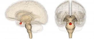
What is the hypothalamus?
The lower part of the human brain is called the hypothalamus. The shape and structure do not have clearly defined boundaries, smoothly transitioning into the tissues of other brain regions.
Consisting of many small cell nuclear groups (to date, only thirty-two nuclear pairs have been studied), the hypothalamus generates impulses that reach other areas of the brain along special pathways. The formed nerve impulses work to control the respiratory, digestive, and circulatory systems. It is from the uninterrupted operation of the hypothalamus that the body regulates the process of maintaining water-salt balance, normalizes body temperature, and during the hot season and with increased physical activity, active sweating occurs in order to cool the skin and prevent overheating.
In addition to transmitting nerve impulses, the hypothalamus produces releasing factors - hormonal substances. Thanks to their production in the postpartum period, the woman “starts” the production of milk in the glands; childbirth, due to hormonal changes, ensures full contractile movements of the uterus to move the fetus along the birth canal, etc.
The hypothalamus performs its function only due to the close relationship of this part of the brain with the pituitary gland, which is the main organ of the endocrine system and continuously links together the work of the two systems.
The hypothalamus is divided into several zones:
Trophotropic
Located in the near zones of the hypothalamus and maintaining a constant internal environment, it normalizes the functioning of the heart and blood vessels during the rest period, and removes decay products of human activity. The main effect occurs through the parasympathetic department. When this zone is stimulated, the production of sweat and saliva increases, the heart rate slows down, blood pressure decreases, etc.
Ergotropic
Responsible for the ability of the human body, regardless of age, to adapt to changing environmental conditions, carrying out adaptation of the body and implemented through the sympathetic department.
The result of the functioning of the ergotropic system and the influence of the parasympathetic nervous system on cardiac activity: blood pressure, blood glucose levels, increased heart rate, and respiratory function increase. The ergotropic zone is located in the posterior part of the hypothalamus.
The parasympathetic division of the autonomic nervous system controls:
- lacrimation;
- pupil enlargement/decrease;
- production of saliva.
It also affects the functioning of internal organs (bronchopulmonary system, cardiac muscle, gastrointestinal tract, etc.).
How do the sympathetic and parasympathetic nervous systems work? Features and main differences
The sympathetic and parasympathetic nervous system, its structure is unique and individual for each person. The activity of the departments will largely depend on age and health status. The neural impulse moves in a strictly specified direction. This movement is based on a reflex arc in the form of a closed chain of neurons. The work of the departments can be seen using a clear example: receptors on the skin detect an irritant in the form of cold, transmit the received data along nerve fibers to the central nervous system, where it is analyzed and evaluated. After which it is these two main departments that make the decision on the impact on the emerging stimulus. In our example, this is a person’s need to warm himself so as not to get too cold and protect himself from the cold. The impulse is transmitted along the nerve endings to the central section, and from there to the peripheral, where the impact occurs as a result of the stimulus - the skin becomes “goosey”, active muscle contraction occurs, aimed at warming the whole body under the influence of cold.
What is the parasympathetic nervous system responsible for?
The human body tends to adapt to stimuli that surround us at almost every step. Moreover, adaptability in each specific case will look different and have the opposite character. For example, the summer heat would have an adverse effect on the human body, if not for its ability to cool down due to increased sweating; in the cold season, sweat production stops and the mechanism of maximum heat retention in the cells is activated. Therefore, the functions of the sympathetic and parasympathetic nervous systems are different and opposite, aimed at the body’s ability to turn on or off a certain “option” at a certain moment.
Differences between the sympathetic and parasympathetic nervous systems.
The sympathetic and parasympathetic nervous system, as a result of its work, activates different “effects”.
Sympathetic:
– dilation of the pupils when exposed to a stimulus (bright light or darkness), protrusion of the eyes;
– narrowing of blood vessels;
– decreased secretion of saliva, which acquires increased viscosity and thickness;
– increased frequency of contractions of the heart muscle;
– noticeable increase in blood pressure;
– rapid breathing;
– dilatation of the bronchi with a simultaneous decrease in excretory function;
– deterioration of intestinal function;
– spontaneous ejaculation or its stimulation;
- "goose pimples".
Parasympathetic
The parasympathetic nervous system performs functions, consider them in the table:
| Eyes | Constriction of the pupil |
| Excretory function | Increased salivation. |
| Heart | Reducing blood pressure and reducing the number of heart beats. |
| Bronchopulmonary system | The amount of secretion in the bronchi increases, breathing becomes calm and measured, the bronchi narrow |
| Intestines | Peristalsis increases, spasmodic pain may appear |
| Digestive system | There is a noticeable increase in secretion production in the digestive gland |
| Genitourinary system | Spontaneously occurring stimulation of the clitoris in women and the onset of erection in men |
In general terms, the activity of the parasympathetic part of the autonomic nervous system leads to a general “excitation” of human organs and vital systems, intensifying their work due to the influence of irritating factors.
As can be seen from the data in the table, the sympathetic and parasympathetic nervous systems differ from each other in terms of the “options” they perform. However, despite all their differences, the autonomic nervous system - sympathetic and parasympathetic - are closely interconnected. However, some functions of the human body are subject to the influence of only the sympathetic department and vice versa. For example, only the sympathetic department is responsible for the work of blood vessels, glands that reproduce sweat, and the adrenal glands, while the parasympathetic department of the nervous system has no effect.
Distinctive features and parameters
Nature has wisely distributed the functional responsibilities of the sympathetic and parasympathetic divisions of the autonomic nervous system - according to the location of their nuclei and fibers, as well as their purpose and responsibility. For example, the central neurons of the sympathetic segment are located exclusively in the lateral horns of the spinal cord. In the parasympathetic, they are localized in the trunk of the hemispheres.
Distant, effector neurons in the first case are always located on the periphery - present in the paravertebral ganglia. They form various plexuses, the most important of which is the solar one. It is responsible for the innervation of intra-abdominal organs. Whereas parasympathetic effector neurons are located directly in the organs they innervate. Therefore, responses to impulses sent to them from the brain occur faster.
Differences can also be observed in functional characteristics. Vigorous human activity requires activation of the heart, blood vessels, and lungs - the activity of sympathetic fibers increases. However, in this case, digestion processes are inhibited.
At rest, the parasympathetic system is responsible for the innervation of the intracavitary organs - digestion, homeostasis, and urination are restored. It’s not without reason that after a hearty lunch you want to lie down and sleep. The unity and indivisibility of the nervous system lies in the close cooperation of both departments.
The parasympathetic nervous system increases the tone of some organs, but decreases others.
In a person who has no health problems, does not suffer from nervous breakdowns, or anorexia, the parasympathetic and sympathetic nervous systems, despite their differences, work on the principle of balance. Normally, there is a slight predominance of one of the systems with a certain effect on the body.
The predominant activity of the parasympathetic part of the autonomic nervous system is called vagotonia, and the sympathetic part is called sympathicotonia.
Due to age-related changes occurring in the human body, the role of the sympathetic and parasympathetic nervous system can change noticeably, reducing the performance of their functions.
Let’s say that in adolescence, the activity of all organs and systems is significantly increased than in senility. The functional significance of the sympathetic and parasympathetic nervous systems is different. For example, the activity of the sympathetic system is expressed by shine in the eyes, enlarged pupils, increased blood pressure, and a high probability of difficulty during bowel movements.
Organization of the human nervous system
Nerve cells cover the entire body, forming an extensive network of fibers and endings. This system, on the one hand, unites every cell of the body, forcing it to work in one direction, and on the other hand, it integrates a particular person into the environment, balancing his needs with external factors. The nervous system ensures the normal processes of digestion, respiration, blood circulation, the formation of immunity, metabolism, etc. - in a word, everything without which normal life activity is unthinkable.
The effectiveness of the nervous system depends on the correct formation of the reflex - the body's response to irritation. Any impact, be it external changes or internal imbalance, triggers a chain of impulses that instantly affect the body, and it, in turn, forms a response. Thus, the human nervous system forms the unity of the tissues, organs and systems of the human body with each other and with the outside world.
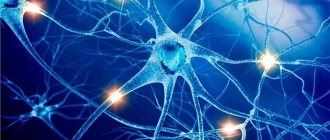
The entire nervous system consists of millions of nerve cells - neurons, or neurocytes, each of which has a body and several processes.
The classification of neuron processes depends on what function it performs:
- the axon sends a nerve impulse from the body of the neuron to another nerve cell or the final target of the chain - a tissue or organ that must perform a certain action;
- The dendrite receives the sent impulse and leads it to the body of the neuron.
Due to the fact that each nerve cell is polarized, the chain of nerve impulses never changes direction, falling into the right direction. In this way, each nerve impulse moves forward, initiating the work of muscles, internal organs and systems.
Types of nerve cells
Before considering the nervous system as a whole, it is necessary to understand what functional units it consists of. The NS includes:
- Sensory neurons. Located in nerve ganglia that receive information directly from receptors.
- Interneurons are an intermediate link, thanks to which the received impulse is transmitted from sensitive neurons further along the chain.
- Motor neurons. They act as initiators of a response to a stimulus, transmitting a signal from the brain to the muscles or glands, which normally should perform their assigned function.
It is according to this scheme that any response of the human body to an external or internal stimulus signal, which acts as an impetus for a specific action, is built. As a rule, the passage of a nerve impulse takes a few fractions of a second, but if this time is delayed or the chain is interrupted, this indicates the presence of a pathology of the nervous system and requires serious diagnosis.