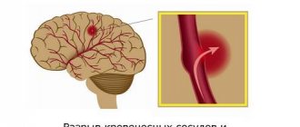
Today, the majority of patients of general practitioners are patients with vascular pathology, including neurological problems. But modern therapists do not know neurology well, and the therapist does not have enough time for a high-quality survey, examination and analysis of clinical data. Consequently, a clinic doctor must be able to identify situations when it is not necessary to deal with the patient on his own, but, without wasting time, refer him to a neurologist.
The outstanding neurologist of our time, academician Alexander Moiseevich Vein, said that it is necessary to engage in interdisciplinary medical practice, and not divide a person into parts that fall into the field of view of narrow specialists. Today there are no narrow-profile diseases, diseases of any particular organ or system. A person is one and is a complex organism, all parts of which are interconnected, therefore treatment requires a systemic, holistic approach.
These thoughts have come to life: general practitioners have appeared, they deal with comorbid patients, most of whom are represented by patients with vascular pathology, including neurological problems. However, as noted by the outstanding internist, Academician of the Russian Academy of Sciences V.S. Moiseev, today the situation is such that “modern therapists do not know neurology well for the reason that it has distanced itself, moved away from therapy, and set off on its own.”
Often in the clinic you can see patients visiting one office after another in the hope of getting the necessary help, and doctors who, experiencing diagnostic difficulties, do not know where to refer patients and where to start. Among the reasons for this situation, one can highlight the lack of sufficient time for a high-quality survey, examination and analysis of clinical data, and the impossibility, due to objective circumstances, of discussing problems with colleagues.
This leads to the prescription of numerous tests and often unnecessary medications, which ultimately lengthens the time for making a correct diagnosis. Consequently, a clinic doctor must be able to identify situations when it is not necessary to deal with the patient on his own, but, without wasting time, refer him to a neurologist.
There are 15 main neurological syndromes, upon identification of which the therapist should refer the patient to a neurologist:
- ✅ Back pain
- ✅ Temporary loss of consciousness
- ✅ Sudden dizziness
- ✅ Acute vestibular syndrome
- ✅ Sudden weakness in a limb or facial asymmetry
- ✅ Speech disorders
- ✅ Pain and numbness in the face
- ✅ Unsteadiness, swaying
- ✅ Memory impairment
- ✅ Numbness
- ✅ Sleep disorders
- ✅ Head or neck posture problems
- ✅ Difficulty writing
- ✅ Involuntary movements
- ✅ Tremor
BACKACHE
Back pain is one of the most common situations when visiting a doctor. In Russia, patients traditionally turn to therapists, general practitioners and neurologists with this complaint. In foreign countries, there are different statistics, since back pain in the vast majority of cases is not a neurological problem. In Fig. Figure 1 presents an algorithm that reflects the necessary actions of a doctor in this syndrome. According to the algorithm, when a patient comes to an appointment with back pain, first of all it is necessary to exclude the so-called “red flags” that pose a threat to the health and life of the patient, which requires urgently referring the patient to a specialized specialist to provide specialized medical care.
"Red flags":
- ✅ connection of back pain with previous injury;
- ✅ irradiation of pain in the leg, feeling of numbness of the toes, weakness of the limb, the appearance of urinary and fecal incontinence;
- ✅ combination of pain with fever, leukocytosis, lymphadenopathy, acceleration of ESR, increased levels of C-reactive protein;
- ✅ unmotivated weight loss, anemia, fever;
- ✅ the appearance of pain at a young age, its duration and inflammatory nature: increased at rest, especially at night and in the morning, decreased during physical activity (warm-up);
- ✅ a history of diseases that may also be accompanied by acute back pain.
It is necessary to approach the diagnosis of the underlying disease very carefully, and it should begin not with an x-ray examination, not with a diagnosis of “osteochondrosis,” but with an assessment of the existing clinical symptoms.
For example, pain only at night, increased body temperature, anemia, accelerated ESR are manifestations of dangerous diseases, often leading to death. A thorough study of complaints, medical history, examination of the patient in combination with data from standard laboratory tests - a clinical blood test, including the mandatory determination of ESR, and a urine test - remain the basis for the initial diagnosis of a patient with back pain.
If “red flags” are excluded, a “multi-domain screening approach” is proposed, which involves assessing the patient’s pain and condition to create a personalized treatment program based on biopsychosocial analysis. In this case, to successfully diagnose the cause of chronic or recurrent pain, it is not enough to limit yourself to performing an X-ray examination, MRI or blood test, but it is necessary to conduct an in-depth analysis of the pain and characteristics of the social status of each individual patient.
The domains developed at the Department of Nervous Diseases of the First Moscow State Medical University are designed to help the clinic doctor quickly cope with the task in a short time for an outpatient appointment. I. M. Sechenov. This is a system of short questions and tests characterizing the pain pattern, which ultimately contributes to the selection of adequate therapy.
Currently, 10 domains have been proposed for a personalized therapy program: 5 for assessing the pain phenotype and 5 for assessing the patient’s social status.
To correctly select a drug or combination of drugs, the doctor’s task is to clarify the participation (“contribution”) of each domain in the formation of the sensation of pain.
For example, if we come to the conclusion that:
- Pain is associated with inflammation , then the drugs of choice are non-steroidal anti-inflammatory drugs (NSAIDs), because they act on the mechanisms of inflammation, especially acute ones.
- In chronic inflammation, chondroprotectors that act on cytokines activated during chronic pain come to the fore.
- For muscle spasms, muscle relaxants are needed.
- For myofascial syndrome, a set of measures is indicated: blockade of trigger points, dry puncture, taping, massage, manual therapy.
- Central sensitization has a very complex mechanism and is the hypersensitivity of central sensory neurons (allogenia, hyperalgesia, hyperpathia). In pharmacotherapy of this domain, drugs such as pregabalin, gabapentin are used, which close calcium channels, reduce the release of pain mediators, and reduce the excitability of central sensory neurons.
- The neuropathic domain (radiculopathy) is represented by pain associated with compression/damage of nerve fibers.
To diagnose this domain, it is important to have a certain clinical picture in combination with objective symptoms identified during examination.
Characteristics of pain associated with radiculopathy:
- ✅ burning, shooting character, reminiscent of an “electric shock”, with irradiation to the foot;
- ✅ combination of pain with other neurological symptoms (for example, tingling sensation, numbness, muscle weakness);
- ✅ the spread of pain along the corresponding dermatomes is typical.
Objective signs revealed during examination of the patient:
- ✅ neurological symptoms: hypoesthesia (decreased various types of sensitivity: temperature, pain, tactile, etc.);
- ✅ provocation of pain using motor tests (for example, the Lasegue symptom, which is considered positive if pain occurs when slowly lifting an outstretched leg with the patient in the supine position, associated with tension of the sciatic nerve).
Assessing the components of the patient’s social status domain, such as psychosocial factors, duration and quality of night sleep, physical activity, cognitive functions, the presence of comorbid pathology, especially if corrected, also helps eliminate chronic pain syndrome.
Thus, a personalized pain treatment program provides an individual approach.
Astheno-neurotic syndrome: diagnosis
Astheno-neurotic syndrome can develop in both children and adults. It is usually very difficult to diagnose due to the presence of somatic complaints that come to the fore. Teenagers often complain of headaches, pain in the heart, fatigue, and drowsiness.
In adults, the symptoms of asthenia are usually much more pronounced and varied. Manifestations of the disease are usually observed more intensely in the afternoon, and reach a maximum in the evening. Weakness and apathy, lack of desire to do any work and decreased concentration are noted. When performing exercises that require mental effort, difficulties in making decisions, absent-mindedness, irritability, and memory impairment are noted.
Emotional stress leads to increased symptoms, which can ultimately provoke the development of depression, neuroses, and neurasthenia.
In addition to the main symptoms, disorders of the autonomic nervous system may come to the fore, such as:
- tachycardia, palpitations, interruptions;
- blood pressure surges;
- increased sweating, feeling of chills and heat;
- decreased appetite, impaired digestive function;
- headache, dizziness;
- decreased libido.
Differential diagnosis
Astheno-neurotic syndrome often accompanies the initial stages of such somatic diseases as: Arterial hypertension, coronary heart disease, angina pectoris, angina pectoris; cerebrovascular accidents; atherosclerosis and others.
Therefore, we carry out differentiated diagnostics to exclude somatic pathology.
If you notice the above symptoms, then you should not delay. Make an appointment with our specialist and undergo differential diagnostics. Diagnosis of asthenia includes a comprehensive examination, the purpose of which is to identify the pathology that caused the disease.
TEMPORARY LOSS OF CONSCIOUSNESS
Short-term loss of consciousness can be caused by many reasons, which are within the competence of different specialists: cardiologists, therapists, neurologists, endocrinologists, gynecologists, etc., however, neurological diseases can be a common cause of fainting.
Thus, a mandatory indication for consultation with a neurologist is attacks of loss of consciousness, accompanied by signs characteristic of epilepsy (sudden onset, convulsions, tongue biting, loss of urine, disorientation).
A transient ischemic attack (TIA) may manifest as a short-term loss of consciousness. It should be noted that convulsive twitching can occur in patients with vasovagal syncope that occurs against the background of emotional stress and orthostatic load. Such patients do not need to consult a neurologist.
Considering the fact that loss of consciousness before finding out its cause poses a threat to the patient’s life, a mandatory consultation with a neurologist is necessary.
The most common reasons for visiting a pediatric neurologist
Neurological monitoring of the child is carried out immediately after birth: already in the first month, the baby is examined by a doctor who collects data on the nature of the pregnancy, checks basic reflexes, evaluates muscle tone, the size of the fontanelles, etc. At one month of age, neurosonography (ultrasound of the child’s brain) is also performed.
Next, the neurologist consults the child at 3, 6, 9 and 12 months, assessing the dynamics of his development. After a year, a child does not consult a neurologist so often, but it is recommended to visit a doctor at 3, 6, 7, 10, 14 years of age and then until he reaches 18 years of age.
The most common complaints with which parents turn to a neurologist are:
- poor sleep: the child has difficulty falling asleep, wakes up easily and often, sleep is intermittent, restless
- increased tone in the arms or legs
- tremor - the child’s chin or limbs periodically tremble
- frequent and profuse regurgitation
- convulsions with increased temperature and so-called afebrile convulsions (at normal or slightly elevated body temperature)
- increased excitability of the child
- frequent headaches, irritability, fatigue
- decreased attention, memory, poor performance at school
- nervous tics, coordination problems
- sharp pain (for example, in the back)
- developmental delays of various types
- lack of normal socialization of the child (for example, no one communicates with him at school)
- poor sleep in children over 1 year old, etc.
SUDDEN Dizziness
Referral to a neurologist requires sudden onset of dizziness in combination with focal neurological disorders, such as ataxia, deafness, vertical or rotational nystagmus, etc., which may be a manifestation of TIA or stroke.
It should be noted that isolated dizziness without the listed neurological disorders is a symptom of more than 80 diseases and is an interdisciplinary problem dealt with by doctors of various specialties: therapists, otolaryngologists, otoneurologists, neurosurgeons, etc. This is also due to the fact that the word “dizziness” » The patient often understands completely different sensations, such as staggering and unsteadiness.
ACUTE VESTIBULAR SYNDROME
As a rule, it is a manifestation of peripheral dizziness and is associated with vestibular neuronitis, but in rare cases this syndrome (dizziness, nausea, vomiting, gait instability (dysbasia)) can be a manifestation of a stroke.
Therefore, adults with sudden-onset acute vestibular syndrome should have the HINTS test performed without attempting to self-administer any symptomatic medications. If it turns out to be positive, immediate neuroimaging and consultation with a neurologist is required to rule out stroke.
SUDDEN LIMB WEAKNESS AND FACIAL ASYMMETRY
Sudden or increasing facial asymmetry in combination with arm weakness and speech impairment first of all requires the exclusion of a stroke. To confirm the diagnosis when identifying the earliest signs of the disease, it is enough to carry out very simple tests:
- ❗ Does your face look unusual? — Ask the patient to smile.
- ❗ Does this sound strange? — Ask the patient to repeat the phrase.
- ❗ One hand falls down? —Ask the patient to raise both arms.
If a stroke is suspected, it is necessary to urgently refer the patient for neuroimaging and to a neurologist.
Asymmetry of one half of the face can be observed with damage to the facial nerve (neuropathy), which occurs suddenly and may be accompanied by other symptoms: the palpebral fissure on the affected side does not close, water and liquid food pour out of the mouth, lacrimation and impaired taste are observed.
It is very important to immediately refer the patient to a neurologist, because treatment started later than 3 days from the onset of the disease may be ineffective, which threatens not only facial asymmetry, but also ophthalmological problems (conjunctivitis, keratitis and even loss of vision). Slowly progressive damage to the facial nerve is observed with cancer of the brain (for example, with a tumor of the cerebellopontine angle).
Main neurological pathologies in children
Hyperexcitability is a set of symptoms that include movement disorders, sleep disturbances, tremors (shaking), increased muscle tone, frequent regurgitation, etc. It is believed that hyperexcitability is a consequence of perinatal pathology of the central nervous system or previous diseases. Often, parents may think that the child is simply capricious and fail to pay due attention to the manifestations of hyperexcitability. The pathology needs treatment (massage, exercise therapy, physiotherapy), as it can lead to negative consequences in the future.
Psycho-speech development delay (PSRD) - children who lag behind their peers in speech and emotional development should be observed by a specialist. Often the child does not understand the words addressed to him, reacts inadequately to them, and cannot study at school. ZPRD can occur in children who were born very quickly (lightning-fast labor), spent a long anhydrous period during the birth process, in children with hydrocephalus, etc. Children with mental retardation disorders need special classes with teachers.
Speech development disorders , which include alalia (immaturity of speech as a result of damage to brain structures), dysarthria (a disorder as a result of which speech becomes incomprehensible), dyslexia (impaired written speech), stuttering, etc. Children are sent to classes with a speech therapist and psychologist, in most cases they are shown training in special programs.
Attention deficit hyperactivity disorder (ADHD) - children suffering from this syndrome are very excitable, active, have difficulty focusing and concentrating on something, and have difficulty learning. The exact reasons for the formation of ADHD are still not clear; it is believed that a hereditary factor plays a role, and damage to the central nervous system may play a role. It has been observed that the brains of hyperactive children work productively for about 15 minutes, and then “accumulate energy” for about 3-7 minutes. To correct ADHD, psychotherapy methods are used; in some cases, it is recommended to give medications that improve brain function.
Epilepsy is a disease of the brain in which “flares” and increased activity of neurons occur in its cells. As a result, a person begins an epileptic seizure, which is accompanied by convulsions, loss of consciousness, etc. Unfortunately, epilepsy cannot be completely cured, but the child’s condition can be significantly improved through the selection of balanced drug therapy. It is also recommended to adjust the diet; sometimes children are prescribed a massage.
Migraines are severe headaches that are localized in a specific area of the head. Hereditary factors play a major role in the occurrence of migraines in children; stress, prolonged use of the computer, overexertion, fasting, etc. also play a role. Treatment consists of relieving pain (medically), as well as eliminating the cause of the pain. A psychotherapist, reflexologist and other specialists can work with the child.
SPEECH DISORDERS
They can have different manifestations: aphasia, alalia, bradyllia, dysarthria, dysorthography, dysgraphia, dyslexia, dyslalia, etc. In adults, they almost always indicate various neurological diseases. Thus, a sudden speech disorder may be associated with a transient ischemic attack or be an early sign of a stroke, and a progressive one may be a manifestation of amyotrophic lateral sclerosis. Therefore, in the case of this serious neurological syndrome, it is imperative to refer the patient to a neurologist.
PAIN AND NUMBITY IN THE FACE
If the facial pain is intense, manifests itself in attacks lasting from 3 to 30 seconds, occurs during conversation, or is provoked by touch, then the probable diagnosis is trigeminal neuralgia (trigeminal neuralgia).
This independent neurological problem is often associated with compression of the vessels of the trigeminal nerve root. In elderly patients, with hypersensitivity of the skin in the temporal region, chewing disorders and accelerated ESR, one can suspect temporal arteritis, which is often confused with migraine. It is necessary to perform a blood test to evaluate ESR and refer the patient to a neurologist to exclude this pathology.
It should be noted that a normal ESR does not exclude the diagnosis of giant cell arteritis. Pain and numbness in the face in combination with other neurological disorders (facial asymmetry, eye movement disorders, etc.) require urgent neuroimaging to exclude cancer.
CHILDREN'S NEUROLOGICAL SYNDROMES
Listed below are the most common nervous system health problems that a pediatric neurologist can diagnose.
Syndrome of increased neuro-reflex excitability
It manifests itself in increased restlessness of the child, disruption of the sleep-wake cycle, when the child is restless at night and sleepy during the day. Grandmothers used to say about this: “I confused day with night.” There may be pronounced tremor of the arms, legs, chin, and even shuddering may occur during prolonged crying. During the examination, the pediatric neurologist will also pay attention to the revival of the baby’s reflexes in the neurological status.
General oppression syndrome
Such a child, on the contrary, is “very calm”, but more often due to lethargy and weakness, his general range of movements is reduced, he reacts little to his surroundings, sleeps a lot, sucks poorly, often does not gain weight, his crying is weak, he often has to feed wake up. This syndrome is more often observed in premature babies, and must be taken into account by parents and a pediatric neurologist. More often it is more complex than the previous syndrome.
Hypertension-hydrocephalic syndrome
Caused by an increase in intracranial cerebrospinal fluid pressure. This is a serious neurological problem, it is characterized by certain complaints and clinical manifestations.
The child becomes restless, sleeps poorly, the large fontanel bulges, is tense, the subcutaneous veins of the head become dilated and become noticeable in the form of a network, vomiting may appear, including a fountain, the size of the head circumference increases, and the stitches come apart. If parents suspect similar symptoms in their baby, they should immediately contact a neurologist, and probably even a neurosurgeon, which may require surgical intervention.
Hydrocephalic syndrome without cerebrospinal fluid hypertension
It occurs with communicating (external) hydrocephalus, when the pressure of the cerebrospinal fluid in the skull does not increase. It is also manifested by an increase in the growth of head circumference more than the average age norms, divergence of the seams, enlargement of the fontanelles, their bulging, the appearance of Graefe’s symptom (when the child seems to “goggle his eyes”), the saphenous veins dilate and can be seen on the face in the area of the eyes, bridge of the nose, forehead like a bluish mesh; when a child cries, the veins swell and become more visible. A pediatric neurologist can detect nystagmus, strabismus, and so-called “pyramidal signs” in a child. Additional research methods will help clarify the diagnosis, such as: fundus examination by an ophthalmologist, neurosonogram, computed tomogram of the brain, which a pediatric neurologist can prescribe if necessary.
Convulsive syndrome
This is a fairly serious problem in the child’s health, which is manifested by attacks of involuntary movements in the muscles of the face, limbs, freezing in certain positions or fixation of the gaze and lack of reaction to the environment, etc., including loss of consciousness, stopping breathing and baby's heartbeat. This is a life-threatening condition; it requires calling an ambulance, emergency measures and, as a rule, hospital treatment. Even if the attacks are short and the child very quickly returns to his normal rhythm of life after them, these conditions are not the norm; they require identification of the cause, and sometimes long-term treatment. You need to understand that seizures, even short-term ones, destroy neurons (brain cells), which should ensure the baby’s growth and development. Any suspicion of attacks or the attacks themselves should be recorded, if possible, filmed and shown to a pediatric neurologist.
At the age of 4 months, a pediatric neurologist can identify the following syndromes in children, the so-called recovery period.
Cerebroasthenic syndrome
If, against the background of normal physical and neuropsychic development, a child becomes emotionally restless, his mood changes frequently and unmotivated, motor restlessness, difficulty falling asleep, shuddering when falling asleep, periodic tremors of arms and legs may appear, sleep becomes superficial, its duration decreases - this is manifestations of this syndrome. It can also occur in a child after suffering a serious acute illness or injury.
Syndrome of vegetative-visceral disorders
It is no secret that all functions of the human body are controlled by the central nervous system. The autonomic nervous system is responsible for the functioning of internal organs, through the so-called autonomic-visceral reactions. In children at birth, it is formed, but has not yet matured, so some of the reactions of autonomic control are disrupted.
This is manifested by thermoregulation disorders (unstable body temperature), the appearance of dysfunction of the gastrointestinal tract in the form of regurgitation, rumbling in the abdomen, increased or, conversely, decreased frequency of stool passage, poor passage of gases, a change in the color of the skin with the appearance of cyanosis, vegetative vascular spots, lability of the cardiovascular and respiratory systems may be noted (parents may record a disturbance in the heart rhythm or breathing rhythm with episodes of breathing disturbance in the form of a short-term stop, which is especially pronounced during sleep). You, as parents, must inform your pediatrician and pediatric neurologist about all such complaints, even if it only seems so to you.
After all, no one knows your child better than you, and specialists will have to understand them. Such complaints may, on the one hand, indicate the immaturity of the autonomic nervous system, which will “ripen” with age, and on the other hand, such complaints may begin with diseases of various organs that will seriously impair the baby’s health and only a doctor can make this differentiation.
Movement disorder syndrome
It is characterized by impaired muscle tone, muscle strength and, as a consequence, a delay in the stages of psychomotor and speech development.
The following are responsible for the formation of movements in the body: the pyramidal system, the extrapyramidal system, the cerebellum, subcortical structures and a certain area of the cerebral cortex, as the highest coordinator. The child’s muscle tone can be disrupted either in the direction of increasing or decreasing or forming muscle dystonia. Also, in parallel with muscle tone, reflexes can also change in the direction of increasing or decreasing them, but at the same time muscle strength necessarily decreases.
Altered muscle tone, decreased muscle strength and altered reflexes in a child disrupt the natural course of neuropsychological and motor development, so he begins to lag behind his peers.
Remembering that the state of muscle tone, muscle strength and reflexes (there are a lot of them) can be assessed by a pediatric neurologist. Only after this, as well as after conducting the necessary set of examinations, will he be able to correctly diagnose and prescribe comprehensive treatment for your baby.
Disturbances in muscle tone and changes in tendon reflexes can be minor and, with a properly designed individual rehabilitation program, can be restored, even if this requires more than one course of rehabilitation treatment. The main thing is that the child will continue to develop. But there are also more serious disorders of muscle tone and tendon reflexes, which are always a consequence of deep brain damage and are markers of such a serious disease as cerebral palsy
.
Typically, the diagnosis of cerebral palsy is made by a pediatric neurologist at 1 year of age.
, and before that, it goes under the guise of the same diagnosis as movement disorder syndrome, but the neurologist can add the phrase that the child is at risk for cerebral palsy.
You can sign up for a consultation and find out more from the administrator by calling 8 (495) 356-30-03.
INSTABILITY, STABLISHING (DYSBASIA)
Sudden onset and increasing instability and staggering is a dangerous signal that may indicate a TIA or stroke, which requires urgent referral of the patient to a neurologist.
Neurological symptoms also include a gradually increasing unsteady gait, indicating alcoholic polyneuropathy, B12-deficiency anemia and normal pressure hydrocephalus. Such patients also need consultation with an appropriate specialist. For complaints of instability, a postural response test after short jolts is recommended.
NUMBNESS
As a rule, a complaint of numbness of the limbs does not cause serious concern to the doctor, however, this symptom may be a manifestation of the following neurological diseases:
- ✅ epilepsy (with repeated episodes of short-term numbness lasting up to 2 minutes);
- ✅ tunnel syndrome (with constant numbness combined with pain in one place);
- ✅ polyneuropathy (with constant numbness in the legs and arms like “socks” and “gloves”), including Guillain-Barre polyneuropathy in case of increasing numbness in the legs (hours - days) combined with weakness.
Thus, if you complain of numbness, the patient must be referred to a neurologist.
How does post-Covid syndrome manifest?
Signs of post-Covid syndrome can be divided into several groups. Let's look at each of them in detail.
Symptoms of general health problems
The main signs of impaired general well-being after coronavirus include:
- attacks of weakness. Weakness can be so severe that a person is forced to remain in bed for several weeks;
- a sharp decrease in exercise tolerance. Even a little activity leads to complete exhaustion of physical strength;
- disruption of the rhythms of life. Insomnia, excessive drowsiness, sleep inversion (waking at night, sleeping during the day) may develop;
- muscle pain. With a coronavirus infection of any form, there is always a significant decrease in protein mass, which negatively affects the condition of the muscles.
Psycho-emotional problems
Today we can draw conclusions that coronavirus negatively affects the psycho-emotional health of people. With post-Covid syndrome, the following may be observed:
- depressed mood. Almost all patients who have had coronavirus are in a minor mood. They develop despondency, depression, and melancholy. In some cases, depressed mood can lead to suicidal thoughts;
- unstable emotional state. Manifested by sudden mood swings, low self-control of behavior;
- panic attacks. People experience attacks of severe anxiety in combination with other symptoms: high blood pressure, shortness of breath, nausea, dizziness.
There are currently known cases in which a severe disturbance of the psycho-emotional state after coronavirus has resulted in suicide.
Symptoms associated with respiratory complications
Such signs can occur even if there was no damage to the respiratory system during the acute phase of coronavirus. These include:
- feeling of lack of air;
- tightness in the chest, inability to take a deep breath;
- bronchospasms.
Bronchospasms are characterized by a decrease in the lumen of the bronchi. With this complication, hypoxia develops - a lack of oxygen in the body.
Symptoms associated with complications of the respiratory system can last from several days to several months.
Neurological manifestations
The coronavirus is able to penetrate the central nervous system, affecting neurons and glial (auxiliary) cells. The main neurological manifestations of post-Covid syndrome include:
- intense headaches. The pain syndrome can be constant or in the form of a migraine - a paroxysmal, recurring headache;
- violation of thermoregulation. Some people have a low-grade fever (37–37.5 degrees) for a long time after COVID-19, while others have a low temperature (up to 36 degrees);
- chills, especially in the evening. Accompanied by a feeling of cold, muscle tremors. At the same time, body temperature can remain normal;
- visual impairment. A person may experience black spots before the eyes, blurred vision, photophobia;
- paresthesia is a sensitivity disorder. Manifested by a sensation of burning, tingling, crawling on the surface of the skin;
- disturbance of smell and taste. Such symptoms can last up to several months.
Another common complication is a malfunction of the vestibular apparatus, which is responsible for the ability to navigate in space and maintain balance. If the coordination system is disrupted, the gait becomes unsteady, a person can crash into any obstacle or fall out of the blue.
Symptoms associated with damage to the cardiovascular system
A distinctive feature of COVID-19 is its pronounced impact on the cardiovascular system. In every fifth patient, the infection causes arrhythmia, acute or chronic heart failure.
With sweating syndrome, the following may be observed:
- blood pressure disorder. Both high blood pressure and prolonged hypotension can develop. In addition, orthostatic collapse often develops. The condition is characterized by insufficient blood flow to the brain when there is a sudden change in body position. With orthostatic collapse, a person’s blood pressure quickly drops, vision becomes dark, dizziness and fainting occur;
- polymorphic dermal angiitis. It is caused by an inflammatory process that develops in the walls of blood vessels. It manifests itself in the formation of nodules, blisters, plaques, hemorrhages, blisters, bruises, and dark spots on the skin. Polymorphic dermal angiitis is one of the complications of thrombovasculitis;
- heart rhythm disturbance. After coronavirus, a person may experience arrhythmia, tachycardia, or a slow heart rate for a long time.
Many patients with post-Covid syndrome note that when they remain in an upright position for a long time, they experience weakness, dizziness, and cold sweat. Such signs are a consequence of a drop in blood pressure.
Gastrointestinal tract disorders
The consequences of COVID-19 often include disruptions to the digestive system:
- decreased intestinal motility, which slows down the movement of food through the gastrointestinal tract;
- stool disorder. Both constipation and diarrhea may develop;
- loss of appetite.
This triad of symptoms often causes dysbiosis - changes in the composition of the normal intestinal microflora. In an advanced state, dysbiosis can lead to decreased immunity, severe anemia, and allergic reactions.
Symptoms associated with disruption of other organs and systems
In addition to the listed pathological signs, the consequences of COVID-19 may be:
- decreased immunity;
- inflammatory processes of the urinary system;
- malfunction of the sexual sphere. Women often experience menstrual irregularities;
- endocrine diseases;
- allergic reactions.
It has been proven that coronavirus primarily affects organs affected by chronic diseases.
At the same time, during the post-Covid syndrome, hidden diseases that a person did not even suspect about often worsen. Therefore, people who have recovered from coronavirus infection need to be very careful
take care of your health and immediately contact a specialist if you experience any unusual symptoms.
DISORDERS IN HEAD OR NECK POSTURE
Poor posture of the head and neck, difficulty writing and involuntary movements not associated with injuries and/or musculoskeletal pathology are dystonia syndromes. This very interesting group of diseases associated with damage to the central nervous system is characterized by continuous involuntary muscle contractions, often causing “twisting” of the affected part of the body and the formation of pathological postures. More recently, patients with torticollis (a form of focal dystonia) were diagnosed with “osteochondrosis” and subsequently prescribed appropriate ineffective therapy. Currently, this pathology can be corrected, so patients with dystonia must be referred to a neurologist.
Treatment of post-coronavirus syndrome
Patients with respiratory manifestations of post-coronavirus syndrome are prescribed the following therapy:
- glucocorticosteroids - with the development of organizing pneumonia, the persistence of pronounced systemic inflammatory changes;
- antitussive drugs of peripheral and central action - in the presence of persistent chronic dry cough;
- antibacterial, mucolytic drugs - with the development of secondary bacterial complications.
In some cases, after additional examination according to indications, inhalation therapy may be prescribed.
Prescription of prophylactic anticoagulant therapy with new oral anticoagulants or low molecular weight heparins plays an important role in the prevention of thromboembolic complications in high-risk patients.
A significant role in developing tactics for managing patients with post-coronavirus syndrome is played by comprehensive rehabilitation, which includes:
- rehabilitation of respiratory function (breathing exercises, exercises using breathing simulators);
- rehabilitation of muscle dysfunction (dosed physical activity);
- rehabilitation of neurological, psychological and cognitive functions (psychotherapy, drug treatment);
- nutritional rehabilitation;
- prevention of exacerbations of concomitant diseases.
How post-coronavirus syndrome is treated at the Rassvet clinic
At the Rassvet clinic, patients diagnosed with post-coronavirus syndrome undergo a comprehensive examination to determine the optimal treatment tactics for both the underlying disease and concomitant pathologies. Based on the diagnostic results, a treatment plan is developed, including drug therapy, dietary recommendations, breathing exercises, and a physical activity plan.
During treatment, regular assessment of its effectiveness is carried out with the study of clinical, laboratory and (if indicated) radiological data.
Author:
Akulkina Larisa Anatolyevna pulmonologist
DIFFICULTIES WHEN WRITING
Difficulty writing—“writer’s cramp”—is another form of focal dystonia. “Writer’s cramp” occurs only during active writing, after writing a few words or lines. In this case, the fingers are constrained and take an unusual position, writing becomes impossible. The violation may be limited to only one hand. A similar pathology occurs when printing. Today, this complex central regulatory disorder is treatable. Difficulty writing is also common in Parkinson's disease.
Thus, patients are referred to a neurologist in whom hand position problems when writing and handwriting problems are not associated with musculoskeletal pathology.
TREMOR
Tremors are rhythmic, rapid contractions of the muscles of the trunk or limbs of an involuntary nature. Tremor occurs when autonomic support is disrupted, metabolic disorders and damage to the central nervous system. It is necessary to distinguish between essential (“autonomous”) tremor, which is more common in clinical practice, and tremor in Parkinson’s disease.
Essential tremor increases with movement, and tremor in Parkinson's disease, on the contrary, is stronger at rest and disappears (or significantly decreases) with the onset of activity. In addition, in Parkinson's disease, tremor may initially seem to be an isolated symptom and usually begins in the hands. Later, a head tremor develops. It is necessary to carry out differential diagnosis of these conditions, since they have different natures and, accordingly, different approaches to treatment. Tremor can occur in practically healthy people due to anxiety, emotional and physical stress. A patient who has persistent tremors in the arms and/or legs should be referred to a neurologist.
Thus, the main neurological syndromes and screening tests that have a high diagnostic value are structured and presented, which will help to competently route the patient to a specialized specialist, which will significantly reduce the time the patient spends in the corridors of the clinic and the time required for the doctor to make the correct diagnosis and prescribe adequate therapy .
This will also contribute to the fulfillment of A. M. Wein’s dream: “I would like the slogan of medicine to be improving the quality of life.”
A lumbar puncture is a diagnostic and sometimes therapeutic procedure performed to obtain CSF for biochemical, microbiological, and cytological tests or, less commonly, to relieve intracranial pressure. The procedure is often performed in acute conditions, in order to exclude the presence of dangerous diseases such as meningitis, encephalitis, subarachnoid hemorrhage, or for therapeutic purposes in the case of pseudotumor syndrome or increased intracranial pressure syndrome.
Back in the 2nd century AD. The Greek physician Galen hypothesized the existence of cerebral ventricles containing gas, the so-called mental pneuma. Only in the 19th century, F. Magendie [1] clearly demonstrated the existence of CSF by puncturing the cistern magna in animals, thereby proving the existence of a subarachnoid space that connects to the IV ventricle. In 1891 Quincke [cit. according to 2] performed the first lumbar puncture in humans, and in 1898 A. Bier [3] first described post-puncture syndrome (PPS).
PPS can complicate all types of dural puncture (lumbar puncture, rachis anesthesia, or epidural anesthesia). It is more common in young people [4], women [5, 6] with low or normal body weight [7], in patients with a previous history of primary headaches (migraine, tension headache) [5, 8], and also anxiety states [9]. The incidence of diagnostic lumbar puncture is 15–40%, rachianesthesia is 7.5%, and this procedure is more often used in obstetric practice or for other indications [7]. The incidence of accidental dural puncture during epidural anesthesia ranges from 0–2.6%, while the incidence of PPS reaches 70% [10, 11].
Clinical case.
A 33-year-old female patient was admitted to the Headache Center (Paris) on 01/28/14 with a headache that began on 12/30/13. A woman of Sri Lankan origin, mother of 3 children, last birth on 12/30/13 naturally with epidural anesthesia. Immediately after giving birth, the patient began to complain of a severe compressive headache localized in the frontal region, radiating to the occipital and cervical regions. At the beginning, she noted a decrease in headaches when lying down. The treatment methods proposed by the gynecologist with painkillers (aspirin, non-steroidal anti-inflammatory drugs) and triptans had little effect, the patient was discharged home under the supervision of a family doctor.
Subsequently, the headache became constant, losing the orthostatic component, which led to insomnia with frequent awakenings due to pain. For this reason, the patient turned to the Headache Center of the Lariboisiere Clinic in Paris (almost 1 month after the onset of the headache). The headache intensity on the visual analog pain scale reached 8/10 points. The pain was diffuse, radiating to the cervical region. The patient's body temperature and blood pressure were normal, general and neurological examination data were within normal limits, without meningeal signs. The patient did not complain of tinnitus, hypoacusis, or tinnitus.
CT scans of the brain were performed without and with contrast (Fig. 1),
Rice. 1. CT scan of the brain without contrast (left) and with arteriovenous contrast (right). Axial slice. Arrows indicate subdural hematomas 3-4 mm thick. angio-CT of the vessels of the brain and neck, venous sinuses, which revealed subdural hematomas on both sides up to 3 mm thick on the left and 4 mm on the right.
MRI of the brain after a bolus injection of gadolinium contrast agent (Fig. 2)
Rice. 2. MRI of the brain with bolus gadolinium contrast. Axial slice. Arrows indicate the following: a - pachymeningeal accumulation of contrast (upper arrow), small lateral ventricles (middle arrow); the sagittal sinus is dilated and clearly visible (lower arrow); b — bilateral subdural hematomas (left and right). bilateral subdural hematomas and pachymeningeal contrast accumulation as a consequence of CSF hypotension were detected. The venous sinuses were permeable and without signs of thrombosis, and the cisterns of the base of the brain were not enlarged, the sagittal sinus was dilated and well visualized.
Taking into account the clinical picture of post-puncture intracranial hypotension due to epidural anesthesia, complicated by bilateral subdural hematomas, the patient was hospitalized in the neurology department. The patient maintained a horizontal position for 48 hours and was offered caffeine treatment, which turned out to be ineffective.
After 2 days, on January 30, 2014, the patient underwent filling of the epidural space with autologous blood (PEB) with a volume of 30 ml at the level LII-LIII. The injection was stopped when headache and back pain occurred, indicating a sufficient volume of blood injected into the epidural space. Subsequently, the patient remained in a supine position for 3 hours. When moving to a vertical position, the patient did not complain of headaches and was discharged with recommendations to continue treatment at home, in particular taking a combination of paracetamol with caffeine 2-3 g/day.
After another 1 month, on 02/03/14, the patient returned to the Headache Center due to a recurrence of diffuse headaches radiating to the back of the head, tinnitus, phonophobia, decreasing in the lying position, as well as due to the appearance of back pain. The neurological status was unremarkable. Computed tomograms of the brain and angiograms performed on 02/03/14 showed partial resorption of subdural collections, without thrombosis of the cerebral veins. MRI of the patient's spinal cord is unremarkable.
Taking into account that the patient is the mother of 3 young children and it apparently was quite difficult for her to fully comply with the recommendations for the prevention of PPS, we assumed that a new leak of CSF had occurred. In view of this, it was decided to perform a second procedure of filling the epidural space with autologous blood in a volume of 15 ml at level LII-LIII. After the procedure, the patient's condition improved. Taking into account the fact that the patient had concomitant manifestations of anxiety-depressive syndrome, droppers with amitriptyline at a dosage of 75 mg/day were administered for 3 days. The course of the disease was favorable, with the disappearance of headache and other associated symptoms. Thus, the patient was given the following clinical diagnosis: 7.2.1. Headache as part of post-puncture syndrome, complicated by bilateral subdural hematomas.
Later, on 05/06/14, after 3 months, at the control examination the patient did not complain of headaches, and according to the control MRI of the brain after a bolus injection of gadolinium contrast agent (Fig. 3)
Rice. 3. MRI with gadolinium contrast. Axial slice. The MRI picture is without any features. There were no pathological manifestations, the disappearance of subdural accumulations and contrasting of the meninges was noted.
Pathophysiology of PPS.
CSF circulation is carried out from the choroidal plexus of the lateral ventricles towards the subarachnoid space through the base cisterns and convexital grooves, and is resorbed into the venous system at the level of granulations of the arachnoid membrane (Pachionian granulations) through the mechanism of the valve system. A very small amount of CSF is resorbed into the brain vessels by a diffusion mechanism.
The hole in the dura mater created by the needle, in the case of slow scarring, leads to leakage of CSF into the epidural space [12]. This leakage may provoke an imbalance between the amount of CSF produced and the amount lost, inducing intracranial hypotension [13].
Intracranial hypotension manifests as orthostatic headache, which is relieved by lying down. Pain is caused by prolapse of brain structures, accompanied by associated tension on pain-sensitive structures (meninges, venous plexuses, and basal arteries) and dilation of the venous plexuses to compensate for lost CSF volume. According to the Monroe-Kelly theory [14], the volumes of the cranial cavity and brain do not change and CSF hypovolemia is compensated by an increase in the volume of the vascular component [15]. Brainstem edema (vasogenic edema) is associated with decreased venous outflow due to sinus “stenosis” (ampullary of Galen, or right sinus). Cochleovestibular symptoms (tinnitus, hypoacusia, dizziness) can be explained by changes in endolymph pressure, dependent on volume, CSF pressure [16]. Involvement of cranial nerves, including VI, is caused by downward displacement of the brain [10].
Clinical picture.
The main manifestation of PPS is an unusual headache, which occurs in the first 5 days after dural puncture [10, 17, 18]. The nature of the onset of headache is progressive or acute, sometimes of an explosive type, as with a thunderclap headache, which, however, is less common. Localization is variable, most often occipital, frontal, temporal or orbital, and may change during the development of headache (in >50% of patients) [6, 10]. Patients may remain upright for no more than a few minutes, and head movements or Valsalva maneuvers, coughing, defecation, and yawning are likely to aggravate headaches [19]. In 70-85% of cases, headache is associated with other signs or symptoms that involuntarily increase when the patient is upright and decrease when lying down [10] (Table 1).
Table 1. Clinical symptoms of PPS and their pathophysiological mechanisms [11]
Changes in the nature of primary headaches, especially the loss of the orthostatic component with the development of disturbances of consciousness, focal deficits or seizures, can be caused by compression of the diencephalon due to cerebellar ptosis, hematoma or cerebral venous thrombosis. The incidence of severe complications in PPS remains uncertain [10].
Criteria for diagnosing headaches in PPS according to the International Classification of Headaches ICHD-3 (2013) [18]:
A. All headaches meeting criterion C.
B. Performing a puncture of the dura mater.
C. The occurrence of headache in the first 5 days after the puncture.
D. Headache does not meet other ICHD-3 diagnostic criteria.
Radiological clinical picture.
In the case of orthostatic headache, it is important to perform an MRI of the brain and/or spinal cord with a bolus of gadolinium contrast to identify complications of PPS associated with direct signs of intracranial hypotension: leakage of CSF through a hole in the dura mater [20], contrasting of the meninges, craniocaudal displacement of the head brain, giving a pseudo-Chiari effect, subdural hematoma or hygroma, decreased ventricular volume and dilation of the venous sinuses. There are very few publications on MRI in PPS, so the duration of the onset and resolution of complications in accordance with clinical manifestations is still unknown. R. Gordon et al. [20] reported the disappearance of contrast accumulation within 2 weeks after treatment with PPS. A brain scan with bolus contrast does not confirm PPS, but may reveal subdural accumulations: subdural hematoma, hygroma, or cerebral vein thrombosis (Table 2).
Table 2. Radiological evidence of CSF hypotension on bolus contrast-enhanced MRI [20]
Adverse factors and prevention of PPS.
The risk of developing PPP predominates in adolescents and young adults; the frequency of its development is 3-4 times higher in patients aged 20-29 years than in patients aged 50-59 years [10]. Exceptionally, PPS occurs in older people (<4% aged >60 years) [22], as well as in children [23].
A history of an episode of PPS is considered a risk factor (RF) for its recurrence after a lumbar puncture [22]. PPS is 2 times more common in women than in men, especially in the presence of anxiety disorders [9], low body weight (obesity is a protective factor) [5, 24] and pre-existing primary headaches [8].
It became obvious that certain mechanical parameters of the instrumentation and procedures for performing lumbar puncture remain very important and their non-compliance can lead to the occurrence of PPS: large caliber of G20/22 needle, “sharp” shape of the tip, removal of the needle without inserting a mandrel, large volume of evacuated fluid and etc. (Table 3).
Table 3. Risk factors for PPS according to the recommendations of the American Academy of Neurology [30, 31] Note. 1 - at least one randomized controlled trial; 2 - at least one well-performed controlled trial without randomization or at least one well-performed quasi-experimental study. Our experience shows that the use of a G25/26 needle, with a “conical” shape and a direction of 30° relative to the axis of the spinal column, significantly reduces the risk of developing PPS [10].
In 1902, the first methods of preventing PPS were published, which consisted of forcing the patient to lie down for the first 24 hours after the puncture [24, 25]. This fact of a forced situation was refuted by the results of a control study in 1981, which demonstrated the complete ineffectiveness of this approach [26].
Treatment of PPS.
According to various authors, without specific treatment, 72% of patients with PPS recover within 7 days without any intervention [7], and according to other authors, 82% of patients recover within 6 months [27].
Epidural space filling with autologous blood (EBSA) or “dural gap filling” is currently the most effective treatment method in case of failure of conservative treatment of PPS. Its effectiveness compared to conservative treatment has been demonstrated in many randomized trials [28, 29]. The effectiveness of PEPA varies, according to studies, in the range of 77-96%, and in case of failure of the first PEPA (<10% of cases) [30], the procedure can be repeated several more times. Some researchers suggest performing PEPA as a prophylaxis after a previous lumbar puncture or spinal anesthesia, namely in the first 48–72 hours in order to prevent complications [31–33].
PPS is iatrogenic in most cases, but can be avoided by using preventive measures such as the use of fine double pencil needles (G25 or G26) with a mandrel [10]. In many situations, the use of double needles (G25 or G26) is prohibited for doctors who do not have specialized training, since the lack of necessary skills creates a risk of possible complications.
Monitoring of patients with PPS is necessary to prevent its chronicity and timely treatment. Filling the epidural space with autologous blood remains the only effective method of treatment, which is carried out 48 hours after puncture of the dura mater, including in case of severe complications (subdural hematoma, hygroma or cerebral vein thrombosis).
The authors declare no conflict of interest.
*e-mail https://orcid.org/0000-0001-5192-1350

