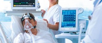
Electroencephalography (EEG) is a method that is used to study the state of the human brain and is based on recording its electrical activity. This examination allows you to detect the spread of pathological processes and signs of epilepsy.
The average duration of the procedure is about an hour, but it is highly informative and makes it possible to track functional changes in the brain, the dynamics of the disease, and evaluate the impact of the therapy.
Since the procedure does not cause any pain or discomfort, EEG can be called one of the most accurate and gentlest methods of examining the brain.
EEG principle
The human brain consists of millions of special cells called neurons. Each of them generates its own electrical impulse. Within individual areas of the brain, impulses must be consistent. They can also strengthen each other or make each other weaker. Their strength and amplitude are not stable and are constantly changing.
Content:
- How EEG works
- Historical reference
- Significance of EEG
- Benefits of an Electroencephalogram
- Indications for the procedure
- Contraindications
- How to prepare for an EEG
- Performing an EEG
- Purpose of EEG video monitoring
- Conclusion of electroencephalography
This is the bioelectrical activity of the brain. To register it, special electrodes are applied to the intact scalp, which will pick up vibrations, amplify them and record them in the form of special curves, so-called waves. The latter, depending on their shape, frequency and amplitude, are divided into five types: α- (alpha), β- (beta), δ- (delta), θ- (theta) and μ- (mu) waves. Each of the waves reflects the work of a specific part of the brain and is named by the first letter of its Latin name.
Their registration in real time is the essence of encephalography.
Decoding the electroencephalogram
At the Yusupov Hospital, the electroencephalogram is deciphered by experienced neurologists, leading experts in the field of neurophysiology. Study results that are difficult to interpret are discussed at a meeting of the expert council with the participation of candidates and doctors of medical sciences, doctors of the highest category. Neurologists collectively make decisions regarding the diagnosis and management of the patient.
The human EEG contains alpha, beta, delta and theta rhythms. They have different characteristics and reflect certain types of brain activity. An electroencephalogram is a recording in computer memory or on paper. The curves that are recorded during the study are analyzed by a neurophysiologist. It evaluates the rhythm of waves, frequency and amplitude, identifies characteristic elements, recording their distribution in space and time. Then all the data is summarized and reflected in the conclusion and description of the electroencephalogram. The conclusion is based on the analysis of EEG curves taking into account the clinical signs of the disease. It reflects the main characteristics of the electroencephalogram and includes 3 mandatory parts:
- description of the activity and type of EEG waves;
- conclusion according to the description of the electroencephalogram and its interpretation;
- determining the correspondence of clinical signs with EEG results.
In the process of deciphering the electroencephalogram, the doctor must take into account the basal rhythm, the level of symmetry in the electrical activity of the neurons of the brain of the right and left hemispheres, the activity of the commissure, and changes in the electroencephalogram against the background of functional tests. Doctors at the Yusupov Hospital make a final diagnosis only taking into account the presence of clinical signs of the disease that worry the patient.
In order to register an electroencephalogram, make an appointment with a neurologist by calling the Yusupov Hospital. After recording the EEG, the doctor will make a conclusion and give recommendations for treating the disease.
Historical reference
One of the founders of the electroencephalography method is considered to be the physiologist and psychiatrist from Germany Hans Berger. In 1924, using a device for measuring small currents called a galvanometer, he was the first to perform a procedure resembling an EEG recording.
Later, a special device called an encephalograph was created to conduct electroencephalography. Today, there are stationary encephalographs, which make it possible to conduct research exclusively in a special room, and portable ones, which are designed for long-term monitoring and can be moved.
It is noteworthy that initially EEG was considered exclusively as a method for identifying mental disorders in a person. Only over time it became clear that the technique also makes it possible to detect deviations not related to psychiatry.
Equipment
To record EEG, devices called Electroencephalographs are used. They consist of an electrode part, an amplifier system, and a recording device. Electrodes come in different types: cup and bridge. They are made from electrically conductive carbon or metal with a silver chloride coating. Such a coating is necessary so that a constant potential does not accumulate on the electrode, which causes polarization of the electrode. This results in interference. Non-metallic electrodes are least polarized.
To ensure accurate registration, parallel common-mode amplifiers with a notch filter are used. This allows you to combat network interference. In terms of their quality, amplifiers now allow recording without an electrically insulated chamber and without grounding.
Recording device. Initially, writing instruments with a paper tape feed were used as a recorder. They differed in ink devices and devices with a thermal pen. But consumables were quite expensive. Nowadays computer technology is used as a recording device. With the advent of computer technology, it became possible not only to record EEG on a non-paper medium, but also to carry out additional mathematical processing of EEG. This increased the resolution of the method.
The application of electrodes is also carried out in various ways. The international system adopted as the standard is the 10 - 20 system. Electrodes are applied as follows. Measure the distance along the sagittal line from Inion to Nasion and take it as 100%. At 10% of this distance from Inion and Nasion, the lower frontal and occipital electrodes are installed, respectively. The rest are placed at an equal distance of 20% of the distance inion - nasion. The second main line runs between the ear canals through the crown.
The lower temporal electrodes are located at 10% of this distance above the auditory canals, and the remaining electrodes of this line are located at a distance of 20% of the length of the biauricular line. The letter symbols indicate, respectively, areas of the brain and landmarks on the head: O - occipitalis, F - frontalis, A - auricularis, P - parietalis, C - centralis, T - temporalis. Odd numbers correspond to the electrodes of the left hemisphere, even numbers - to the right.
According to Young's system, frontal electrodes (Fd, Fs) are located in the upper part of the forehead at a distance of 3 - 4 cm from the midline, occipital electrodes (Od, Os) - 3 cm above the inion and 3 - 4 cm from the midline. The segments of the lines Od - Fd and Os - Fs are divided into three equal parts and central (Cd, Cs) and parietal (Pd, Ps) electrodes are installed at the division points. At the horizontal level of the upper edge of the auricle, the anterior temporal (Tad, Tas) are installed along the frontal line Cd - Cs, and the posterior temporal (Tpd, Tps) are installed along the frontal line Ps - Pd.
The advantage of the 10 - 20 system is a large number of electrodes (from 16 to 19 - 24), but this system requires more sensitive equipment, because the interelectrode distance is small and the potential is weak. Young's system provides sufficient distance and all electrodes are evenly distributed over the surface of the head, but the degree of localization during abduction is insufficient.
The method of removing potential can also be different. The generally accepted system is monopolar recording. In this case, the electrodes on the head are active and record changes in potential relative to an indifferent electrode (most often located on the earlobes). Bipolar recording detects the change in potential between two electrodes located at different points on the surface of the scalp.
All the indicated methods and techniques have their advantages and disadvantages. Therefore, in international practice, mandatory recording according to the 10-20 system is established, both in monopolar and bipolar modes. During computer recording, registration using the 10-20 system is allowed with further digital conversion of the EEG according to the selected bipolar scheme.
Significance of EEG
EEG is a highly informative diagnostic method that allows:
- assess the nature of brain dysfunction and how severe they are;
- determine the localization of the pathological focus;
- clarify information obtained during other diagnostic procedures (for example, computed tomography);
- monitor how effective the therapy is;
- identify areas of the brain in which epileptic activity is present;
- assess brain function in the periods between attacks;
- identify the causes of panic attacks and fainting;
- study the sleep-wake cycle.
It should be taken into account that if a person has seizures, the study will be informative only if it is carried out after about a week.
Regional EEG features
The dominant rhythm is the rhythm of potentials that predominates in a given section of the curve and, when visually analyzed, is distinguished by the greatest periodicity and regularity, and by frequency analysis, by the greatest amplitude. Occipital, parieto-occipital and temporo-occipital regions . The dominant alpha rhythm is clearly expressed, biphasic, sinusoidal, suppressed upon opening the eyes. The appearance in the posterior parietal and parietal region of a rhythm with a frequency of 20-26 Hz, in a state of rest, can be considered as irritation of the cortex.
Parietal-central and anterior 2/3 of the temporal region . The alpha rhythm predominates, but without clear dominance. In addition to it, the following are recorded: 1) theta rhythm (4-7 Hz), with an amplitude less than the alpha rhythm. When opening the eyes it changes; 2) rolandic (arc-shaped) rhythm (^-rhythm) with a frequency of 8 - 12 Hz, arch-shaped, does not change when the eyes open, and even intensifies. Disappears with contralateral physical activity. 3) Frequent extrarolandic frequency of 16 - 30 Hz, with an amplitude of 10 - 30 μV, does not change with afferent influences ((31-rhythm).
The anterior parts of the hemispheres are the precentral and frontal regions. Frequent rhythms are intensified, alpha is almost not visible. The theta rhythm is reduced compared to the central regions.
That. The background pattern of the EEG is a complex organized wave process consisting of spindles of alpha rhythm modulated to varying degrees, against the background of low-amplitude high-frequency activity such as the beta rhythm. This pattern appears in the posterior regions. In more oral areas, elements of slow-wave activity with a background beta rhythm appear.
Theoretically, the origin of the basic EEG pattern is derived from the biophysical premise that each cell is a small impulse generator. But the central nervous system cannot be perceived as a collection of different centers, which in turn consist of individual, elementary (even if interconnected by processes of excitation and inhibition) impulse generators. The nervous system is a complex, balanced, flexible system, the function of which is determined primarily by morphological and dynamic connections. This is confirmed by the great compensatory capabilities of the NS.
Phylogenetically, the oral ganglion of the worm developed into the olfactory brain, which was later supplemented by the visual brain and limbic cortex to organize behavioral reactions. With increasing complexity of afferent impulses, the thalamic system is organized. As movements become more complex, a subcortical extrapyramidal system is formed. The last to form is the bark. In parallel with the emergence of new structures, the organization of the system also becomes more complex. To ensure all the variety of connections, their flexibility and constancy, the system must have energy and information supply. An additional, undifferentiated system is organized - the reticular formation. Consequently, the main functional structures that determine brain activity are the cortex, subcortical regions and the reticular formation.
The correct phase interaction of systems at rest and during physiological changes comes to the fore in the formation of the basic pattern of electrical activity. Let us examine the principle of connections between the cortex, subcortex and trunk. In order for the cerebral cortex to analyze something and make decisions, it must receive information. According to P.K. Anokhin, a complex self-regulating system functions through circular connections with an active feedback response: information -> processing -> action -> information; the circle closes. If we take the normal organized EEG pattern, we see the alpha rhythm in the form of spindles in the posterior regions (and its motor counterpart, the mu rhythm in the central regions) and the predominance of slower rhythms in the frontal regions. All this is superimposed on the fast rhythms of the cortex.
The relationship of rhythms, regardless of amplitude values, is mathematically assessed by coherence, cross-correlation and phasicity. According to the wave theory (Grindel O. M. et al.), built on the basis of analysis of a large amount of data, all EEGs were divided into two large types according to the nature of the connections: wave and impulse (20%). The wave type, being more common, determines the balance and constancy of cyclic processes, which is consistent with the principle of active feedback (according to P.K. Anokhin). Coherence is maximum in the frontal regions across all wavelength ranges and minimal in the occipital regions. Considering that coherence determines the degree of connection, we can assume that in the occipital regions a large number of separate sources are formed, and in the frontal regions they are united by a single organizing force. Let's try to explain the processes as follows.
Afferent impulses arrive at the thalamus, where they are switched and, after certain processing, move to the cortex (generally accepted idea). According to the theory of dynamic localization of functions in the cortex (Pavlov I.P.), impulses functionally arrive in different sections, which leads to the emergence of many centers for processing heterogeneous information. The combination of centers gives a combination of impulses that manifests itself in the back of the head (not surprising since the bulk of the information comes to the visual centers, in addition, heterogeneous information from other affectors comes to the temporo-occipital regions) (Krol B. M.). This information is quite non-specific in a state of quiet wakefulness. The sendings are pulsed, which is consistent with the trigger function of the thalamus (other sendings will not lead to the formation of centers, taking into account the functional refractoriness of the latter). Impulsivity is expressed in the modulation of the alpha rhythm in the occipital regions and the phase mismatch of the spindle envelope in different leads (visible by eye when assessing the curve). Similar processes occur in the central sections, where the afferent and efferent (motor) analyzers meet. Thanks to this connection, the degree of process mismatch is less. Next comes the complex process of perception and analysis of irritation “on the spot.” In the frontal regions, all information is integrated and a single action is formed. This leads to the emergence of a single center in the frontal regions, but one with a slower wave function. Next, the information goes to the subcortical structures and is implemented by the system in the form of voluntary reactions. The wave circle of information closes and a new one begins, which also determines the degree of modulation.
The EEG pattern changes during functional tests. During functional tests, there is an increase in the activity of certain structures. The following loads are used: eye opening, flash of light, hyperventilation, photostimulation, phonostimulation.
Test "Opening the eyes" . When the eyes are opened on the EEG, the alpha rhythm disappears and is replaced by fast rhythms (activation reaction). At the same time, the speed of onset of the reaction, the degree of inhibition of the alpha rhythm, and the persistence of activation are assessed (according to our data, it is noted that on average the reaction lasts 20 - 25 s, then elements of the alpha rhythm appear). After closing the eyes, normally, a recoil reaction occurs, which manifests itself in a temporary increase in the basic rhythm. At the same time, the latency of restoration of the basic rhythm, the degree and persistence of the recoil reaction are assessed. With this test, the reactivity of the cortex, the persistence of excitation processes in the cortex, and the severity of subcortical tone are assessed. This test is more physiological than the activation reaction to a flash of light and carries more information. (But the reaction to a flash of light can be used when examining comatose patients). The processes occurring during the activation reaction can be functionally represented as follows. Opening the eyes significantly increases the flow of impulses into the cortical regions, which leads to increased differentiation of the cortex. This manifests itself on the EEG as an activation reaction with desynchronization (external desynchronization) due to fast rhythms. Mathematically, the coherence gradient increases, but in general the coherence remains at the basic level because physiological activation does not disrupt the connection system.
Hyperventilation test . During the test, the patient breathes heavily, focusing more on exhalation. Hyperventilation is carried out for 3 minutes. During examination, during special examinations, 5-minute hyperventilation is performed. On the EEG, during the test, an increase in the alpha rhythm occurs with its slight slowdown and redistribution to the anterior sections. The degree of modulation decreases. Physiologically, hyperventilation reduces the partial pressure of CO2 in the blood. This leads to the activation of nonspecific subcortical structures and increases the flow of nonspecific, synchronizing impulses into the cortex. When the subcortical parts are overexcited, island-wave activity occurs on the EEG (normally occurs with hyperventilation for more than five minutes).
Photostimulation . It is carried out in two versions: rhythmic and trigger. With rhythmic photostimulation, flashes of light are given rhythmically at a certain frequency. Various frequency ranges are used. With rhythmic stimulation, a reaction of rhythm assimilation occurs. A rhythm appears on the EEG that matches the frequency of the stimulation rhythm. With spectral analysis, it is possible to identify not only the assimilation of rhythm by the main harmonic (stimulation frequency), but also subharmonics, as a rule, by frequencies even to the main stimulation frequency. Normally, rhythm changes in people are expressed to varying degrees. But more often medium and fast rhythms are acquired, without pronounced asymmetry, mainly in the posterior or central sections. Based on the degree of rhythm assimilation, compliance with the frequency of harmonics, and symmetry, one can assess the degree of trigger function of the thalamus and the mobility of processes in the cortex. Trigger stimulation is carried out by delivering light stimulation at the frequency of the main EEG rhythm. For this purpose, special synchronizing devices are used.
Benefits of Electroencephalography
Today, electroencephalography is widely used in neuropathological practice. It makes it possible to clarify a huge number of problematic situations that are associated with the diagnosis and differentiation of neurological diseases. One of the undeniable advantages of encephalography is the fact that it not only helps to identify certain problems, but also helps to distinguish true disorders from hysterical manifestations or simulation.
In addition, the procedure is not as expensive as examination using a tomograph or other similar devices. EEG equipment is available in most hospitals.
The procedure does not have any negative impact on human health and condition. The patient remains fully functional. The study can be carried out even on patients in serious condition, children and adults of any age, since it does not cause deterioration of the condition, discomfort or pain.
What is electroencephalography
Electroencephalography or EEG is one of the basic ways to assess the functioning of the nervous system. The procedure is based on monitoring the activity of the cerebral cortex during its functioning. Pulses are recorded using sensors that are placed on the subject’s head.
The electrodes are placed in such a way as to monitor the activity of all parts of the brain.
The result of the examination is an encephalogram - this is a paper tape with a set of curved lines reflecting brain activity during the examination. The results are deciphered, as a rule, using a computer program, and in ambiguous situations - with the involvement of a neurologist-neurophysiologist.
Thanks to the technique you can:
- find out how the brain functions as a whole: normal or pathological;
- detect affected areas;
- establish the severity of damage and their type;
- clarify the suspected diagnosis;
- decide on the treatment method;
- monitor ongoing therapy.
Indications for the procedure
Today, electroencephalography is widely used in the practice of neurologists to solve a number of problems.
EEG is recommended:
- for prolonged insomnia and other sleep disorders, including apnea, somnambulism and talking during sleep;
- during a convulsive attack;
- for diseases of the thyroid gland;
- with signs of developmental disorders in children or mental disorders in adults;
- after recently experienced traumatic brain injury;
- in case of pathological changes in the vessels of the neck and head, brain tumors detected during ultrasound;
- with frequent migraines, complaints of regular dizziness, feeling of constant fatigue;
- for panic attacks, autism, Asperger's syndrome, stuttering, nervous tics, delayed speech and mental development in children;
- for meningitis, encephalitis, after a stroke and micro-stroke;
- after neurosurgical operations.
Pathologically altered brain
Pathological manifestations on the EEG are slow rhythms - theta and delta. The lower their frequency and higher the amplitude, the more pronounced the pathological process. Slow wave activity appears in the following pathological processes:
- dystrophic diseases;
- demyelinating and degenerative brain lesions;
- compression of brain tissue;
- liquor hypertension;
- the presence of some inhibition, deactivation phenomena, a decrease in the activating influences of the brain stem.
Unilateral local slow-wave activity is a sign of local damage to the cerebral cortex. Flashes and paroxysms of generalized slow-wave activity in waking adults appear in the presence of pathological changes in the deep structures of the brain. High-frequency rhythms (beta-1, beta-2, gamma rhythm) are also a criterion for pathology. Its severity is greater, the more the frequency is shifted towards high frequencies and the more the amplitude of the high-frequency rhythm is increased. The high-frequency component of the EEG occurs due to irritation of brain structures (irritation of brain centers).
Polymorphic slow activity with an amplitude below 25 μV is often considered as a possible activity of a healthy brain. If its index is more than 30% and its occurrence is not a consequence of successive indicative reactions (in the absence of a soundproof chamber), then the presence of polymorphic slow activity in the EEG indicates a pathological process in the deep structures of the brain. The dominance of low-amplitude polymorphic slow activity may be a manifestation of activation of the cerebral cortex, but it may also be a sign of deactivation of cortical structures. In order to carry out differential diagnosis, neurophysiologists at the Yusupov Hospital carry out functional exercises.
High-frequency asynchronous low-amplitude activity may be a consequence of processes of excitation of areas of the cortex or the result of an increase in activating influences from the reticular system. Pathological images of the electroencephalogram (sharp waves, spike, peak, slow spike, complexes) are a manifestation of synchronous discharges of huge masses of neurons in epilepsy.
Contraindications
It is noteworthy that there are no absolute contraindications for electroencephalography. If a person suffers from convulsive attacks, is diagnosed with coronary heart disease, hypertension, or mental disorders, an anesthesiologist must be present during the diagnosis.
The procedure should be postponed if there are open wounds, traumatic injuries, postoperative sutures or any signs of an inflammatory process in the area where it is necessary to install electrodes. Also, the study is not carried out for patients with ARVI.
Why is an electroencephalogram prescribed, contraindications
EEG of the brain is used for additional examination in disorders of a neurological, mental and neuropsychiatric nature. It is also often recommended when undergoing routine medical examinations, especially for professional suitability.
The doctor gives a referral for an EEG if there are complaints about:
- constant pain in the head;
- regular severe headaches;
- dizziness and loss of orientation in space;
- fainting;
- problems falling asleep;
- convulsions;
- shallow night sleep with frequent awakenings.
If certain diseases are suspected, the doctor also prescribes electroencephalography. For example, with stuttering and mental retardation, with epilepsy, vegetative-vascular dystonia, suspected tumors in the cerebral hemispheres.
For problems with blood vessels and poor circulation, after head injuries and concussions, if inflammatory processes or degenerative changes in the brain are suspected, electroencephalography is also indicated.
There are no serious contraindications for diagnosis using this method. But in certain cases, the examination can be difficult and uninformative:
- with strong psychomotor agitation of the patient;
- for mental illness in the acute phase;
- in the presence of open wounds on the head.
How to prepare for an EEG
It should be noted that there are no special restrictions that must precede the procedure. However, there are a number of rules that are recommended to be followed to ensure that the examination is successful and informative.
First of all, inform your doctor if you are taking any medications. You may need to stop taking them for a while or change the dosage.
At least twelve hours before the procedure, or even better, a day before, eliminate from your diet foods that contain caffeine, carbonated drinks, chocolate and cocoa, as well as foods that contain them, foods with energy components, for example, taurine. You should also avoid products with sedative effects.
Wash your hair before the procedure. It is not recommended to use additional styling products (oils, gels, balm, varnishes, etc.) as this may affect the quality of contact of the electrodes with the skin.
If the main purpose of the procedure is to detect seizure activity, you need to sleep before the study.
In order for the result to be as reliable as possible, the patient should avoid stressful situations and refrain from driving for twelve hours before the procedure.
Eating is recommended two hours before the procedure.
If the patient is prescribed EEG sleep monitoring, the night before should be sleepless. Immediately before the procedure, the subject will receive a special sedative, which will enable him to fall asleep during the electroencephalography.
If a child is to undergo an examination, parents should first psychologically prepare him for the manipulations, explaining that there will be no pain or discomfort. You can practice putting on a pool cap under the pretext of playing pilots or astronauts, teach your child to breathe deeply, showing him how to do it by personal example. On the eve of the procedure, the child should wash his hair without using any additional styling products. Before leaving the house, the baby should be fed and calmed down. Just in case, parents are advised to take with them tasty food and drink, and a favorite toy that will help calm and distract the baby.
Please note that if the above rules are not followed, the EEG result may not be very accurate or very informative. In this case, the procedure will have to be repeated.
Preparation requirements
Preparation for an EEG of the brain for a good result should always include a visit to a neurologist who can examine a sick child or adult and determine his indications and contraindications for this procedure. As recent scientific studies show, preparation for an EEG greatly influences the reliability of the results obtained, and therefore the recommendations cannot be ignored. When recording EEG in young children, you have to put up with inevitable interference
Parents should closely monitor their child during the period leading up to the diagnostic procedure. At the same time, it is important to devote time to the psychological component of proper preparation through a conversation about the upcoming visit to a medical facility.
Preparing for an EEG brain scan includes the following tips:
- The patient should stop taking medications that affect the brain. This group of medications includes antiepileptic medications, anxiolytics and any sedative medications. You should stop taking these medications 3-5 days before the upcoming test after consulting with your doctor.
- You should not drink drinks that can stimulate or depress the central nervous system, including coffee, alcohol, strong tea, etc. Many doctors advise giving up chocolate.
- Any patient should thoroughly wash the scalp, as dirty hair leads to disruption of the conduction of electrical current, which can negatively affect the results. To combat this problem, it is recommended to use a special EEG gel. The gel allows you to increase electrical conductivity and ensure better contact of the electrodes with the skin.
- For the reasons stated above, it is prohibited to use gels, balms, mousses and hair sprays due to their effect on electrical conductivity.
- You should eat well before the procedure. Low blood glucose levels can negatively affect brain activity.
- Smoking patients should definitely stop smoking 6-10 hours before the test, as nicotine leads to spasm of the cerebral arteries.
You must stop smoking before the procedure
- If the patient has hairpins, clips or any other accessories on her hair, they should be removed before going into the room for an EEG.
- During the process of taking an electroencephalogram, you must remain completely calm and under no circumstances worry. During the study, it is necessary to follow all recommendations of the attending physician. This is especially true if the patient needs to undergo an EEG with functional tests associated with flashes of light or sharp sound stimuli.
It is not difficult to prepare a patient for a routine EEG. As a rule, compliance with the above recommendations allows you to eliminate distortions in the results and obtain reliable data necessary to determine further diagnostic and treatment tactics.
If the patient was unable to prepare for the study, then it is best to reschedule it, since in this case it may not be informative.
Sometimes, the procedure may be prescribed to the patient during sleep. Preparation for an EEG of the brain in this case includes a mandatory lack of sleep in the last 24 hours, since the patient must quickly move on to dreams.
Electroencephalography is a good method for assessing the activity of individual areas of the central nervous system. Doctors know well how to prepare for an EEG of the brain so that the results are as informative as possible and the procedure is safe for the patient. To this end, you need to visit your doctor and take into account a number of tips regarding lifestyle changes 1-2 days before the scheduled procedure. If all recommendations are followed, electroencephalography allows you to make an accurate diagnosis, as well as select additional diagnostic methods indicated for a particular patient.
Performing an EEG
Electroencephalography is performed in a special room, which is completely isolated from light and sound. The patient sits in a chair or is asked to lie on the couch. A special cap with electrodes is first placed on his head. During the procedure, the patient is alone in the room, contact with doctors is maintained using a camera and microphone. If the diagnosis is performed on a child, one of the parents remains in the office.
Before starting the procedure, the patient is asked to close and open his eyes several times to adjust the equipment. During the diagnosis, the eyes must be closed. If during the procedure the patient needs to change position or visit the restroom, he can inform the doctors about this, after which the diagnosis will be suspended.
It is extremely important that the patient lies with his eyes closed and does not move during the procedure. In the event that a person opens his eyes slightly or moves, the doctor makes a corresponding note, since these actions must be taken into account when deciphering the electroencephalogram.
After the resting EEG is recorded, so-called “stress tests” are performed. Their goal is to test how the brain will react to situations that are stressful for it.
So, a hyperventilation test can be performed. The patient is asked to breathe frequently and deeply for three minutes. Photostimulation with a stroboscopic light source can also be used. It flashes frequently, and this allows you to evaluate how the brain reacts to bright light.
Stress tests can trigger convulsions or an epileptic seizure. The doctors who conduct the study have the appropriate skills to provide emergency care to the patient if necessary.
After the study is completed, the physician should remind the patient to resume taking medications that were stopped the day before the EEG.
The total duration of the procedure is from forty minutes to two hours.
EEG recording methods
The examination is based on recording bioelectrical parameters. Brain activity can be recorded in one of four ways:
| Routine method | EEG with deprivation | Long recording | Night EEG |
| It is used to detect hidden violations. The doctor asks the patient to perform several actions: · breathe deeply; · blink your eyes; · move your lips. At the same time, during the procedure, bioelectric parameters are recorded for 15 minutes. | This method is used if the routine method does not provide comprehensive results. EEG deprivation is partial or complete deprivation of sleep at night. The patient is not allowed to sleep at all or wakes up a couple of hours before the end of normal sleep. | This method records cortical activity during sleep. The procedure is carried out if there are concerns that negative changes occur precisely in the “sleeping” state. | This method is considered the most informative of all. Research begins at a time when a person is just getting ready for bed. Recording continues while you fall asleep. Readings are recorded during sleep and upon awakening. When necessary, the doctor uses electrodes and video recording equipment. |
Checking your brain activity overnight is called EEG monitoring. This option for the procedure requires the use of additional equipment, so the examination is carried out strictly in a hospital.
Purpose of EEG video monitoring
One of the types of electroencephalography is EEG video monitoring. This is a long-term recording of electroencephalography, usually lasting several hours, which can be performed during sleep. The duration of the procedure in each specific case is determined by the attending physician and the personnel of the laboratory that conducts the examination.
EEG video monitoring is prescribed if a short standard procedure does not reveal pathologies, but they are present.
Also, this type of examination allows you to evaluate the EEG during wakefulness and sleep.
Many patients are interested in the question of whether they should necessarily sleep during the study. The answer to this question cannot be unambiguous, because it depends on the specific situation. So, for example, if the reason for the examination is a tic that bothers the patient while awake, there is no need to sleep during the examination.
At the same time, EEG video monitoring during sleep sometimes helps to identify conditions that neither the patient nor his loved ones may even be aware of.
The peculiarity of this procedure is that it can be carried out not only during the day, but also at night. In the event that sleep EEG is required, night monitoring is more rational. Not everyone can fall asleep without problems during the daytime.
At the same time, we should not forget that carrying out a multi-hour procedure in a completely isolated room can be extremely tiring for the patient, especially if we are talking about a child. Most pathologies can be detected during a relatively short recording of a conventional EEG.
Best materials of the month
- Coronaviruses: SARS-CoV-2 (COVID-19)
- Antibiotics for the prevention and treatment of COVID-19: how effective are they?
- The most common "office" diseases
- Does vodka kill coronavirus?
- How to stay alive on our roads?
Also, overnight EEG video monitoring is much more expensive.
EEG price in Moscow
EEG is carried out both in public medical institutions and in private clinics.
In budgetary treatment and preventive institutions, the cost of conducting research is rubles. Private medical centers in Moscow, for example, "NIARMEDIC", "SM-Clinic", "Dobromed", "Mental Health" and others offer this diagnosis for rubles.
How we save on supplements and vitamins: probiotics, vitamins intended for neurological diseases, etc. and we order on iHerb (use the link for a $5 discount). Delivery to Moscow is only 1-2 weeks. Many things are several times cheaper than buying them in a Russian store, and some goods cannot be found in Russia in principle.
SOURCES: https://diametod.ru/funkcionalnaya/podgotovka-eeg-golovnogo-mozga https://diagnostinfo.ru/drugie/efi/podgotovka-k-eeg.html https://medsins.ru/eeg/zachem- iv-kakih-sluchayah-delaetsya-eeg-golovnogo-mozga.html
Additional EEG Analysis Methods
Currently, visual analysis remains the main method of EEG analysis. Additional analysis methods include calculating the power spectrum using the fast Fourier transform. The power spectrum shows the severity of the rhythm of a given frequency. The power spectrum is visually presented in the form of averaged curves, distribution of power spectra by epoch, and spectral mapping.
Another additional method is coherence calculation. Coherence shows the degree of similarity of oscillatory processes at two different points, regardless of their amplitude representation. It has been established that the average coherence value is constant and reflects the degree of stability of connections in the system.
Recently, another processing method has been used. This is the localization of sources of pathological activity using the Multi-step Dipole Localization method. Through numerous calculations, a mathematical model of the probable location of the source of a given wave is created. This model is compared with the amplitude distribution of the same waves on the scalp. For localization, only those EEG slices are used that have a given probability of convergence between the computational model and the scalp recording. A probability of 0.95 or more is considered reliable.
