Coordination
Coordination
- the process of coordinating the work of muscles necessary for the successful performance of a motor function. With the development of a motor skill, the coordination of movements changes, including the development of the inertia of moving organs. At first, control occurs through active static fixation of these organs, then through short-term physical impulses directed at a certain moment to the desired muscle. At the final stages of coordination development, inertial movements are used. In an already established dynamically stable movement, balancing of all inertial movements occurs automatically, without producing additional correction impulses.
Coordination of movements
Coordination of movements
given to a person so that he can perform precise movements and control them. If there is a lack of coordination, this indicates changes occurring in the central nervous system. Our central nervous system is a complex, interconnected formation of nerve cells located in the spinal cord and brain. When we want to make any movement, the brain sends a signal, and in response to it, the limbs, torso or other parts of the body begin to move. If the central nervous system does not work coherently, if deviations occur in it, the signal does not reach the target or is transmitted in a distorted form.
Functions and tasks
Quite a difficult topic to discuss, since the brain does almost everything that you do (or controls these processes).
We need to start with the fact that it is the brain that performs the highest function that determines the intelligence of a person as a species - thinking. It also processes signals received from all receptors - vision, hearing, smell, touch and taste. In addition, the brain controls sensations in the form of emotions, feelings, etc.
It is impossible not to mention that all movements of the human body are also controlled by the brain - even if these are reflex reactions that we are not always aware of.
Causes of impaired coordination of movements
There are many reasons for impaired movement coordination. These include the following factors:
- physical exhaustion of the body;
- exposure to alcohol, narcotic and other toxic substances;
- brain injuries;
- sclerotic changes;
- muscle dystrophy;
- ;
- ;
- catalepsy is a rare phenomenon in which the muscles weaken due to an explosion of emotions, say, anger or delight.
Lack of coordination is considered a dangerous deviation for a person, because in such a state it costs nothing to get injured. This often accompanies old age, as well as previous neurological diseases, a striking example of which in this case is stroke. Impaired coordination of movements also occurs in diseases of the musculoskeletal system (poor muscle coordination, weakness in the muscles of the lower extremities, etc.) If you look at such a patient, it becomes noticeable that it is difficult for him to maintain an upright position and walk.
People with such ailments move uncertainly, their movements show laxity, too much amplitude, and inconsistency. Having tried to outline an imaginary circle in the air, a person is faced with a problem - instead of a circle, he gets a broken line, a zigzag. Another test for incoordination is to ask the patient to touch the tip of the nose, which also fails. Looking at the patient’s handwriting, you will also be convinced that his muscle control is not all right, since letters and lines creep on top of each other, becoming uneven and sloppy.
Hindbrain, midbrain and diencephalon
The purpose of the lesson is to provide general topographic information about the position of the brain in the skull, its structure and functions, to reveal the role of the medulla oblongata, midbrain, diencephalon and cerebellum in the implementation of unconditioned reflexes and to find out their significance, to conduct anti-nicotine and anti-alcohol propaganda, to provide information on the prevention of injuries in children.
It is advisable to divide the lesson into three parts: in the first, repeat the structure and functions of the spinal cord and clarify the understanding of basic terms; in the second - show the parts of the brain on a table or on filmstrip frames and conduct tests to determine the characteristics of the reflexes of the main parts of the brain; in the third - repeat the information received in class using dummies and planar preparations of the brain.
When explaining new material, it is convenient to use the “Structure and Functions of the Brain” filmstrip, which contains instructional footage that helps the teacher conduct functional tests with students that demonstrate the reflexes of various parts of the brain. As an example, we can see how the teacher of experimental school No. 82 of the APN in the village of Chernogolovka, Noginsk district, Moscow region, N. G. Pimanova, uses this filmstrip in class.
She uses the first fragment of the mentioned filmstrip to repeat previously studied material about the brain. Considering the structure of the cranium and membranes of the brain, the teacher reveals the importance of cranial fluid, arteries feeding the brain, and simultaneously provides additional material about the blood supply to the brain and complications that can occur with hypertension. N.G. Pimanova uses this material for anti-nicotine propaganda, emphasizing that smoking tobacco causes an increase in blood pressure, promotes the deposition of salts in the walls of brain vessels, and this leads to an increase in their fragility and the possibility of a stroke.
The teacher uses the filmstrip frames containing questions primarily to check how clearly students differentiate concepts about the gray and white matter of the brain, the cortex and nuclei of the brain, the neural pathways of the brain and nerves. She invites students to show these formations in a drawing, find them on a planar preparation after viewing the first fragment of a filmstrip, and teaches them to use definitions when identifying objects.
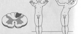
During the demonstration of the second fragment, the teacher conducts the functional tests included in this filmstrip. She begins her study of the medulla oblongata by comparing it with the spinal cord. The teacher draws students' attention to the morphological similarity: in the medulla oblongata, the gray matter lies inside the white matter, but does not form gray columns, as in the spinal cord, but is located in separate nuclei in the bottom of the fourth ventricle. Then it establishes functional similarities: reflexes are caused by irritation of certain parts of the body (stretching the tendon of the quadriceps femoris muscle causes a knee-jerk reflex of the spinal cord, irritation of the root of the tongue causes a swallowing reflex of the medulla oblongata). The teacher emphasizes that both reflexes are unconditional. They arise involuntarily only when the corresponding parts of the body are irritated (Fig. 32, A, B).
Rice. 32. Experiments to determine the functions of the spinal cord and brain: A - swallowing reflex; B - knee reflex; B - experiment revealing cerebellar reflexes involved in the inhibition of movements that continue by inertia; D — finger-nose test, which reveals the participation of the cerebellum in the performance of precise, targeted movements that require the coordinated work of many muscles; D - tonic reflexes of the midbrain, restoring body position in case of loss of balance; E - reflexes of the diencephalon, ensuring the maintenance of a certain posture between movements
Exercise 1.
To prove that the unconditioned swallowing reflex of the medulla oblongata cannot be carried out without irritation of the root of the tongue, the reflexogenic zone of this reflex.
The experiment is carried out frontally. The teacher shows a frame of the filmstrip, where it is proposed to make several swallowing movements in a row. The phrase located under the picture: “Why did you stop? Have you forgotten how to swallow?!” - shown after the experiment.
The experiment can be performed without a filmstrip. Students, at the teacher’s command, make several swallowing movements in a row at a fast pace and make sure that in the absence of an irritant (in this case, saliva), it is impossible to make a swallowing movement. When an irritant is exposed to the root of the tongue, the act of swallowing occurs involuntarily, and a person can swallow an inedible object.
N. G. Pimanova usually dwells in detail on the frame, where she explains why children should not be given small objects to play with (screws, nuts, buttons, balls). Babies often put things in their mouths and may involuntarily swallow small objects. A characteristic feature of N. G. Pimanova’s teaching is the close connection of theoretical issues with the facts of everyday life; she carries out hygienic education unobtrusively, as if in passing, and convinces students without unnecessary moralizing.
It is convenient to present the structure and function of the second part of the hindbrain - the cerebellum - in textbook terms: talk about the consequences of removing the cerebellum in experimental animals, and then show how the functions of the cerebellum are performed normally in humans in functional tests.
Task 2.
Observe the coordination of muscle work carried out by the cerebellum when performing the cerebellar finger-nose test.
This experiment can be carried out frontally. At the teacher’s command, eighth-graders close their eyes, extend their hand forward with their index finger extended and touch the tip of their nose with its tip (Fig. 32, D). The teacher draws the students' attention to the fact that this purposeful movement was carried out quickly and smoothly, although more than 30 muscles were involved in it. The sequence of muscle contractions when performing movements is programmed by parts of the brain, and in particular the cerebellum, which controls the sequence and order of operation of the nerve centers of the spinal cord that control the muscles. After all, each muscle must engage in work on time, develop a strictly defined effort and exit from work on time. In this case, a person has to overcome external resistance, inertial forces and the influence of other factors. The cerebellum receives impulses from many receptors, processes them, and thanks to its activity, the response occurs taking into account these external forces.
Task 3.
Observe how movements arising due to inertia are eliminated thanks to the functions of the cerebellum.
The subject is called to the board. The teacher grabs his hand, bent at the elbow, by the forearm and invites him to pull his hand towards himself, overcoming the experimenter’s resistance. After the subject performs this action with sufficient effort, the experimenter suddenly releases the subject's hand so that it moves by inertia (Fig. 32, B). However, this movement does not occur: the subject makes a small jerk and stops: thanks to the function of the cerebellum, the movement that arose by inertia is inhibited.
Damage to the cerebellum during intoxication is a fact well known to students. It can be used for anti-alcohol propaganda. For this purpose, students can be asked the following questions: 1. Why is a drunk person, trying to take one step, forced by inertia to take several steps in the same direction? (Unconditioned reflexes carried out with the participation of the cerebellum are impaired and it is not able to slow down movements that continue by inertia.)
2. Why do drunk drivers turn their car sharply, press the brake pedal sharply, and what can this lead to? (The movements that have begun cannot end on time, they continue due to inertia, resulting in excessively sharp turns, sudden pressing of the brake pedal. Such a driver can run into an obstacle, and with excessive braking, the car can skid and even overturn.) These facts can be used for the purpose of legal education, explaining why in all countries it is strictly forbidden for a drunk person to drive a tractor, car, or airplane, and why it was necessary to create special indicators to detect and neutralize a drunk person sitting behind the wheel.
The work of the midbrain can be demonstrated in two functional tests: the first makes it possible to determine the role of tonic reflexes of the midbrain in maintaining body balance, the second makes it possible to show the participation of the midbrain in the orientation reflex.
Task 4.
Observe the midbrain reflexes that help maintain body balance.
The subject is called to the board. He is asked to take an unstable position: stand so that one foot touches the toe of the heel of the other foot located in front. (The feet should be in a straight line - one in front, the other behind. The arms are folded, the elbows are brought closer together. The subject is warned that if he loses balance, he cannot move his legs.)
The subject stands motionless for some time, but muscle tension (their tone) continuously changes. This can be noticed by observing the continuous swaying of the torso. Over time, these swaying becomes more and more noticeable. You can observe how, when you lose balance, the midbrain reflexes intensify, restoring the correct position of the torso (Fig. 32, E). At the end of the experiment, significant deviations of the body from an unstable equilibrium position occur. To compensate, the subject is forced to balance with his hands or put his leg aside, violating the instructions. The stimulus that causes this reflex is the deviation of the body from the vertical axis, the response is a compensatory movement. Students' attention can be drawn to the complexity of this reflex compared to the reflexes of the medulla oblongata. A reflex reaction is caused by any deviation, and the final effect can be achieved by completely different movements. This feature can be shown even more clearly using the example of the orienting reflex, which is also carried out with the participation of the midbrain.
Teacher N.G. Pimanova, whose experience has already been referenced, begins a conversation about the orientation reflex with an analysis of frame 26 of the slide “Structure and Functions of the Brain.” There's a cat on the windowsill. Someone scared her. She arched and hissed. This stimulus caused an orienting reflex in the child. He turned his head towards the cat, his eyes searching for the source of the sound. However, after perceiving this frame, some students develop an unreasonably narrow idea that distorts the essence of the matter: they believe that the orienting reflex is “when the cat meows, the child will turn his head.” The teacher explains the narrowness of this judgment, emphasizes that the orienting reflex is caused by any new stimulus, and puts on a new experiment.
Task 5.
Prove that the orienting reflex occurs in response to any new stimulus and is manifested in movements towards this stimulus.
The teacher taps a pencil on the table. Many students look up at the teacher: a new stimulus has caused an orienting reflex. The teacher draws the students' attention to the fact that the stimulus affected the organ of hearing. Continuing the experiment, the teacher walks along the rows and puts his hand on the shoulder of one of the distracted students: the tactile stimulus causes the same indicative reaction - turning the head towards the stimulus. Conclusion: the orientation reflex can be caused by any stimulus - auditory, visual, tactile. It is only important that the stimulus is new. It can be shown that when repeated, the stimulus loses its novelty and the orienting reflex no longer appears. You can also pay attention to the generality of this reaction: the movement is made in the direction of a new stimulus, regardless of where this stimulus is located - in front, behind, right, left.
At the end of the lesson, we should consider the functions of the diencephalon: regulation of sleep and wakefulness, maintaining a constant internal environment, postural fixation reflexes. Having explained these complex functions using the frames of the filmstrip “Structure and Function of the Brain,” you can offer students the following exercise:
Task 6.
Determine which reflex of the diencephalon can be detected when executing the “Freeze” command.
The teacher invites students to stand up and then gives the command “Freeze.” Students in the class freeze in different positions, which makes it possible to observe the postural reflex of the diencephalon.
The command “Freeze” caused a traffic stop. At the same time, movements in many joints of the body had to be blocked. If this had not happened, the students would have fallen, like a table lamp whose hinges were not secured. From the topic “Musculoskeletal System,” students know that bones are secured in joints when opposing muscles contract at the same time. Consequently, when the movement stops, all the muscles of the opposite action of all joints simultaneously contract, fixing the new body position (Fig. 32, E).
At the end of this lesson or during the next one, during a survey, it is useful to use brain models or planar preparations to find the main parts of the brain and fill out the following table:
Table 18. Unconditioned reflexes of the brain
Symptoms of impaired coordination of movements
The following symptoms of impaired coordination of movements exist:
- Unsteady movements - This symptom occurs when the muscles of the body, especially the limbs, weaken. The patient's movements become uncoordinated. When walking, he sways a lot, his steps become abrupt and have different lengths.
- Tremor - shaking of the hands or head. There is a strong and almost imperceptible tremor. In some patients it begins only during movement, in others - only when they are motionless. With severe anxiety, the tremor increases; shaky, uneven movements. When the muscles of the body are weakened, the limbs do not receive a sufficient basis for movement. The patient walks unevenly, intermittently, the steps are of different lengths, and he staggers.
- Ataxia is caused by damage to the frontal parts of the brain, cerebellum, and nerve fibers transmitting signals through the channels of the spinal cord and brain. Doctors distinguish between static and dynamic ataxia. With static ataxia, a person cannot maintain balance in a standing position; with dynamic ataxia, it is difficult for him to move in a balanced manner.
Cerebral cortex treatment
Sarclinic uses proprietary methods to restore the functioning of the cerebral cortex. Treatment of the cerebral cortex in Russia in adults, adolescents, children, treatment of the cerebral cortex in Saratov in boys and girls, boys and girls, men and women allows you to restore lost functions. In children, the development of the cerebral cortex and brain centers is activated. In adults and children, atrophy and subatrophy of the cerebral cortex, disruption of the cortex, inhibition in the cortex, excitation in the cortex, damage to the cortex, changes in the cortex, pain in the cortex, vasoconstriction, poor blood supply, irritation and dysfunction of the cortex, organic damage, stroke, detachment are treated , damage, diffuse changes, diffuse irritation, death, underdevelopment, destruction, diseases, perinatal encephalopathy and post-resuscitation encephalopathy, cerebral palsy, minimal cerebral dysfunction, delayed speech development, delayed psychomotor development, FMR, birth trauma. Cells and neurons of the cortex restore their work, structure and function are restored. The structure and activity of the cerebral cortex is restored, polypeptides and their content return to normal, the cortex and subcortex begin to work. In children, age-appropriate maturation of the cerebral cortex occurs. Unfortunately, under certain pathological conditions, death occurs, the death of cells in the cerebral cortex; in this case, it is not possible to restore the functioning of the cortex. On the website sarclinic.ru you can ask a doctor a question online for free. If the cerebral cortex is damaged, then with proper and adequate treatment it is possible to restore its functions.
Sign up for a consultation. There are contraindications. Specialist consultation is required.
Text: ® SARCLINIC | Sarclinic.com \ Sarlinic.ru Photo: © MedusArt / Photobank Photogenica / photogenica.ru The people depicted in the photo are models, do not suffer from the diseases described and/or all coincidences are excluded.
Related posts:
Human brain, child brain, sections, structure, brain treatment
I'm afraid to speak, logophobia: treatment of logophobia for stuttering, the child is afraid to speak
Dysmorphophobia, treatment in Saratov, how to treat dysmorphophobia
Foville syndrome (ALF Foville) alternating pontine
Mobius syndrome, treatment of Mobius syndrome, congenital facial palsy, Poland syndrome
Comments ()
Motor coordination test
Unfortunately, many people have poor coordination. If you want to test yourself, we offer you a very simple test.
- 1. To do this you need to do the exercise while standing. Try pushing your toes and heels together while your eyes are closed.
- 2. Another option to test your coordination is to sit on a chair and lift your right leg up. Rotate your leg clockwise, and at the same time draw the letter “b” with your right hand, imitating its silhouette in the air, starting from the “tail” of the letter.
- 3. Try placing your hand on your stomach and stroking it clockwise while tapping your head with the other hand. If, as a result of the test, you completed all the tasks the first time, this is an excellent result. We congratulate you! You have good coordination. But if you were not able to immediately perfectly perform all of the above, do not despair!