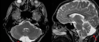
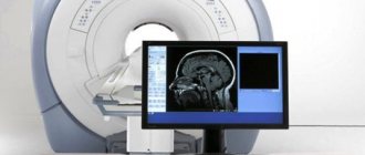
The brain is a complex organ consisting of gray and white matter, blood vessels, nerve fibers, membranes, and venous sinuses. To study anatomical structures, one diagnostic method is not enough. A computed tomography scan can be performed if hard tissue pathology is suspected. Verification of soft tissue changes is best done using magnetic resonance imaging (MRI). Oncological search is carried out comprehensively using both procedures.
Residents of large megalopolises in Russia (Moscow, St. Petersburg) have access to the services of public and private clinics, the addresses of which can be found on the global network. When choosing an establishment in St. Petersburg, you need to focus on reviews from previous clients of the centers. The addresses are distributed on the global network and in the media. In the city, more than a hundred clinics are equipped with radiation diagnostic methods.
Magnetic resonance imaging scanners are open and closed, low- and high-field. Gradation is carried out based on the structure of the electromagnet location zone.
Equipment for performing computed tomography can be step-by-step, spiral (multispiral). The separation is based on the principles of the scanning procedure.
A radiation diagnostics doctor will be able to determine the type of examination and choose the right type. It is also necessary to take into account the harm to health and the diagnostic benefits of the method of studying the brain. Read the article for details.
CT scan of the brain - what will it show?
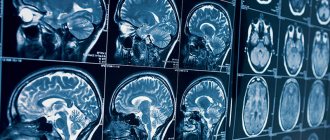
X-ray radiation, which underlies the functioning of computed tomography, detects dense formations with a diameter of more than 1 mm. After intravenous administration of an iodine-containing drug, radiology doctors are able to use tomograms to identify vascular anomalies, aneurysms (wall dissection), hemorrhages in the brain parenchyma, and strokes (hemorrhagic, ischemic).
Contrast CT scan of the brachiocephalic arteries shows changes in thin sections. During one scan, from 200 to 500 tomograms are obtained, performed at 1 mm (or other interval). Through consistent study, the specialist determines the pathology. Analysis takes a long time.
Innovative software for CT machines allows for spatial reconstruction of the brain. The spatial model shows the smallest anatomical details. 3D reconstruction is used by neurosurgeons and oncologists before planning surgery.
Preparation for angiography
Computed tomography of the head and neck does not require complex preparation. To reduce side effects from contrast contrast, a light snack (sandwich, yogurt or fruit) is advisable before leaving home. Breastfeeding mothers need to express and store milk, since after angiography you cannot immediately put the baby to the breast.
CT of the head and neck vessels requires blood test results for creatinine. In some diagnostic clinics, a rapid test is performed right on the spot. You can find out about the availability of such a service when making an appointment for an examination. The patient must inform the doctor about the medications he is taking: a number of medications cannot be combined with iodine-containing drugs. In agreement with the specialist, they are canceled or replaced at least 12 hours before the procedure.
Drawing blood for creatinine testing
CT scan of the brain - why do it?
Clear medical indications have been developed, on the basis of which you can determine when to do a computer examination of the brain:
- Speech disorders;
- Prolonged headaches, dizziness;
- Suspicion of micro- and macro-strokes;
- Identification of the causes of increased intracranial pressure;
- Muscle cramps;
- Tumors;
- Cerebral edema, displacement of structures due to hydrocephalus.
CT angiography (vascular contrast) is performed to study intracerebral blood supply, identify hemorrhagic foci, if MRI is contraindicated. The main purpose is to detect small cancerous tumors.
CT brain perfusion provides information about tissue oxygen saturation and gas diffusion through nerve sheaths.
CT or MRI of the brain - what to do
Magnetic resonance scanning is used to study structures containing water. The brain consists mostly of soft tissue, which makes magnetic resonance examination a popular method for verifying nosological forms. The study determines vascular malformations (pathological joints between arteries and veins), congenital anomalies without contrast. The procedure is harmless and can be performed on children who are able to remain still for 30-40 minutes (duration of the examination). Combined MRI and MSCT of the brain are done when tumors are suspected, metastases are determined, and perifocal structures around the cancer focus are studied.
When is it better to do an MRI of the vessels of the brain and neck:
- Diagnosis of inflammatory processes – abscess, encephalitis, meningitis;
- Mental disorders;
- Stuttering, convulsions;
- Sudden loss of consciousness.
- When is spiral tomography preferable:
- Determination of the state of the ventricles;
- Verification of fluid accumulations;
- Diagnosis of cystic cavities;
- Identification of areas of accumulation of cerebrospinal fluid (CSF).
Difficulties arise with prescribing a diagnostic procedure for suspected stroke. In the ischemic variant, it is better to use MRI, which records changes in the physicochemical properties of gray and white matter. If clinical symptoms indicate a hemorrhagic form, it is more rational to prescribe CT.
MSCT
Indications for MSCT
MSCT is preferable for:
- suspected stroke
- traumatic brain and associated injuries
- patient's serious condition
- the need to study the skeletal apparatus
- assessment of atherosclerotic plaques and internal walls of blood vessels
- presence of contraindications to MRI
For other pathologies, the decision to scan remains with the patient. It should be noted that MSCT scanning is more accessible.
Contraindications to MSCT
Contraindications for CT:
- pregnancy at any stage of gestation
- not recommended for children under 14 years of age
- not recommended for existing myeloma and recent ionization examinations
- the patient is overweight
- inability to assume a supine position
Contraindications for contrast enhancement:
- uncompensated thyrotoxicosis
- history of severe reactions to an iodine-containing drug
- renal failure with a significant decrease in clearance
- with caution in severe bronchial asthma
Contraindications for cardiac CT:
- an irregular rhythm that cannot be made regular with medication (for example, permanent atrial fibrillation)
- inability to hold your breath for 15 seconds
How is MSCT performed?
Preparation for MSCT with intravenous contrast
The MSCT procedure is carried out on an empty stomach; you cannot eat or drink for at least 4 hours; in addition, when examining the heart, you cannot smoke or drink coffee for 4 hours. Also, due to the fact that when scanning the heart and coronary vessels, the heart rate should not exceed 65 beats/min, take 50 mg of metaprolol or any other beta blocker 40 minutes before the scan. When examining the intestines, the procedure is carried out before fluoroscopy with barium, or after its complete removal from the body; if it is necessary to contrast intestinal loops, drink a liter of oral contrast within an hour before the procedure; if you need to examine the large intestine in detail, then add 0.5 liters the night before. When performing MSCT cholangiography, you must drink at least 1.5-2 liters of still water an hour before the examination.
If there is a history of allergy to contrast, premedication (with prednisone/antihistamines), hydration, and only low-osmolar non-ionic contrast are used.
Carrying out the procedure
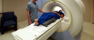
Before the procedure, it is necessary to provide the doctor with a direction indicating the purpose of examining a certain area and the conclusion of the examinations already carried out. Next, the patient lies down on the table deck, contrast is injected intravenously with an injector, the table moves through the device and scanning is performed. If necessary, you will need to hold your breath for up to 15 seconds. Depending on the area being examined, MSCT lasts from 10 to 40 seconds. Most of the time is spent deciphering MSCT images, since their number is often measured in thousands.
How many times can MSCT be done per year?
The answer to this question, how often an MSCT examination can be done, depends on the area being examined. For example, with MSC scanning of the head, the radiation dose is on average 1.4 mSv, for the chest and abdominal cavity - 2 times more, pelvis - 2.5 times more. The safe radiation dose for the population is 1 mSv per year (equivalent to two x-rays of the lungs), if necessary, it can be increased to 5 mSv per year. As you can see, the permissible number of studies for the head is 3 per year, for other areas - 1 time per year (all this taking into account the absence of other ionizing procedures). If research is still necessary, it is recommended to use a safe and equally high-precision imaging method - MRI (in the absence of contraindications).
Is MSCT harmful to a child and is MSCT dangerous during pregnancy?
Children are especially susceptible to radiation exposure (and with MSCT, the radiation dose can reach up to 12 mSv versus 0.5 mSv with conventional radiography), they have a greater risk of developing cancer. This happens because the tissues of a child’s (and even more so the unborn) body are in a phase of continuous growth, and, therefore, are more susceptible to DNA changes. Also, the chance of developing a tumor after MSCT is much higher in a child than in an adult, because there is actually more time for a tumor to develop in children than in adults and the elderly. To reduce the risk of consequences, it is highly not recommended to perform MSCT on children until the process of active tissue growth has completed (up to 14 years). During pregnancy, MSCT is an absolute contraindication and can be performed only if there is an absolute risk to life and it is impossible to urgently perform an alternative non-ionizing scan (MR imaging).
Computed tomography of the vessels of the brain and neck with contrast - how it is performed
Contrast examination is used to visualize the vessels that tightly envelop the brain. Examination of the cerebral arterial network and neck (carotid, vertebral, brachiocephalic artery) allows us to identify the causes of pathological symptoms (dizziness, headaches, short-term loss of consciousness).
Native scanning does not show the soft tissue component. After an intravenous injection of an iodine-based enhancing drug, specialists are able to study the arterial and venous network of the brain. The appearance of pathological circulation indicates neoplasms that have their own capillaries. Atypical vessels appear in the early stages, when the lesion is several millimeters in size. Innovative MSCT of the brain has the ability to verify lesions with a diameter of 0.5 mm. When determining where to do a CT scan, you need to take into account the specifics of the devices.
Using the bolus contrast method, radiology doctors are able to monitor the structure of the vessels of the neck and brain as the contrast spreads.
The CT perfusion method involves studying small capillary networks after the contrast agent has spread through them. The study is used for partial visualization of soft tissue components when it is impossible to use a magnetic resonance procedure. The contrast is excreted by the kidneys after 1.5 days. After CT angiography, women should not breastfeed.
What you need to know when going for a CT scan
To undergo MSCT, you need a referral from a doctor, which would indicate the area and purpose of the study. When referring for MSCT with bolus contrast, it is also necessary to have a report from an allergist with permission to conduct the study or recommendations for preliminary preparation. If you do not have a referral, you can go to any of the Family Doctor clinics by making an appointment with a specialist or general practitioner. An experienced specialist will examine you, give his recommendations and write out a referral for MSCT.
Depending on the area of study, you may need to prepare for MSCT. For example, MSCT of the liver and gallbladder is done in the morning strictly on an empty stomach. MSCT of the kidneys or pelvic organs can be done during the day, with a light breakfast allowed. MSCT of the brain, sinuses, chest organs, bones and joints can be done at any time, since no special preparation is required for these studies.
For a more accurate, individual selection of the MSCT program and targeted image reconstruction, it is advisable to have with you all medical documents reflecting the history of the disease. Take with you all the results of past studies you have (X-ray, ultrasound, ultrasound, CT, MRI, PET). Your entire archive will be returned to you in full along with the MSCT results.
During the examination, you will need, as directed by your doctor, to remain still and hold your breath for 10-20 seconds.
Sign up for diagnostics Do not self-medicate. Contact our specialists who will correctly diagnose and prescribe treatment.
MR angiography of the neck - types, indications
Anomalies, aneurysms, and malformations determine the clinical symptoms of dizziness and unexplained headaches. To verify these nosologies, MRI is performed, and if contraindications are performed, contrast-enhanced MSCT is performed.
The value of both studies is determined individually after analyzing the patient's medical history. A special type is cisternography of the brain, which allows one to estimate the size of the intracerebral ventricles.
For example, with an unsteady gait and double visual objects, vestibulopathy is suspected. Nosology is best determined using MRI of the brain and neck. The presence of metal objects is a contraindication. An alternative is contrast tomography of the brain and neck.
When do you need MRI and MSCT?
During an oncological search, doctors need comprehensive information about the consistency of tissues, the location of anatomical structures, visualization of additional shadows, and the condition of blood vessels (presence of plaques, blood clots, malformations).
The norm in the study of white and gray matter is rare. Specialists often find minor pathological abnormalities that do not require treatment. MRI of the brain and neck is indispensable for verification:
- Aneurysmal dilatations;
- Multiple sclerosis;
- Alzheimer's and Huntington's diseases.
The diagnosis of a tumor is more rational than its formation after a person undergoes an MRI-MSCT complex. To prescribe methods for examining the brain and neck, clear indications are required.
If an MRI shows cancer, there is no reason to despair. The final conclusion is formed only after a systemic examination and the use of all necessary diagnostic methods. High-field MRI and CT are the basis of oncological diagnostics. Radiation imaging is informative and highly specific.
When choosing a clinic in St. Petersburg where you can do MSCT, you should pay attention to the availability of important software modes:
- Selective fluid exclusion (Flair);
- Visualization of the total diffusion of molecules;
- 3D modeling;
- Assessment of perfusion parameters;
- Mapping of nerve centers;
- Determination of physicochemical composition (MRI).
The full complex will allow you to obtain maximum information about the structure of brain structures.
What are CT and MRI?
Both computed tomography (CT) and magnetic resonance imaging (MRI) are two effective hardware-based research methods that allow, without instrumental intervention, to obtain a clear, detailed picture of the state of the brain and the vessels that supply it. The basic principle of both examination methods is to obtain anatomically accurate images, on the basis of which doctors of various specialties then determine the diagnosis and prescribe therapy. There are three basic tomographic protocols, the same for both MRI and CT:
- native brain tomography;
- brain tomography with contrast;
- angiography of cerebral vessels.
The main differences between CT and MRI are:
- in the method by which the images are obtained;
- in the list of contraindications;
- in possible complications;
- in the price of tomography.
Radiation imaging of the neck – when is it performed?
An MSCT of the neck can be done if there are symptoms of intervertebral hernias or compression of the vertebral artery by unstable vertebrae. Neurologists can determine whether a nerve fiber is pinched in the cervical spine. Radiation imaging is used to assess the degree and nature of cerebral circulatory disturbances. The methods identify nosology at an early stage of development, when there are no symptoms.
MRI of the brain and neck is performed to identify or exclude blood clots, atherosclerotic deposits, and blockage of the lumen of the vessel.
Procedures are not used for mass examinations. Scientists have only theoretically substantiated the need for MSCT and MR imaging for early verification of atherosclerotic lesions of the heart arteries. The widespread spread of harmful radiation and the high cost of procedures are limited.
Computer scan of the neck and brachiocephalic trunk
The maximum number of cases of insufficiency of cerebral blood supply occurs in the brachiocephalic complex, which includes the carotid, vertebral, subclavian arteries, and brachiocephalic trunk.
Atherosclerotic deposits in the wall and a blood clot clog the lumen of the vessel. Non-contrast MSCT of the brain reveals abnormalities, helps assess the degree of microcirculation impairment, and visualize enlarged lymph nodes.
Late detection of BC damage is highly likely to lead to disability or death.
The use of multispiral tomography of the brain with contrast detects aneurysms, pathological anastomosis between arteries and veins (malformations). The study helps determine the volume and prevalence of death of brain parenchyma (atrophy), and areas of lack of oxygen supply (ischemia). Information is necessary for specialists when planning surgical interventions to install vascular stents, perform embolization, and remove aneurysmal dilatations.
What MSCT of the brachiocephalic arteries shows:
- Ischemic areas;
- Areas of narrowing of the arteries that do not manifest clinical symptoms;
- Discirculatory encephalopathies;
- Anomalies in the course and structure of blood vessels;
- history of stroke;
- Neoplasms of the neck;
- Thromboembolism.
The information content of spiral scanning is beyond doubt, but there are contraindications:
- Kidney failure (with contrast examination);
- Excessive secretion of thyroid hormones;
- Allergy to iodine;
- Carrying a child;
- Decompensated diabetes.
Vascular stenosis is the leading pathology leading to ischemic strokes. To prevent nosology, surgeons introduced endoscopic revascularization operations. Their use requires early diagnosis using MRI and MSCT of the brain.
Long-term persistence of ischemia due to lack of blood supply contributes to the expansion of the capillary areas located in front of the block. Tissues receive more oxygen, which leads to the development of hyperperfusion syndrome. If such areas are detected on computer angiography, an additional examination is prescribed - CT or MR perfusion, which evaluates the parameters of blood passage through the capillaries. The choice of method is determined by diagnostic tasks.
The option of choice is MSCT perfusion - a method with a quick analysis of the passage of blood through the capillaries without loss of tomogram quality.
Indications
A CT scan of the brain is possible with or without a contrast agent. Computed tomography without contrast is indicated for the following conditions:
- Head injuries. This group of indications includes bruises, concussions, and damage to the bones of the skull.
- Dental examination. The dentist may prescribe a computed tomography scan to determine developmental disorders of the maxillofacial area. In addition, research is necessary before implantation.
- Acute cerebrovascular accident. If acute cerebrovascular accident is suspected, a CT scan of the brain is performed to confirm the diagnosis.
- Diseases accompanied by loss of consciousness. Syncope, accompanied by convulsive syndrome, requires a computed tomography scan of the brain.
A CT scan of the brain using a contrast agent is called angiography. It allows you to most accurately examine pathological formations in the brain area. This research method has the following indications:
- Determination of the area of stenosis. Angiography allows you to determine the area of vascular narrowing, as well as the degree of stenosis.
- Diagnosis of cerebral aneurysms.
- Suspicion of ischemic stroke.
- Checking for vascular patency. Using tomography, obstruction is diagnosed due to thrombus formation or blockage of the lumen of the vessel by an embolus.
MSCT angiography of the aorta – what it shows
The procedure determines the narrowing and expansion of the aorta located below the neck. The vessel is large, so serious blockage is accompanied by hypoxia of the organ complex - heart, brain.
MSCT angiography shows even minor areas of narrowing of the aorta and arteries. The study is characterized by intravenous administration of a contrast agent excreted by the kidneys. Enhancement with iodine-containing products causes restrictions - it cannot be done for people with kidney disease.
Doctors have developed clear criteria that limit the capabilities of multislice tomography:
- Blood creatinine level is more than 130 µmol per liter;
- Excess thyroid hormones;
- Hypersensitivity to iodine;
- Planning for radioiodine scintigraphy.
If any of the described criteria are present, contrast MSCT of the brain is not performed. The procedure can cause disability or death of a person if prescribed unreasonably.
CT scan of the neck and larynx with contrast is not an alternative to MRI and is prescribed for people with contraindications to the procedure.
MRI of the pituitary gland - what it shows
The pituitary gland is a soft tissue gland located in the area of the sella turcica. A tumor in this zone, an adenoma, leads to damage to the entire endocrine sphere. A computer scan does not show a neoplasm, but with its help it is possible to determine changes in the bone structure (expansion of the bed, calcification).
The pituitary gland is studied using MRI. The procedure shows intrasellar, laterosellar, intrasellar formations. Verification of lesions is achieved with dynamic contrast. The study is based on assessing the accumulation of contrast in the area of the corpus callosum, cerebral ventricles, and sellar zone.
Radiologists identify microadenomas by analyzing the accumulation curve at the initial stage. A pathological focus can be suspected by assessing contrast enhancement of the neck area immediately after injection.
For large formations of the fossa turcica, the features of the contrast accumulation curve during the peak are studied. For example, craniopharyngioma can be traced only 10 minutes after intravenous injection of the drug. MSCT of the brain does not have similar capabilities, but if the patient has metallic inclusions, there is no alternative to computed tomography.
MSCT coronary angiography
The heart is a moving organ. So that the vessels of the heart, the so-called. coronary arteries can be clearly distinguished; a special program for synchronizing the image with the heart rhythm is required. Synchronization with ECG ensures minimal heart motion and optimal image quality.
Along with assessing the condition of the coronary bed, multislice computed tomography of the coronary arteries (MSCT CA) allows one to study valve structures (cusp calcification, anomalies of valve development, vegetations), reveals myocardial lesions (scars, aneurysms, hypertrophies, ruptures), the condition of the cavities of the heart and pericardium. Additional information is provided by determining the systolic function of the myocardium with identifying areas of impaired contractility. Modern MSCT devices have the opportunity to study myocardial perfusion and its viability.
The “gold standard” for diagnosing coronary artery diseases is the so-called. invasive coronary angiography, in which, however, it is possible to evaluate only the lumen of the arteries. With atherosclerotic damage to blood vessels, one can see a narrowing or expansion of the lumen, but cannot in any way assess the structure and structure of the so-called. atherosclerotic plaques. For this purpose, an invasive method is used - intravascular ultrasound (IVUS). An important value of CA MSCT is the possibility of morphological assessment of the plaque, without the use of invasive intravascular examination. The accuracy and comparability of measurements of the degree of narrowing of the arteries during multislice computed tomography of the coronary arteries (MSCT CA) with IVUS data was noted. In addition, a special computer program makes it possible to accurately assess the functional significance of the narrowing of the coronary artery, which until recently was only possible using invasive methods.
Multislice computed tomography of the coronary arteries
- Cost: 16,000 rub.
More details
The 3D model obtained from image reconstruction is indispensable in identifying anomalies in the development of coronary arteries, anomalies in the development of the heart and other large vessels of the heart, and arteriovenous fistulas. MSCT of the coronary arteries provides important information for the interventional cardiologist in the presence of complete blockage of the lumen of the coronary arteries, the so-called. chronic occlusions, allowing to obtain additional data necessary to perform recanalization of the affected arteries. Thus, multislice computed tomography of the coronary arteries (MSCT CA) combines the capabilities of several diagnostic techniques: coronary angiography (CAG), echocardiography, cardiac MRI, and IVUS. Without exaggeration, we can say that, based on the totality of data, the MSCT CA technique for studying the heart and its vessels has no equal.