How the study is performed
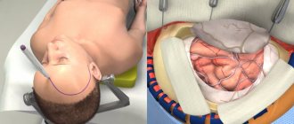
Cerebral angiography is performed in the operating room of the cath lab department. The patient's head is gently fixed to the table with a special tape passing through the frontal area. This is necessary to eliminate head movements during the examination and obtain a high-quality “unblurred” image.
In the department, before transportation to the operating room, premedication is carried out using a mild sedative and an antiallergic drug.
The puncture site in the area of the femoral artery through which the examination is carried out must first be shaved the day before and treated with an antiseptic in the operating room. The examination is performed under local anesthesia. Through a small incision with a diameter of 2.0-3.0 mm, a catheter is inserted into the mouth of the arteries of the brain, through which contrast is injected, and the image is recorded on a computer. If there is pathology of the cerebral vessels, this will be reflected in the resulting image. After completion of the study, a pressure aseptic bandage is applied to the puncture site. For 12 hours after the examination, it is necessary to remain in bed in a position with the lower limb extended on the side where the intervention was performed. After completing the study, it is recommended to drink at least 0.5-0.7 liters of clean water to ensure rapid removal of the contrast from the body.
How is vascular angiography done?
CT examination of blood vessels (angiography) is performed as prescribed by a doctor if there are indications, so the very first thing you need to do is visit your doctor. During this visit, the specialist will explain the purpose of the study and prescribe the necessary laboratory tests, including the mandatory determination of kidney function. If renal function cannot be assessed in advance, patients can have a rapid test done at our center.
If there is a concomitant pathology that may complicate diagnosis, consultation with specialists may be required before prescribing CTA. You should also tell your doctor if you are taking medications on a regular basis, because some medications must be stopped before the test.
Before the manipulation, you need to rest; if you have increased anxiety, you can take mild sedatives. You should have a light snack, but not overeat - the contrast is best tolerated after eating a light meal about a couple of hours before the procedure.
The study is carried out in a specialized equipment room in compliance with all rules of asepsis and antisepsis. The procedure takes place in several stages:
- The doctor or laboratory assistant instructs the patient, tells how the procedure will take place and answers questions;
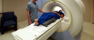
- The patient is placed on the retractable tomograph table in a comfortable position; special cushions are placed under the head and arm to make lying comfortable throughout the entire scan;
- The healthcare worker treats the skin with an antiseptic drug in the area of vascular access, usually the elbow or forearm;
- The X-ray contrast agent is administered using a special device - an injector, which is synchronized with the operation of the tomograph and ensures that the contrast enters the bloodstream at a certain speed;
- The contrast is distributed along the vascular bed and a series of images is simultaneously taken;
- Once completed, the laboratory assistant helps the patient stand up and clarifies how long it will take to prepare the results.
Due to the fact that contrast is injected into the cubital vein, the way carotid artery angiography is done is no different from CT angiography of cerebral or limb vessels. Angiography is usually not accompanied by any unpleasant sensations and no more than 30 minutes pass from the start of the introductory briefing to the end of the scan.
If the examination is planned for a nursing woman, then on the eve of the examination it is necessary to express a sufficient amount of milk, because it is impossible to feed the child after a computed tomography scan for 2 days. You will need to pump at least twice after the procedure.
Features of the study of vessels of various locations
When prescribing examinations of vessels of various locations, there are some nuances that you should be aware of.
Examination of the vessels of the abdominal cavity and small pelvis
Indications for CT angiography of abdominal and pelvic vessels
- Anomalies in the development of blood vessels and organs;
- Neoplasm of vascular origin;
- Arterial hypertension presumably of renal origin;
- Signs of vascular obstruction.
When examining the renal artery, excretory urography is simultaneously performed, through which the functional abilities of the kidneys are assessed.
How to prepare for the test
Before the study, be sure to inform your doctor about the presence of:
- allergies to strawberries, shellfish, seafood or iodine-containing substances;
- history of bleeding of unknown origin;
- an allergic reaction to a contrast agent in the past;
- pregnancy.
Before the study, it is necessary to exclude food and liquid intake for 4-8 hours. Before the examination, you need to remove all jewelry, decorations and costume jewelry to prevent their contours from interfering with the resulting image.
Preparatory actions
The examination lasts about 30 minutes.
No special preparation is required for a diagnostic procedure using a magnetic resonance imaging scanner. If during this diagnostic procedure it is necessary to administer contrast, then the following actions must be performed:
- testing for allergies to iodine-containing drugs;
- performing urine and blood tests;
- taking an ECG;
- consultation with a therapist.
It is advisable to have on hand the results of other diagnostic methods performed previously to examine the brain (if any were performed). Including the results of previous MRA procedures. If for any indication the examination must be carried out under general anesthesia, then you need to notify the anesthesiologist about any allergies to medications. In addition, before anesthesia, a 6-8 hour fast is required.
To ensure that the examination results are as accurate as possible, it is not recommended to consume alcoholic beverages or blood thinning medications a week before the diagnostic procedure.
Why is the research being done?
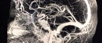
Cerebral angiography is used to detect or exclude pathologies of the blood vessels of the brain, such as:
- abnormal blood vessels (vascular malformation);
- aneurysms;
- narrowing of the arteries of the brain;
- vasculitis;
The study is also used for:
- confirmation of the presence and location of a brain tumor;
- assessment of the head and neck artery before surgery;
- determining the presence of blood clots in the vessels of the brain, which may cause a stroke.
In some cases, this procedure is used to clarify the diagnosis after abnormalities have been identified according to MRI or CT. Contrast that extends beyond the outline of a blood vessel may be a sign of internal bleeding. Narrowing of the arteries may indicate cholesterol deposits, spasms, or hereditary diseases. Abnormally developed blood arteries may be associated with brain tumors, intracranial bleeding, aneurysm (bulging of the artery wall), or arteriovenous malignancy.
Cerebral angiography is also performed to prepare for x-ray surgery to eliminate malformation, aneurysm or thrombosis of cerebral vessels.
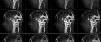
Indications for MR angiography
Doctors prescribe magnetic resonance angiography of blood vessels on a tomograph if the patient exhibits the following symptoms:
- memory impairment;
- noise in ears;
- blurred vision;
- chronic migraine;
- sleep disturbance;
- dizziness,
- disorder of consciousness;
- intracranial pressure;
- weakening of attention;
- headache;
- brain contusion;
- before carrying out operations.
The main advantages of MRI of neck and brain vessels
In addition to the high accuracy of the study, MRI of neck vessels and MRI of cerebral arteries have a whole list of advantages. These include:
- Absolute harmlessness and non-toxicity of the medical device for the health of the patient.
- No contact with surgical or any other medical equipment.
- Maximum ease of assessment for the patient.
- A narrow list of contraindications.
- The ability of a tomograph to detect deviations from the norm and different types of diseases at the initial stage of their development.
- The ability to be tested repeatedly without a long recovery period.
You need to know that examination of the main organ in the human body using resonance equipment is almost always done together with an MRI of the neck and its parts. This technique is required because the filling of blood vessels in the brain is directly dependent on stable blood flow in the arteries below. If the neck was subject to trauma or injury, then the described case may lead to a lack of oxygen supply, which could very well lead to death.
With the help of the ECP, our clients can apply for an appointment with a treating specialist - without leaving the comfort of their home! All you need to do is visit our official website, where you can find a lot of useful information: price lists of clinics, verified addresses of medical organizations and the number of available places for the coming days. Also, with the help of our company, you have the opportunity to get acquainted with truthful and honest reviews of real patients of medical centers and make a choice of clinic based on the assessment. Our managers are always ready to listen to you and give detailed answers to your questions.
Prescriptions for MRI of neck and brain vessels
Often, doctors prescribe an MRI of the brain arteries to patients who suffer from the following symptoms:
- The presence of regular and severe pain in the head area.
- Constant dizziness and frequent loss of consciousness.
- Reduced level of receptors responsible for the sense of smell (vision and hearing).
- The likelihood of having malignant or benign neoplasms.
- The appearance of numbness or difficulties in performing motor functions of the upper extremities.
- Characteristic clicking noises when turning.
- Damage and injury.
- Displacement of intervertebral discs.
If the described conditions are present, the patient can undergo an MRI absolutely free of charge if he has a compulsory medical insurance policy. More detailed information is provided by the treating specialist, because each procedure is designed to solve a specific problem. Contact the ECD for a quick and instant appointment with the best doctors in the capital! Using our official website, you will receive information about current prices, availability and location of clinics near you. In addition, using the services of our search service, our clients receive favorable discounts and promotional offers that help significantly save their diagnostic budget.
Stages of MRI of neck vessels and MRI of cerebral arteries
The procedure is very simple and easy. Let's take a closer look at what will happen during an appointment with a specialist:
- The patient enters the office with a tomograph installed.
- Fits on a mobile couch.
- The doctor can fix the limbs with special fasteners.
- The patient is placed in the scanner corridor.
- The average time spent in the device's chamber is 60 minutes.
The equipment may produce characteristic noises during operation, so in order to get rid of loud sounds, you are allowed to wear earplugs. Also, during the session, the light in the tunnel is always on and there is a two-way connection, through which the patient can contact the doctor if he feels discomfort. If a scan is scheduled with the introduction of a contrast agent, then before immersion in the tomograph, the nurse injects the drug into the body intravenously.
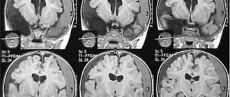
Interpretation of the received report after MRI of the cerebral arteries
The information is processed by a technologist after scanning. Typically, report generation takes up to 60 minutes. In the photographs, the attending physician will be able to see various pathologies, cracks and various modifications. All deviations from the norm will be recorded by the device and displayed on the computer monitor. With the information received, it is worth making an appointment with the doctor with whom you are being treated so that he can prescribe you a timely course of therapy or refer you to a more qualified specialist.
Brief information about the areas studied during MRI of the neck and brain vessels
Probably, few people have thought about the fact that the neck and its entire section, together with the main organ of the central nervous system, play a huge role in the life and functionality of the entire organism. In the first two sections there are centers responsible for regulating the most important systems that allow a person to breathe and move blood through the arteries. In the second case, the organ performs huge and varied parameters, without which it is impossible to exist normally.
To detect neck diseases, doctors often prescribe special testing technologies, such as Doppler or X-ray analysis. But there are situations related to the likelihood of abnormal processes occurring in soft tissues in the patient’s body, then doctors write a referral for an MRI of the neck vessels. Such an examination shows even the smallest and most insignificant changes.