The role of the cerebral cortex
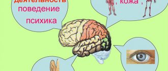
In the posterior central gyrus, behind the central sulcus, there is an area of cutaneous and joint-muscular sensitivity.
Here the signals that arise when touching our body, when it is exposed to cold or heat, or when it is painful are perceived and analyzed. In contrast to this zone, in the anterior central gyrus, in front of the central sulcus, the motor zone is located. It identifies areas that provide movement of the lower extremities, muscles of the trunk, arms, and head. When this area is irritated by electric current, contractions of the corresponding muscle groups occur. Injuries or other damage to the motor cortex lead to paralysis of the body muscles.
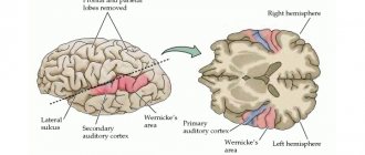
The auditory area is located in the temporal lobe. The impulses arising in the receptors of the cochlea of the inner ear are received here and analyzed. Irritation of areas of the auditory zone causes sensations of sounds, and when they are affected by the disease, hearing is lost.
The visual zone is located in the cortex of the occipital lobes of the hemispheres. When it is irritated by electric current during brain surgery, a person experiences sensations of flashes of light and darkness. When it is affected by any disease, vision deteriorates and is lost.
Near the lateral sulcus there is a taste zone where taste sensations are analyzed and formed based on signals arising in the receptors of the tongue. The olfactory zone is located in the so-called olfactory brain, at the base of the hemispheres. When these areas are irritated during surgery or during inflammation, people smell or taste something.
There is no purely speech zone. It is represented in the cortex of the temporal lobe, the inferior frontal gyrus on the left, and parts of the parietal lobe. Their diseases are accompanied by speech disorders.
The cerebral hemispheres occupy about 80% of the volume of the cranium, and consist of white matter, the basis of which consists of long myelinated axons of neurons. The outside of the hemisphere is covered by gray matter or the cerebral cortex, consisting of neurons, unmyelinated fibers and glial cells, which are also contained in the thickness of the sections of this organ.
The surface of the hemispheres is conventionally divided into several zones, the functionality of which is to control the body at the level of reflexes and instincts. It also contains the centers of higher mental activity of a person, ensuring consciousness, assimilation of received information, allowing adaptation in the environment, and through it, at the subconscious level, through the hypothalamus, the autonomic nervous system (ANS) is controlled, which controls the organs of circulation, respiration, digestion, excretion , reproduction, and metabolism.
In order to understand what the cerebral cortex is and how its work is carried out, it is necessary to study the structure at the cellular level.
Functions
The cortex occupies most of the cerebral hemispheres, and its thickness is not uniform over the entire surface. This feature is due to the large number of connecting channels with the central nervous system (CNS), which ensure the functional organization of the cerebral cortex.
This part of the brain begins to form during fetal development and is improved throughout life, by receiving and processing signals coming from the environment. Thus, it is responsible for performing the following brain functions:
- connects the organs and systems of the body with each other and the environment, and also ensures an adequate response to changes;
- processes incoming information from motor centers using mental and cognitive processes;
- consciousness and thinking are formed in it, and intellectual work is also realized;
- controls speech centers and processes that characterize the psycho-emotional state of a person.
In this case, data is received, processed, and stored thanks to a significant number of impulses passing through and generated in neurons connected by long processes or axons. The level of cell activity can be determined by the physiological and mental state of the body and described using amplitude and frequency indicators, since the nature of these signals is similar to electrical impulses, and their density depends on the area in which the psychological process occurs.
It is still unclear how the frontal part of the cerebral cortex affects the functioning of the body, but it is known that it is little susceptible to processes occurring in the external environment, therefore all experiments with the influence of electrical impulses on this part of the brain do not find a clear response in the structures . However, it is noted that people whose frontal part is damaged experience problems communicating with other individuals, cannot realize themselves in any work activity, and they are also indifferent to their appearance and outside opinions. Sometimes there are other violations in the performance of the functions of this body:
- lack of concentration on everyday objects;
- manifestation of creative dysfunction;
- disorders of a person’s psycho-emotional state.
The surface of the cerebral cortex is divided into 4 zones, outlined by the most distinct and significant convolutions. Each part controls the basic functions of the cerebral cortex:
- parietal zone - responsible for active sensitivity and musical perception;
- the primary visual area is located in the occipital part;
- the temporal or temporal is responsible for speech centers and the perception of sounds coming from the external environment, in addition, it is involved in the formation of emotional manifestations, such as joy, anger, pleasure and fear;
- The frontal zone controls motor and mental activity, and also controls speech motor skills.
The premotor cortex helps us quickly change our behavior.
Long-term learning—such as learning a language, math, or new movements—likely requires cortical plasticity: changes in the structure of the brain, but we often require much more rapid changes, sometimes even after single errors. Professor Lee Miller's team from Northwestern University's Limb Movement Control Laboratory, in a paper published in one of the top neurojournals, Neuron, proposed a neural mechanism for the rapid development of a new motor skill without changing the functional connectivity within or between cortical areas. Lee Miller will be a guest speaker at the BCISamara-2019 conference, which will take place in October, and our portal will definitely talk with the scientist about new developments, but for now let’s talk about his article.
Graphic abstract of the work. Miller et al, Neuron, 2018
As the authors write, one of the most fundamental questions in neuroscience is how the coordinated activity of interconnected neurons produces behavior and how these neurons quickly and flexibly change their “output” during learning to adapt behavior to changing conditions. There is evidence that learning, lasting from several days to several weeks, is associated with persistent synaptic changes in the cerebral cortex (Kleim et al., 2004). However, behavior can also be adapted much more quickly: motor errors can be corrected on a trial-a-trial basis, Thoroughman and Shadmehr, 2000, and sensory associations can be learned even after a single exposure (Bailey and Chen , 1988).
Another fact is that there are limits to the types of rapid motor learning. In a brain-computer interface experiment, monkeys had difficulty learning to control a computer cursor when a new control decoder required them to change the natural conjugate variability between neurons whose activity was sensed by the electrodes (Sadtler et al, 2014). Such covariance (i.e., simultaneously changing activity) structures appear to be associated with synaptic connectivity (pardon the tautology), which does not easily change on time scales of seconds to minutes (Okun et al., 2015). So all this data suggests that when we change our behavior very quickly, changes in the connectome cannot be the main mechanism for this.
What did the authors of the work do? In their experiment, two black macaque monkeys performed a “standard” center-to-edge test: a cursor appeared on a screen at a central point, and then the monkey had to use a robotic arm to guide it to one of eight evenly distributed targets on the edge that appeared in a circle. after the beep. A second was given to reach the target, and the cursor had to be held for another half a second. However, after the first test, special servos applied twisting forces to the manipulator, and the animal was required to adjust its movement in order to complete the task. And the monkeys coped with the task quite quickly.
Cursor movement trajectories during different tasks
At the same time, the authors recorded the activity of two areas of the brain - the dorsal premotor cortex (PMd), in which movement planning occurs, and the main motor cortex (M1), which, in fact, carries out movement.
The researchers put forward four hypotheses about where learning occurs (see figure below), based on the signal path: from the visual cortex to PMd, then to M1 and then to the movement itself. The first hypothesis suggested learning before the premotor cortex, then at the level of “zero planning” in the premotor cortex, then at the level of the PMd-M1 interaction map and at the level of local connectivity. Each hypothesis had its own calculation model.
Four hypotheses put forward by the authors and associated models
Analysis of the data obtained during short-term adaptation to two different tests, which was achieved in one day, showed very interesting results: the authors did not see any changes in either connectivity within each region or in neural associated variability (covariance) between the PMd and PMd areas. M1. It turned out that adaptation to changing conditions occurred in a special “output-NULL” subspace of the premotor cortex, which gradually regulated output to the underlying region of the chain with complete preservation of connectivity.
Text: Alexey Paevsky
Perich, M. G., Gallego, J. A., & Miller, L. E. (2018). A Neural Population Mechanism for Rapid Learning. Neuron.doi:10.1016/j.neuron.2018.09.030
Literature:
Kleim, J. A., Hogg, T. M., VandenBerg, P. M., Cooper, N. R., Bruneau, R., and Remple, M. (2004). Cortical synaptogenesis and motor map reorganization occur during late, but not early, phase of motor skill learning. J. Neurosci. 24, 628–633.
Thoroughman, K. A., and Shadmehr, R. (2000). Learning of action through adaptive combination of motor primitives. Nature 407, 742–747.
Bailey, C. H., & Chen, M. (1988). Morphological basis of short-term habituation in Aplysia. J. Neurosci. 8, 2452–2459.
Sadtler, P. T., Quick, K. M., Golub, M. D., Chase, S. M., Ryu, S. I., Tyler-Kabara, E. C., Yu, B. M., and Batista, A. P. (2014). Neural constraints on learning. Nature 512, 423–426.
Okun, M., Steinmetz, N., Cossell, L., Iacaruso, M.F., Ko, H., Bartho´, P., Moore, T., Hofer, S.B., Mrsic-Flogel, T.D., Carandini, M., and Harris, K. D. (2015). Diverse coupling of neurons to populations in sensory cortex. Nature 521, 511–515.
First and second signaling systems
The role of the cerebral cortex in improving the first signaling system and developing the second is invaluable. These concepts were developed by I.P. Pavlov. The signaling system as a whole is understood as the entire set of processes of the nervous system that carry out perception, processing of information and the response of the body. It connects the body with the outside world.
The first signaling system determines the perception of sensory-specific images through the senses. It is the basis for the formation of conditioned reflexes. This system exists in both animals and humans.
In the higher nervous activity of man, a superstructure has developed in the form of a second signaling system. It is peculiar only to humans and is manifested by verbal communication, speech, and concepts. With the advent of this signaling system, abstract thinking and generalization of countless signals from the first signaling system became possible. According to I.P. Pavlov, words turned into “signals of signals.”
The emergence of the second signaling system became possible thanks to complex labor relationships between people, since this system is a means of communication and collective work. Verbal communication does not develop outside of society. The second signaling system gave rise to abstract (abstract) thinking, writing, reading, counting.
Words are perceived by animals, but completely differently from people. They perceive them as sounds, and not their semantic meaning, like humans. Therefore, animals do not have a second signaling system. Both human signaling systems are interconnected. They organize human behavior in the broad sense of the word. Moreover, the second changed the first signaling system, since the reactions of the first began to largely depend on the social environment. A person has become able to control his unconditioned reflexes, instincts, i.e. first signaling system.
Functions of the cerebral cortex
Familiarity with the most important physiological functions of the cerebral cortex indicates its extraordinary importance in life. The cortex, together with the subcortical formations closest to it, is a department of the central nervous system of animals and humans.
The functions of the cerebral cortex are the implementation of complex reflex reactions that form the basis of higher nervous activity (behavior) of a person. It is no coincidence that it received the greatest development from him. The exclusive properties of the cortex are consciousness (thinking, memory), a second signaling system (speech), and a high organization of work and life in general.
Features of the structure of the cerebral cortex
The anatomical structure of the cerebral cortex determines its characteristics and allows it to perform the functions assigned to it. The cerebral cortex has the following number of distinctive features:
- neurons in its thickness are arranged in layers;
- nerve centers are located in a specific place and are responsible for the activity of a certain part of the body;
- the level of activity of the cortex depends on the influence of its subcortical structures;
- it has connections with all underlying structures of the central nervous system;
- the presence of fields of different cellular structure, which is confirmed by histological examination, while each field is responsible for performing some higher nervous activity;
- the presence of specialized associative areas makes it possible to establish a cause-and-effect relationship between external stimuli and the body’s response to them;
- the ability to replace damaged areas with nearby structures;
- This part of the brain is capable of storing traces of neuronal excitation.
The large hemispheres of the brain consist mainly of long axons, and also contain in their thickness clusters of neurons that form the largest nuclei of the base, which are part of the extrapyramidal system.
As already mentioned, the formation of the cerebral cortex occurs during intrauterine development, and at first the cortex consists of the lower layer of cells, and already at 6 months of the child all structures and fields are formed in it. The final formation of neurons occurs by the age of 7, and the growth of their bodies is completed at 18 years.
An interesting fact is that the thickness of the cortex is not uniform over its entire length and includes a different number of layers: for example, in the area of the central gyrus it reaches its maximum size and has all 6 layers, and sections of the old and ancient cortex have 2 and 3 layers. x layer structure, respectively.
The neurons of this part of the brain are programmed to restore the damaged area through synoptic contacts, so each of the cells actively tries to restore damaged connections, which ensures the plasticity of neural cortical networks. For example, when the cerebellum is removed or dysfunctional, the neurons connecting it with the terminal section begin to grow into the cerebral cortex.
analyzer, closure apparatus of conditioned reflex connections and working device. Weakness of the closure function of the cortex and trace manifestations can be observed in children with severe mental retardation, when the formed conditioned connections between neurons are fragile and unreliable, which entails learning difficulties.
The cerebral cortex includes 11 areas consisting of 53 fields, each of which is assigned its own number in neurophysiology.
Regions and zones of the cortex
The cortex is a relatively young part of the central nervous system, developing from the terminal part of the brain. The evolutionary development of this organ occurred in stages, so it is usually divided into 4 types:
- The archicortex or ancient cortex, due to the atrophy of the sense of smell, has turned into the hippocampal formation and consists of the hippocampus and its associated structures. With its help, behavior, feelings and memory are regulated.
- The paleocortex, or old cortex, makes up the bulk of the olfactory area.
- The neocortex or new cortex has a layer thickness of about 3-4 mm. It is a functional part and performs higher nervous activity: it processes sensory information, gives motor commands, and also forms conscious thinking and human speech.
- The mesocortex is an intermediate version of the first 3 types of cortex.
The cerebral cortex has a complex anatomical structure and includes sensory cells, motor neurons and internerons, which have the ability to stop the signal and be excited depending on the received data. The organization of this part of the brain is built according to the columnar principle, in which the columns are divided into micromodules that have a homogeneous structure.
The basis of the micromodule system is made up of stellate cells and their axons, while all neurons react equally to the incoming afferent impulse and also send an efferent signal synchronously in response.
The formation of conditioned reflexes that ensure the full functioning of the body occurs due to the connection of the brain with neurons located in various parts of the body, and the cortex ensures synchronization of mental activity with the motor skills of organs and the area responsible for analyzing incoming signals.
Signal transmission in the horizontal direction occurs through transverse fibers located in the thickness of the cortex, and transmit the impulse from one column to another. Based on the principle of horizontal orientation, the cerebral cortex can be divided into the following areas:
- associative;
- sensory (sensitive);
- motor.
When studying these zones, various methods of influencing the neurons included in its composition were used: chemical and physical stimulation, partial removal of areas, as well as the development of conditioned reflexes and registration of biocurrents.
The associative zone connects incoming sensory information with previously acquired knowledge. After processing, it generates a signal and transmits it to the motor zone. In this way, it is involved in remembering, thinking, and learning new skills. Association areas of the cerebral cortex are located in proximity to the corresponding sensory area.
The sensitive or sensory area occupies 20% of the cerebral cortex. It also consists of several components:
- somatosensory, located in the parietal zone, is responsible for tactile and autonomic sensitivity;
- visual;
- auditory;
- taste;
- olfactory.
Impulses from the limbs and organs of touch on the left side of the body enter along afferent pathways to the opposite lobe of the cerebral hemispheres for subsequent processing.
Neurons of the motor zone are excited by impulses received from muscle cells and are located in the central gyrus of the frontal lobe. The mechanism of data receipt is similar to the mechanism of the sensory zone, since the motor pathways form an overlap in the medulla oblongata and follow to the opposite motor zone.
The cerebral cortex is formed by several layers of neurons. A characteristic feature of this part of the brain is a large number of wrinkles or convolutions, due to which its area is many times greater than the surface area of the hemispheres.
Cortical architectonic fields determine the functional structure of areas of the cerebral cortex. All of them are different in morphological characteristics and regulate different functions. In this way, 52 different fields are identified, located in certain areas. According to Brodmann, this division looks like this:
- The central sulcus separates the frontal lobe from the parietal region; the precentral gyrus lies in front of it, and the posterior central gyrus lies behind it.
- The lateral groove separates the parietal zone from the occipital zone. If you separate its side edges, you can see a hole inside, in the center of which there is an island.
- The parieto-occipital sulcus separates the parietal lobe from the occipital lobe.
The core of the motor analyzer is located in the precentral gyrus, while the upper parts of the anterior central gyrus belong to the muscles of the lower limb, and the lower parts belong to the muscles of the oral cavity, pharynx and larynx.
The right-sided gyrus forms a connection with the motor system of the left half of the body, the left-sided one - with the right side.
The posterior central gyrus of the 1st lobe of the hemisphere contains the core of the tactile sensation analyzer and is also connected to the opposite part of the body.
Cell layers
The cerebral cortex carries out its functions through neurons located in its thickness. Moreover, the number of layers of these cells may differ depending on the area, the dimensions of which also vary in size and topography. Experts distinguish the following layers of the cerebral cortex:
- The surface molecular layer is formed mainly from dendrites, with a small inclusion of neurons, the processes of which do not leave the boundaries of the layer.
- The external granular consists of pyramidal and stellate neurons, the processes of which connect it with the next layer.
- The pyramidal layer is formed by pyramidal neurons, the axons of which are directed downward, where they break off or form associative fibers, and their dendrites connect this layer with the previous one.
- The internal granular layer is formed by stellate and small pyramidal neurons, the dendrites of which extend into the pyramidal layer, and its long fibers extend into the upper layers or descend down into the white matter of the brain.
- The ganglion consists of large pyramidal neurocytes, their axons extend beyond the cortex and connect various structures and sections of the central nervous system with each other.
The multiform layer is formed by all types of neurons, and their dendrites are oriented into the molecular layer, and axons penetrate the previous layers or extend beyond the cortex and form associative fibers that form a connection between gray matter cells and the rest of the functional centers of the brain.
ZONES OF THE LARGER HEMISPHERES
Localization of functions in the cerebral hemispheres. The cerebral cortex is divided into main zones, consisting of several cortical fields. Each of these zones performs a certain general function, and its constituent fields are specialized in the implementation of individual elements of this function. However, thanks to the conductive pathways, several zones of the cerebral hemispheres, certain subcortical centers, nuclei of the brain stem and segments of the spinal cord are involved in the implementation of individual links of higher and lower nervous activity.
With the subtle and precise specialization of certain groups of neurons, the brain and spinal cord function as a single unit. Mental functions of the brain are also not limited to individual areas of the cortex, but are the result of the joint activity of large areas of the cerebral hemispheres and subcortical centers.
Rice. 123. Individual changes in the main fields of the neocortex of the cerebral hemispheres in three adults (A, B, C). Numbers are fields according to Brodmann. The motor zone (field 4) is located in the anterior central gyrus along the central sulcus. In the upper quarter of the zone there are motor centers for the leg muscles.
At the top are the neurons that innervate the muscles of the toes, and at the bottom - the thighs and torso. The middle two quarters are occupied by the centers for the arms, above - the center of the scapula muscles, and below - the muscles of the fingers. And finally, in the lower quarter of the anterior central gyrus there are centers of the muscles of the face and speech apparatus. As a result of the historical development of the human brain in the process of work and speech, a particularly large place is occupied by groups of neurons that cause contraction of the muscles of the hand, mainly the thumb, and the muscles of the face, tongue and larynx. They receive centripetal fibers from proprioceptors, entering along the dorsal roots into the spinal cord, where they rise as part of the posterior column of the same side to the nuclei of the tender and wedge-shaped fasciculi of the medulla oblongata. From these nuclei the fibers of the second neurons emerge, forming a medial loop and, after decussation, reaching the nuclei of the optic thalamus of the opposite side. From here, most of the centripetal fibers of the third neurons reach the posterior central gyrus and then enter the anterior central gyrus, and a smaller part enters it directly. Thus, the anterior central gyrus is connected to the posterior central gyrus through fibers passing through the pathways of the cortex. From the motor zone emerge centrifugal motor fibers of pyramidal neurons, which make up the pyramidal pathways; they reach the neurons of the anterior horns of the spinal cord. The motor area causes coordinated movements of skeletal muscles, mainly on the opposite side of the body. It functions together with the subcortical centers - the striatal bodies, as well as the Lewis body, the red nucleus and the substantia nigra. When certain areas of the anterior central gyrus are damaged, voluntary movements of individual muscle groups are impaired. Incomplete damage to the zone causes movement disorders—paresis, and its complete destruction—paralysis. The area of musculocutaneous sensitivity (fields 1, 2, 3, 43 and partially 5 and 7) is located in the posterior central gyrus along the posterior central sulcus. In this zone, the granular layers of the cortex are especially strongly developed, to which centripetal fibers from skin receptors approach, running as part of the same pathways as fibers from proprioceptors. The location of the perceptive groups of neurons is the same as in the motor zone. The largest surface area is occupied by neurons that receive impulses from the receptors of the hand, face, tongue and larynx. Field 7 is more associated with hand sensitivity than other fields. The zone of musculocutaneous sensitivity is not completely delimited from the motor zone, since in fields 3, 4 and 5 there is a combination of granular neurons with giant pyramidal neurons. The motor zone contains approximately 80% of motor neurons, and the musculocutaneous sensitivity zone contains 20%. Each hemisphere receives impulses mainly from receptors on the opposite side of the body, but also from receptors on the same side. Centripetal impulses enter this zone mainly from the lateral and semilunar nuclei of the thalamus optic. With lesions of certain areas of the posterior central gyrus, sensitivity in certain areas of the skin is impaired. The loss of the ability to recognize objects by touch is referred to as tactile agnosia. When the functions of the zone are impaired, disorders of touch, pain and temperature sensations of the skin and muscle-joint sensitivity are observed. Incomplete damage to the zone causes a decrease in reception - hyposthesia, and complete damage - its loss - anesthesia. The frontal zone (fields 6, 5, 9, 10, 11, 44, 45, 46, 47) is located in the frontal lobe in front of the motor lobe. It is divided into premotor and speech motor. The premotor zone (fields 6, 8, 9, 10, 11) regulates the tone of skeletal muscles and coordinated movements of the body, orienting it in space. Field 46 is functionally connected with field 10, which is involved in the execution of motor conditioned reflexes. Centripetal impulses from the internal organs enter the premotor zone and a significant part of the centrifugal vegetative fibers emanate from it. Therefore, damage to the premotor zone causes impaired coordination of movements - ataxia and dysfunction of the cardiovascular, respiratory, digestive and other systems of internal organs. The visual zone (fields 17, 18, 19) is located on the inner surface of the occipital lobe on both sides of the calcarine groove. In humans, it occupies 12% of the total surface of the cortex. Area 17 is located at the occipital pole; it is surrounded by area 18, which surrounds area 19, which borders the posterior limbic region, superior and inferior parietal regions. In field 17, the central field of the visual zone, there are 16 times more neurons than in the central field of the auditory zone (field 41), and 10 times more neurons than in the central field of the motor zone (field 4). This indicates the leading importance of vision in the historical and individual development of man. From the retina, 900 thousand - 1 million centripetal fibers of the optic nerves reach the external geniculate body, in which individual parts of the retina are accurately projected. The centripetal fibers of the neurons of the lateral geniculate body are directed to the visual zone, mainly to the main visual field 17. Other intermediate visual centers involved in the transmission of not visual impulses, but oculomotor ones, are the cushion of the visual thalamus and the anterior tubercles of the quadrigeminal. Before entering the external geniculate body, the fibers of the optic nerve intersect. Thanks to this decussation, the visual pathway heading to the visual zone of each hemisphere contains 50% of the fibers on its side and 50% of the fibers on the opposite side. The visual zone of the left hemisphere receives visual impulses from the left halves of the retinas of both eyes, and the zone of the right hemisphere receives from the right halves of the retinas of both eyes. Therefore, the destruction of one of the visual zones causes blindness in the same halves of the retinas in both eyes - hemianopsia. In the optic nerves, in addition to centripetal fibers, there are also somewhat thicker centrifugal fibers to the muscles of the iris and centrifugal thin sympathetic fibers from the neurons of the subcortical centers. A small part of the centripetal fibers of the optic nerve is not interrupted in the subcortical formations, but is directly sent to the cerebellum and visual areas of the cerebral hemispheres. The destruction of both fields 17 causes complete cortical blindness, the destruction of field 18 leads to loss of visual memory while maintaining vision, which is designated as visual agnosia, and the destruction of field 19 leads to loss of orientation in an unusual environment. The auditory zone (fields 41, 42, 21, 22, 20, 37) is located on the surface of the temporal lobe, mainly the anterior transverse temporal gyrus and the superior temporal gyrus. Area 41, located in the superior temporal gyrus and in the anterior part of the transverse gyrus, is a projection of the organ of Corti of the cochlea. From the organ of Corti, centripetal impulses pass through the spiral ganglion along the cochlear nerve, consisting of approximately 30 thousand fibers. This node contains the first bipolar neurons of the auditory pathway. Next, the fibers of the first neurons transmit auditory impulses to the nuclei of the auditory nerve in the medulla oblongata, where the second neurons are located. The fibers of the auditory nerve nuclei communicate with the nuclei of the facial nerve in the medulla oblongata and the oculomotor nerve in the anterior tubercles of the midbrain. Therefore, when strong sounds occur, the muscles of the face, eyelids, and auricle reflexively contract and eye movements are caused. Most of the fibers of the auditory nerve nuclei decussate in the pons, and the smaller part passes on its side. Then the fibers of the auditory pathway enter the lateral lemniscus, which ends in the posterior tuberosity of the quadrigeminal and in the internal geniculate body, where the third neurons are located - their fibers conduct centripetal impulses to the auditory zone. There are also direct pathways connecting the nuclei of the auditory nerves with the cerebellum and auditory area. Most of the direct cerebellar tracts are formed by the vestibular nerve, and a smaller part by the cochlear nerve, which together make up the common trunk of the auditory nerve. The vestibular apparatus is also projected in the auditory zone. Destruction of field 41 on one side causes deafness on the opposite side and weakened hearing on one’s side, and destruction of field 41 on both sides leads to complete cortical deafness. Destruction of field 22 in the anterior third of the superior temporal gyrus leads to musical deafness - the perception of the intensity of tone, timbre and rhythm of sounds is lost - auditory agnosia. Destruction of areas 21 and 20 in the middle and inferior temporal gyri causes ataxia, a disorder of balance and coordination of movements. The speech-auditory center is also located in the auditory zone. Olfactory and gustatory zones. The olfactory zone is located in the ancient cortex, which receives centripetal impulses from the olfactory cells. In addition to the olfactory function, it also performs a gustatory function and is involved in the activities of the digestive, excretory and reproductive systems. Previously, it was believed that the hippocampus has an olfactory function. It is currently believed that, together with the limbic system, the hypothalamic region of the diencephalon and pituitary gland, the midbrain and medulla oblongata, and especially the reticular formation, the hippocampus is involved in general motor reactions and autonomic reflexes during emotions. The taste zone itself is probably located in area 43, which is located in the lower part of the posterior central gyrus.
The limbic gyrus (posterior area 23 and anterior area 24) and the insular cortex (areas 13 and 14) are involved in higher nervous activity. All zones of the cortex are not isolated, but are interconnected by conducting pathways. Speech centers (fields 44, 45, 46, 39, 40, 42, 22,37). The motor speech center is located in the lower part of the anterior central gyrus in area 44. In most right-handed people, the area of area 44 in the left hemisphere is larger than in the right hemisphere. Field 44 causes the complex contractions of the speech muscles necessary to pronounce words. When this field is destroyed, a person cannot speak, but can produce the simplest contractions of the speech muscles - screaming and singing. This is a motor aphasia, which in some cases manifests itself in the absence of contractions of the muscles of the tongue and other speech muscles. Since in these cases the auditory center of speech is not damaged, the understanding of the speech of others is preserved. When field 44 is damaged, not only oral speech is often impaired, but also internal speech or the ability to formulate thoughts in words without pronouncing them, based on accumulated sound images that have a certain semantic content. At the same time, reading to oneself is difficult, the ability to write freely and under dictation is impaired, but copying letters when writing is preserved. In right-handed people, motor aphasia is observed when the left hemisphere is damaged, and in left-handed people, when the right hemisphere is damaged.
Rice. 129. Localization of speech centers: 1 - motor, 2 - auditory, 3 - visual. In front of field 44 there is field 45, which regulates the construction of grammatically correct combinations of words and singing. When this field is damaged due to loss of memory for pronunciation techniques, singing becomes upset. Facial expressions and gestures, which give speech its expressiveness, are carried out thanks to impulses coming from field 46 to fields 44 and 45, to the fields of the premotor area and to the subcortical centers. The auditory, or sensory, center of speech is located in the posterior part of the left superior temporal gyrus in area 42, which carries out the understanding of a word when hearing it. If the field is destroyed, the ability to understand the meaning of words is lost, but their perception as sounds is preserved - sensory aphasia, or speech deafness. At the same time, due to a lack of understanding of one’s own speech, excessive talkativeness is sometimes observed - logorrhea, or verbal diarrhea. In the rear part of field 22, connections between sound images of words and all perceptual zones in which ideas about objects and phenomena arise are recorded. Therefore, damage to this field also causes sensory aphasia. Fields 39 and 40, located in the parietal lobe next to field 22, carry out understanding of the meaning of combinations of words or phrases. Therefore, their defeat leads to a speech disorder called semantic aphasia. If field 39 is affected, due to the loss of the ability to recognize letters and numbers and understand the meaning of visible written images of words and numbers, the ability to read aloud, write and count is lost. A lesion of field 40 causes loss of the ability to write, since there is no orientation of movements in space and their sequence is disrupted. This lack of ability to produce systematic, purposeful movements (apraxia) does not exclude the ability to correctly perform individual hand movements not related to writing. Consequently, the process of writing in right-handers is carried out by the temporal, inferior parietal and inferior frontal regions of the left hemisphere. When field 37 is damaged, memory loss for words is caused - amnestic aphasia. Thus, the cerebral hemispheres as a whole are involved in the implementation of the speech function, but a special role is played by individual fields of the cortex. In right-handed people, as a result of the preferential development of the functions of the right hand and the right half of the body, the most complex mental functions of the left hemisphere of the brain are especially developed.
Related materials:
Brain phylogeny
The structure of the cerebral hemispheres
Blood supply to the brain
Cerebrospinal fluid