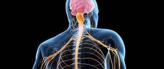
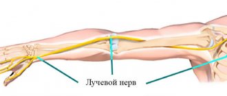
General information
All nerves include a huge number of fibers that are surrounded by connective tissue.
The fiber itself consists of a special process - an axon, which is covered with an ectodermal membrane. They are collected in certain bundles, thus creating tracts in the brain, spinal cord and peripheral nervous system. It is worth noting that the processes are either pulpy or non-pulpous (for example, the nerve endings of the skin). They all differ in the nature of their coverage, as well as in their belonging to a particular nervous system. They are divided into two main groups: those covered with myelin and those without it. In general, the first group predominates in the human body.
Unmyelinated fibers are located in the sympathetic autonomic nervous system.
Let's take a closer look at the structure of the myelin fiber.
Its main components are:
- a cylinder that runs along the central axis;
- directly the sheath of myelin nature, which covers the axial cylinder;
- Schwann membrane.
The main components of the cylinder are neurofibrils. Due to the presence of myelin components in the sheath, the impulse reaction passes through the nerve fiber faster. It is important to note that the cylinder is not completely covered with a shell; there are separate areas of Ranvier on it. It is at this point that the cylinder comes into contact with the Schwann shell. Its cells are of ectodermic origin. It is important to note that these types of membranes are present only in the peripheral nervous system. In the case of complete absence of a shell, the cylinders are called “naked”.
Classification of nerve fibers
All of them are classified into three main groups:
- by impulse transmission speed;
- by transverse diameter;
- by the duration of the action potential.
It is worth noting that the larger their diameter and myelination, the faster the impulse travels through it. There are three varieties:
- Group A. All of them are covered with a membrane, their action potential is the lowest. In turn, they are divided into 4 subspecies: alpha, beta, gamma and delta. These include all receptors of the somatic nervous system, sensory fibers of the skin, thermoregulation, and proprioceptors. All these processes are responsible for human tactile senses.
- Group B. The processes are not completely covered with a myelin sheath; these include components of the autonomic nervous system. These include pain mediators and indicators of the functioning of internal organs.
- Group C. The membrane is completely absent, the speed of impulse conduction is low. These include ANS cells, as well as somatic pain and temperature cells.
Myelin contains phospholipids, cholesterol, basic protein and other useful components.
Thus, the membrane is a unique membrane, thanks to which the nervous system becomes able to quickly transmit impulses. All nerve processes are divided into two main groups: afferent (conduct impulses from tissues to the central nervous system) and efferent (act vice versa).
Nerve fibers and their endings
Nerve fibers are extensions of neurons. Histology determines their classification. Depending on the presence or absence of the myelin layer in oligodendrocytes (lemmocytes) surrounding the fibers, they are divided into:
- myelin;
- unmyelinated.
The myelin sheath is formed by Schwann cells (for peripheral nerves) or oligodendrocytes (for the central nervous system), which are wound around the process of the nerve cell. The areas where there is a border between two adjacent lemmocytes and no myelin layer are called nodes of Ranvier.
The sheath of unmyelinated fibers is also formed by lemmocytes, but they lack a myelin layer.
Depending on the structure, speed of excitation and other functional abilities, the fibers are divided into groups:
A. Represented by myelin fibers. However, this group is graded depending on the diameter of the nerve fiber, and accordingly, the speed of impulse conduction, into four subclasses: α, β, γ, δ. Their characteristics are presented in the table.
| Fiber | Diameter, microns | Excitation propagation speed, m/s | Functions |
| α-fibers | 12-22 | 70-120 | They conduct impulses from the motor zones of the central nervous system to the striated skeletal muscles and from proprioceptors to the nerve centers. |
| β-fibers | 8-13 | 40-70 | They are predominantly represented by sensitive conductors that transmit impulses from various receptors to the structures of the central nervous system. |
| γ-fibers | 4-8 | 15-40 | They transmit excitation from the cells of the spinal cord to the striated muscle fibers. |
| δ-fibers | 1-4 | 5-15 | They are represented mainly by sensitive elements that conduct impulses from tactile, temperature receptors and part of the nociceptors to the structures of the central nervous system |
- B. This type of fiber includes myelinated prenodal autonomic nerves. Their diameter ranges from 1 to 3 microns. The speed of impulse conduction ranges from 3 to 18 m/s.
- C. C fibers are unmyelinated. They have no more than 2 microns in diameter. The excitation propagation speed is also small – from 0.5 to 3 m/s. The vast majority of type C fibers are represented by postnodal sympathetic conductors and nerve fibers that conduct impulses from nociceptors, some thermoreceptors and baroreceptors.
Nerve fibers end in nerve endings. There are three options:
- Effectors (or effectors) are represented by the motor endings of motor neurons;
- Sensory (or receptors) are the terminal parts of the dendrites of afferent neurons;
- Synaptic (places of contact between two neurons), providing interneuron connections.
Important Lecture No. 16. ontogeny of multicellular animals that reproduce sexually
Nervous tissue is a complex system of interconnected elements that have certain properties. Histology, anatomical structure and functions of nervous tissue are closely interrelated. It is the cellular composition that determines its characteristic physiological features. Due to the combined complex interaction of individual structures, the possibility of coordinated work of the whole organism arises.
Rate this article:
- 4.26
Total votes: 149
Myelination of nerve fibers and demyelination
As described above, each process contains an axial cylinder, which is covered with a special myelin sheath. This process is called myelination. Due to the presence of sites of Ranvier, impulse is transferred from one to another. This is what ensures high transmission of excitation along the process towards the nerve.
Areas that are covered with the myelin sheath (pulpous) take part in the metabolic processes of muscle muscles; they have a high level of resistance to the effects of currents of a bioelectric nature.
In Ranvier gaps, impulse reactions are generated and accelerated. Their functions in the autonomic nervous system are assumed by oligodendroglia.
Non-pulpless tissues do not have a myelin sheath in their composition, therefore they are characterized by low insulating ability. In this case, the speed of impulse transmission is significantly reduced due to the fact that when it is transmitted from neurons, it comes into direct contact with the environment. The transmission of impulses for them requires large energy expenditures from the body (unlike pulp-type fibers).
From these two groups of fibers, a large nerve is subsequently formed, which has small bundles at its end. They differ in their main functions. It is important to note that these areas are the final ones in the formation of the interneuronal system.
When the functioning of the myelin sheath is disrupted or damaged, the process of demyelination occurs. This pathology can be caused by the presence of an inflammatory or infectious process in the body, metabolic disorders, ischemic processes in tissues, or the spread of neuroinfection. As a result of this process, the myelin in the sheath is replaced by fibrous plaques. The conductivity of impulse reactions in this case is significantly reduced.
There are two types of dimyelination:
- myelinopathy, which is the result of autoimmune disorders in the body;
- myelinoclasty appears with a genetic predisposition to the process of demyelination.
This process is considered quite dangerous, as it causes serious disturbances in the functioning of the central nervous system. It is very important to diagnose the disease at an early stage in order to provide effective therapy.
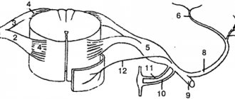
Nerve fibers. Pulpy, pulpless. Structure, function, regeneration, age-related changes.
They consist of a process of a nerve cell covered with a membrane, which is formed by oligodendrocytes. The process of a nerve cell (axon or dendrite) within a nerve fiber is called the axial cylinder.
| Unmyelinated nerve fibers | Myelinated nerve fibers |
| 1. Usually - several axial cylinders located along the periphery of the fiber. | 1. One axial cylinder is located at the center of the fiber. |
| 2. Axial cylinders are, as a rule, axons of efferent neurons of the autonomic nervous system. | 2. The axial cylinder can be either an axon or a dendrite of a neurocyte |
| 3. Oligodendrocyte nuclei are located in the center of the fibers. | 3. The nuclei and cytoplasm of lemmocytes are pushed to the periphery of the fiber. |
| 4. The mesaxons of the axial cylinders are short. | 4. Mesaxon twists repeatedly around the axial cylinder, forming a myelin layer. |
| 5. Na+ channels are located along the entire length of the axial cylinder. Age-related features: 1. Hypertrophy of Schwann cells. 2.Thinning of individual fibers. Regeneration of nerve fibers: (physiological only) | 5. Na+ channels - only at the node of Ranvier. Age-related features: 1.Thinning of the myelin layer. 2. The appearance of muff-like thickenings. 3. Focal demyelination occurs. 4. The number of myelin fibers decreases. After cutting the nerve fiber, degeneration of the axial cylinder occurs distal to the site of damage. Lemmocytes and macrophages phagocytose decay products, cleanse the site of damage, and then multiply and form strands - Bünger's bands. An influx of axoplasm forms on the proximal segment of the axial cylinder—a growth flask is formed (as in embryogenesis). The axial cylinder grows along a path of lemmocytes at a rate of 2-4 mm per day until it reaches the innervated organ. After this, the lemmocytes form a myelin sheath around the newly formed axial cylinder, and the nerve ending is again formed (restored) in the working organ. |
1. Unmyelinated nerve fibers are predominantly part of the autonomic nervous system.
Neurolemmocytes of the sheaths of unmyelinated nerve fibers, arranged tightly, form cords oval nuclei are visible at a certain distance from each other . In the nerve fibers of internal organs, as a rule, in such a cord there is not one, but several (10-20) axial cylinders belonging to different neurons. Such fibers containing multiple axial cylinders are called cable-type fibers.
Electron microscopy of unmyelinated nerve fibers shows that as the axial cylinders are immersed in the cord of non-irolemmocytes, the membranes of the latter bend, tightly envelop the axial cylinders and, closing above them, form deep folds, at the bottom of which individual axial cylinders are located. The areas of the neurolemmocyte shell, brought together in the area of the fold, form a double membrane - mesaxon, on which an axial cylinder is suspended, as it were.
The membranes of neurolemmocytes are very thin, so neither the mesaxon nor the boundaries of these cells can be seen under a light microscope, and the membrane of unmyelinated fibers under these conditions is revealed as a homogeneous strand of cytoplasm, “dressing” the axial cylinders.
A nerve impulse along an unmyelinated nerve fiber is carried out as a wave of depolarization of the cytolemma of the axial cylinder at a speed of 1-2 m/sec.
2. Myelinated nerve fibers in the central and peripheral nervous systems.
They are much thicker than unmyelinated nerve fibers. They also consist of an axial cylinder, “clad” with a sheath of neurolemmocytes (Schwann cells), but the diameter of the axial cylinders of this type of fiber is much thicker, and the sheath is more complex .
In the formed myelin fiber, it is customary to distinguish two layers of the sheath:
1. inner , thicker, myelin layer,
2. outer, thin, consisting of cytoplasm, nuclei of neurolemmocytes and neurolemma.
The myelin layer contains a significant amount of lipids , so when treated with osmic acid it turns dark brown . Narrow light lines are periodically found in the myelin layer - myelin notches, or Schmidt-Lanterman notches . At certain intervals, sections of the fiber devoid of the myelin layer are visible - nodular nodes, or nodes of Ranvier, i.e. boundaries between neighboring lemmocytes.
The length of fiber between adjacent nodes is called an internodal segment.
During development, the axon plunges into a groove on the surface of the neurolemmocyte. The edges of the groove are closed. In this case, a double fold of the plasma membrane of the neurolemmocyte is formed - mesaxon. Mesaxon elongates, concentrically layers on the axial cylinder and forms a dense layered zone around it - the myelin layer. The cytoplasm with nuclei is moved to the periphery - an outer shell or a light Schwann membrane is formed (when stained with osmic acid).
The axial cylinder consists of neuroplasm, longitudinal parallel neurofilaments, and mitochondria. The surface is covered with a membrane - an axolemma, which ensures the conduction of nerve impulses. The speed of impulse transmission fibers is greater than by non-myelinated fibers.
A nerve impulse in a myelinated nerve fiber is conducted as a wave of depolarization of the cytolemma of the axial cylinder, “jumping” (salting) from an interception to the next interception at a speed of up to 120 m/sec.
If only the neurocyte process is damaged, regeneration is possible and proceeds successfully under certain conditions. In this case, distal to the site of injury, the axial cylinder of the nerve fiber undergoes destruction and is resorbed, but the lemmocytes remain viable. The free end of the axial cylinder above the site of damage thickens - a “growth flask” is formed and begins to grow at a speed of 1 mm/day along the surviving lemmocytes of the damaged nerve fiber, i.e. these lemmocytes play the role of a “conductor” for the growing axial cylinder. Under favorable conditions, the growing axial cylinder reaches the former receptor or effector end apparatus and forms a new end apparatus
3. Kidneys. Stages of development in human ontogenesis. Definitive kidney, structure and functions. Types and histophysiology of nephrons. Features of blood supply. Structural organization of the renal filter and its role in urine formation. Phases of urine formation and their structural support. Endocrine apparatus of the kidney. Regeneration, age-related changes.
Development Source: nephrogonotomy/segmental pedicles (upper half of the body)+non-segmental pedicles/nephrogenic cord (lower half of the body)
Structure The kidney is surrounded by a fibrous capsule, which consists of:
· connective tissue,
· smooth myocytes, which apparently contribute to the filtration of plasma in the kidneys and the removal of urine from them. In the parenchyma of the kidneys there are
cortical substance
· medulla
· and intrarenal urinary tract: calyces - small and large, as well as the pelvis. The most striking feature of the cortex is the presence of renal (Malpighian) bodies - round formations with a high concentration of cells. Cortex:
forms the peripheral layer of parenchyma,
· penetrates between clusters of brain matter in the form of the so-called. renal columns. Brain matter
· lies under the cortex in the so-called renal pyramids
· and in addition, penetrates the cortex with thin medullary rays
❖ The pyramids project their apices (papillae) into the renal calyces.
❖ There are 8-12 pyramids in the human kidney.
❖ The calyces are lined with transitional epithelium.
❖ In the human kidney: 8-9 small calyces, they merge into →2-3 large calyces, and those →into the pelvis. The main components of both the cortex and medulla (closely interrelated):
· specific system of blood vessels and
· specific system of epithelial tubules. The renal corpuscle includes:
capillary glomerulus
· the Shumlyansky-Bowman epithelial capsule surrounding it, consisting of two layers. Inner leaf of the Shumlyansky-Bowman capsule
formed by branched epithelial cells - podocytes,
· surrounds each capillary on almost all sides (endotheliocytes of the glomerular capillaries and podocytes are separated by a common basement membrane)
· Indistinguishable under light microscopy. All together these structures make up the so-called. a filtration barrier through which the filtration process continuously occurs - the passage of many components of blood plasma from the capillaries of the glomerulus into the lumen of the Shumlyansky-Bowman capsule - with the formation of primary urine. The outer layer of the capsule is formed by single-layer squamous epithelium. 5 The renal corpuscle contains mesangial cells:
located between those areas of the glomerular capillaries that are not covered by the inner layer of the capsule,
· and there are two types: smooth muscle and macrophage
Produce platelet activating factor
promotes renin incretion
· The main function is phagocytic Nephron = Shumlyansky-Bowman capsule + long unbranched epithelial tubule. The end of the nephron, the place where it enters the collecting duct, is a large epithelial tubule running approximately perpendicular to the surface of the kidney towards the pyramidal papilla.
The nephron (after the Shumlyansky-Bowman capsule) is divided into a number of sections:
· proximal tubule o proximal convoluted tubule o proximal straight tubule
· loop of Henle o descending part (thin tubule) o knee of the thin tubule of the nephron o ascending part (distal straight tubule),
· as well as the distal convoluted tubule (which flows into the collecting duct) o distal straight tubule o distal convoluted tubule Both convoluted sections of the nephron are always located in the renal cortex. The localization of the loop of Henle depends on the type of nephron:
· short cortical nephrons loop - short and entirely located in the cortex (more precisely, in the medullary rays penetrating the cortex).
· long cortical nephrons loop - medium in size and partly located in the cortex, partly in the medulla of the pyramids;
· juxtamedullary nephrons and renal corpuscles lie in the lower (border) layer of the cortex, and the loop of Henle is long and entirely located in the medulla of the pyramids. The nephron wall is formed by single-layer epithelium
Nephron: cortical tubules (convoluted tubules)
| Proximal convoluted tubule | Distal convoluted tubules |
| large tubules with a narrow uneven lumen Cells - cubic in shape: · on the apical surface - brush border · basal striations (↑functional activity) Function of the tubules: · reabsorption of a significant part of water, ions, organic components, which is active (t i.e. requiring energy) and also obligate (not regulated by hormones). | the diameter of the tubules is smaller, and the lumen is wider and more even The epithelium is low prismatic · there is no brush border, · there is basal striation (↓functional activity) Function of the tubules: · active reabsorption of the remaining Na + ions (in exchange for the secretion of K + ions ), stimulated by aldosterone, and passive reabsorption of water through intercellular spaces, stimulated by vasopressin (ADH). one of its sections necessarily adjacent to the renal corpuscle - between the afferent and efferent arterioles |
Tubules of the medulla (straight tubules)
Brain substance:
forms medullary cords that penetrate the cortex
· forms the basis of the renal pyramids. There are three types of straight tubules in the medulla:
| Thin tubules | Distal straight tubules | Collecting ducts |
| small diameter and very thin wall | internal clearance - wide and smooth | By diameter - the largest |
| Epithelial cells: flat, (↓f-c. activity) | epithelium - low prismatic, with basal striations of cells | Epithelium - single layer (cubic at the level of the cortex; high prismatic in the pyramids) |
| Occurs: passive reabsorption of water (reabsorption appears to be independent of ADH) | Function: active reabsorption of electrolytes (stimulated by aldosterone) passive reabsorption of water, stimulated by ADH | Occurs: passive reabsorption of water, regulated by ADH. At the tops of the pyramids→flow into the papillary canals→open into the renal calyces→receive final urine |
1. Pre-kidneys (pronephros). Initially, by the 4th week of development, the buds are formed from 8–10 anterior nephronotomes. But they actually do not function and are quickly reduced.
2. Primary kidneys (mesonephros).
· From 20–25 nephrogonotomes of the trunk region of the embryo, epithelial tubules are formed, while the formation of renal corpuscles is the result of the oncoming growth of the blind ends of epithelial tubules and vessels, which extend from the aorta, break up into smaller branches and then form capillary glomeruli. When both of them meet, the tubules grow at their ends into glomeruli, forming a capsule.
· At their other end, the tubules grow towards the mesonephric duct and flow into it.
· Primary kidneys reach their maximum size by the middle of the 2nd month and function as excretory organs during the first half of intrauterine development.
· Another significance of the primary kidneys is that the epithelium covering them forms the so-called on the medial side. genital ridges are the rudiments of the epithelial part of the gonads.
Functions of nerve fibers
The main function of nerve processes is the transmission of impulse response from neuron to neuron. There are two types of such transfer:
- impulse. It is based on electrolyte and neutrotransmitter mechanisms. As described above, in fibers covered with a myelin sheath the transmission rate is much higher;
- non-pulse. All reactions occur due to axoplasmic flow using axon microtubules. The latter contain a special substance that has a trophic effect on the innervating organ.
During the transmission of an impulse, a transformation of electrical potentials occurs, as a result of which unique molecules are formed - neurotransmitters.
All this formation has unique properties:
- lability (a limited number of impulses can be carried out over a certain time);
- excitability;
- conductivity.
It is believed that the nerve fiber is tireless. This is due to the low cost of ATP during the transmission of the impulse reaction. In the case of unmyelinated fibers, energy is required many times more, and therefore the transmission speed is significantly reduced.