Author Zybina A.M.
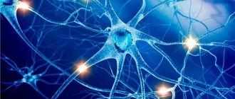
The nervous system integrates the entire organism into a single orchestra, carries out its interaction with the environment, voluntary movements (together with the muscular system), and all manifestations of mental activity. All functions of the nervous system are performed by a network of neurons connected to each other through synapses. Their viability is supported by glial cells.
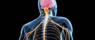
The nervous system is divided according to its anatomical location into central (CNS) and peripheral (PNS). The central nervous system consists of the brain and spinal cord. PNS - made up of nerves (a bundle of nerve cell processes) and nerve ganglia, or ganglia (a collection of neuron bodies) located outside the nervous system.
Based on their functions, the nervous system is divided into somatic (animal, SomNS) and vegetative (autonomous, ANS) divisions. The SomNS controls voluntary contractions of skeletal muscles. The ANS controls the activity of internal organs. It is divided into two divisions: sympathetic (SNS) and parasympathetic (PNS). Both the SomNS and the ANS have both central and peripheral sections.
Structure of the central nervous system
The CNS consists of the brain and spinal cord, each of which has white and gray matter. White matter consists of pathways, myelinated and unmyelinated axons. Myelin is white, which gives the corresponding shade to the tissue. Gray matter consists of neuron cell bodies. It can be located in the nervous system in the form of a tube (spinal cord); nuclei, or ganglia (clusters of neuron bodies in the thickness of the white matter), as well as the cortex (gray matter on the surface of the white matter).
The spinal cord is located in the spinal canal and its mass is 40 g. On its lateral surface, the dorsal roots, carrying afferent (sensitive, to the brain) information, enter from the back, and the anterior roots, carrying efferent (motor, from the brain) information, exit from the front. The section of the spinal cord corresponding to each pair of roots is called a segment. The segments are named according to where the roots exit the spine. The spinal cord has 8 cervical, 12 thoracic, 5 lumbar, 5 sacral and 1 coccygeal segments. In general, the number of spinal cord segments corresponds to the number of vertebrae. Exceptions are the cervical region, where there are 8 segments per 7 vertebrae; and coccygeal, where there is 1 segment for 3-4 vertebrae (Fig. 1).
Rice. 1. Structure and location of spinal cord segments.
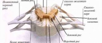
In a cross section of the spinal cord, there is gray matter in the center surrounded by white matter. The gray matter has the shape of a butterfly, in the center of which is the spinal foramen, filled with cerebrospinal fluid (CSF). The butterfly consists of approximately 13 million neurons and has anterior and posterior horns (Fig. 2b, 3). The middle horns are also well defined in the middle sections of the spinal cord. Sensitive (sensory) information to interneurons (interneurons) enters the dorsal horns along the dorsal root. The anterior horns contain motoneurons (motor neurons) that send motor information to the muscles; it is their axons that form the anterior root. The middle horns contain neurons of the central sections of the ANS.
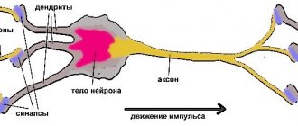
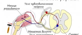
The spinal cord works on a reflex principle. A reflex is a stereotypical response of the body to any (external or internal) influence. The simplest reflex is monosynaptic. To implement it, two neurons are enough. An example of such a reflex is the knee reflex. When the receptor is irritated, the impulse is transmitted along the dendrite to the body of the neuron located in the nerve ganglion near the spinal cord. The axon of this neuron enters the spinal cord through the dorsal roots and forms a synapse with the motor neuron in the anterior horn. The axon of the motor neuron exits through the anterior roots and goes to the effector organ, where it changes the activity of the organ itself (Fig. 50a). The polysynaptic reflex includes an additional link in the form of one or more interneurons between the ganglion and motor neurons. Interneurons can additionally process information, compare it with other stimuli and the internal state of the body, making a decision about how to respond to the stimulus.
Rice. 2. Reflex arc (a) and histological section (b) of the spinal cord.
Rice. 3. Diagram of the structure of a section of the spinal cord.
The white matter of the spinal cord includes pathways. It is divided by the butterfly into anterior, posterior and lateral funiculi (Fig. 3).
In the posterior cords there are ascending tracts through which information is transmitted from the PNS to the spinal cord and further to the brain. In the anterior horns of the spinal cord there are descending tracts through which information goes from the brain to the spinal cord, and from the latter to the PNS. In the lateral horns, the ascending tracts are located posteriorly, and the descending tracts are located anteriorly.
The brain is located in the skull and consists of 5 sections. Its average mass is 1.5 kg and it contains up to 100 billion neurons. There are 12 pairs of cranial nerves (cranial nerves) that arise from the brain.
The medulla oblongata is the junction of the spinal cord and the brain. Its length is approximately 25 mm. In the lower part of the medulla oblongata one can still distinguish a butterfly; in the upper parts the bodies of neurons are collected into nuclei. The IX-XII pairs of the cranial nerves (Fig. 5) depart from the medulla oblongata and the corresponding nuclei lie in it. These nerves control movement and sensation in the throat, tongue, and neck. The medulla oblongata contains the largest center of the parasympathetic nervous system, which, through the X nerve (vagus, vagus nerve), controls the activity of all internal organs. The medulla oblongata contains centers for regulating breathing and vital reflexes, such as sneezing and coughing. Here is the olive kernel, which is responsible for balance. All tracts from the spinal cord to the brain pass through the medulla oblongata.
The hindbrain consists of the pons and cerebellum. The pons serves as a continuation of the medulla oblongata. It contains a lot of white matter that connects the cerebellum to the rest of the brain. This white substance forms a ridge on the underside of the bridge, making it easy to distinguish. The pons, together with the medulla oblongata, form the bottom of the 4th ventricle of the brain (a continuation and expansion of the spinal canal). V-VIII ChMN depart from the bridge. Here lie the auditory and vestibular nuclei, nuclei that innervate sensitivity and facial muscles (including facial muscles). The pons contains the locus coeruleus, which is responsible for regulating sleep.
Rice. 4. Main parts of the brain.
Rice. 5. Cranial nerves. I-olfactory, II-visual, III-oculomotor, IV-trochlear, V-trigeminal, VI-abducens, VII-facial, VIII-vestibular-cochlear, IX-glossopharyngeal, X-vagus, XI-accessory, XII-hyoid.
The cerebellum is well developed in humans due to upright posture and fine motor skills of the hands. This part of the brain is responsible for maintaining posture, balance, motor learning, and some motor reflexes. The cerebellum has a cortical structure. The cerebellar cortex consists of three layers and is divided into two hemispheres by the vermis. Under the cortex there is white matter, among which there are 3 pairs of cerebellar nuclei. To carry out its functions, it receives information from the vestibular apparatus, olive and other parts of the human motor system.
Rice. 6. External structure (a) and histological section (b) of the cerebellar cortex.
The midbrain consists of the cerebral peduncles and the roof (Fig. 7). The aqueduct of Sylvius passes through the center of the spinal cord and connects the third and fourth ventricles. The third and fourth pairs of cranial nerves depart from the midbrain. These nerves control the movements of the eyeballs. The third nerve contains parasympathetic fibers that control pupil width. The midbrain contains elements of the motor system: the red nucleus and the substantia nigra. On the roof of the brain is the quadrigeminal region. The colliculus receives visual information, and the inferior colliculus receives auditory information. This is necessary for the implementation of the orientation reflex.
Rice. 7. Appearance (a) and section (b) of the midbrain.
The medulla oblongata, pons, and midbrain together form the brainstem. The reticular formation runs through the entire trunk, regulating the overall level of brain activity.
The diencephalon consists of the thalamus, hypothalamus, pituitary gland and pineal gland. The third ventricle of the brain is located here. It serves as the origin of the second cranial nerve. The pituitary gland is the gland through which the nervous system controls the humoral one. The pineal gland is also a gland that regulates circadian rhythms. The thalamus filters information entering the cortex and removes unimportant repetitive sensory stimuli (heartbeat, gastrointestinal function, nose in the field of view, touch of clothing, etc.) In addition, the thalamus contains nuclei of the limbic system (forms mood), motor and associative kernels. The hypothalamus controls the activity of the pituitary gland and also regulates the internal state of the body. It contains the centers of hunger, thirst, sexual behavior, pleasure, displeasure, etc. Thus, the main function of the hypothalamus is to maintain homeostasis of the entire organism.
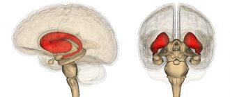
The telencephalon (forebrain) consists of the cerebral cortex and basal ganglia (nuclei). The first and second ventricles of the brain are located symmetrically under the cortex. Its area is about 220 cm2, it forms grooves and convolutions (Fig. 8). It consists of 6 layers. The hemispheres are connected to each other by the corpus callosum - a ridge of white matter. The cerebral cortex processes sensory information, the formation of voluntary movements, memory and higher nervous activity. The first cranial nerve approaches the olfactory bulbs. The basal ganglia are nuclei of gray matter located in the thickness of the white matter. They play an important role in voluntary movements, motor learning and the formation of emotions.
Rice. 8. Structure (a) and histological sections (b, c) of the cerebral cortex.
Nerves, nerve fibers and ganglia.
A nerve is a bundle of fibers, each of which functions independently of the others. The fibers in a nerve are organized into groups surrounded by specialized connective tissue that contains vessels that supply the nerve fibers with nutrients and oxygen and remove carbon dioxide and waste products. The nerve fibers along which impulses travel from peripheral receptors to the central nervous system (afferent) are called sensitive or sensory. Fibers that transmit impulses from the central nervous system to muscles or glands (efferent) are called motor or motor. Most nerves are mixed and consist of both sensory and motor fibers. A ganglion (nerve ganglion) is a collection of neuron bodies in the peripheral nervous system.
The axonal fibers in the PNS are surrounded by the neurilemma, a sheath of Schwann cells that are located along the axon, like beads on a string. A significant number of these axons are covered with an additional sheath of myelin (a protein-lipid complex); they are called myelinated (pulpy). Fibers surrounded by neurilemma cells, but not covered with a myelin sheath, are called unmyelinated (unmyelinated). Myelinated fibers are found only in vertebrates. The myelin sheath is formed from the plasma membrane of Schwann cells, which is wound around the axon like a roll of ribbon, forming layer upon layer. The section of the axon where two adjacent Schwann cells touch each other is called the node of Ranvier. In the central nervous system, the myelin sheath of nerve fibers is formed by a special type of glial cells - oligodendroglia. Each of these cells forms the myelin sheath of several axons at once. Unmyelinated fibers in the CNS lack a sheath of any special cells.
The myelin sheath speeds up the conduction of nerve impulses that “jump” from one node of Ranvier to another, using this sheath as a connecting electrical cable. The speed of impulse conduction increases with thickening of the myelin sheath and ranges from 2 m/s (for unmyelinated fibers) to 120 m/s (for fibers especially rich in myelin). For comparison: the speed of propagation of electric current through metal wires is from 300 to 3000 km/s.
Autonomic nervous system
The ANS includes two divisions, the sympathetic (SNS) and the parasympathetic (PNS). The SNS is activated during a stressful situation. It increases the heart rate, constricts blood vessels, pupils, increases blood flow to the muscles and outflow from the gastrointestinal tract. The center of the SNS is located in the thoracic and lumbar regions of the spinal cord (Fig. 9). The PNS has the opposite effect. It is activated in a calm environment and leads to a rush of blood to the gastrointestinal tract, outflow from the muscles, a decrease in heart rate, pupil dilation, etc. PNS centers are located in the medulla oblongata, some nuclei of the cranial nerve and the sacral spinal cord.
The main difference between the autonomic reflex arc and the somatic one is the presence of another synaptic switch in the ganglion after the spinal cord. Thus, the autonomic reflex begins from the receptor, then the sensitive neuron from the ganglion transmits information to the neuron of the middle horns of the spinal cord (or another center of the ANS). The axon of the autonomic neuron exits through the anterior roots and goes to the ganglion, where it forms a synapse with the ganglion neuron, the process of which goes directly to the effector organ. The nerve fiber running from the spinal cord to the ganglion is called preganglionic. The nerve fiber running from the ganglion to the organ is called postganglionic. The ganglia of the SNS are located next to the spinal cord, so the preganglionic fiber is short and the postganglionic fiber is long. The ganglia of the PNS are located near or in the wall of the organ, so their preganglionic fiber is long and the postganglionic fiber is short. The effector neurotransmitter of the sympathetic nervous system is norepinephrine, and the parasympathetic nervous system is acetylcholine.
Rice. 9. Effects of the SNS and PNS.
Rice.
10. Comparison of the reflex arc of the somatic and autonomic reflex. # Human anatomy
Spinal cord.
Located inside the spinal column and protected by its bone tissue, the spinal cord has a cylindrical shape and is covered with three membranes. In a cross section, the gray matter is shaped like the letter H or a butterfly. Gray matter is surrounded by white matter. Sensitive fibers of the spinal nerves end in the dorsal (posterior) parts of the gray matter - the dorsal horns (at the ends of the H, facing the back). The bodies of motor neurons of the spinal nerves are located in the ventral (anterior) parts of the gray matter - the anterior horns (at the ends of the H, distant from the back). In the white matter there are ascending sensory pathways ending in the gray matter of the spinal cord, and descending motor pathways coming from the gray matter. In addition, many fibers in the white matter connect different parts of the gray matter of the spinal cord.