What is hydrocephalus?
The term hydrocephalus is formed from two Greek words: “hydro” - water and “cephalus” - head (“dropsy of the brain”). In hydrocephalus, there is a significant buildup of cerebrospinal fluid (CSF) inside cavities of the brain called ventricles. An increase in the volume of cerebrospinal fluid (CSF) occurs when the balance between the production of cerebrospinal fluid and its absorption and further removal from the body is disturbed. This may be the result of a deficiency in the circulatory system or excessive production of cerebrospinal fluid. Hydrocephalus can be congenital or acquired. The concept of congenital hydrocephalus means that it appears from the moment of birth. The disease can be provoked by various factors: traumatic brain injury, brain tumor, necrosis, meningitis, etc. The frequency of this disease is high throughout the world. According to the literature, hydrocephalus affects 5-15 children out of every thousand newborns. Treatment consists of diverting CSF outside the cerebrospinal fluid system into body cavities where it can be absorbed (absorbed). An effective operation allows a child to live and develop normally, study in a regular school, and an adult to return to a full, active life. However, patients and their loved ones must be able to recognize the signs and symptoms of postoperative complications so that medical care can be provided in a timely manner.
Rehabilitation after the procedure
Before the operation, the patient should quit smoking and drinking alcohol 2-3 weeks before, and stop taking non-steroidal medications.
Before the operation itself, the patient undergoes a cardiogram examination, fluorography, and submits urine and kcal samples for analysis. Among other things, the doctor may additionally prescribe the following examination procedures:
- MRI and CT, as well as intra-arterial angiography - all these examination methods allow you to determine the exact location of stenosis, blockage of the arteries;
- examination of the condition of the arteries using ultrasound - this method will allow you to diagnose the general condition of the blood vessels of the brain.
If there is a failure in the outflow of cerebrospinal fluid, a patient’s brain may develop a disorder such as hydrocephalus, a disease of complex theology that, in the absence of proper treatment, can lead to the development of severe consequences.
Most often, such irreversible processes, changes and disorders are neurological and mental illnesses.
https://www.youtube.com/watch?v=fANsbdTaYiA
Today, the most common method of treating hydrocephalus is brain shunting - in this case, doctors use a special hose - a shunt made of silicone, which allows fluid to be removed from the ventricles of the brain to another place in the body, for example, to the chest or abdominal cavity, bladder.
Doctors use the following types of shunting for hydrocephalus:
- ventriculoperitoneal intervention;
- ventriculoatrial intervention.
The second method of operation is not so dangerous - the mechanism and system of shunts themselves are more complex in their structure, and are equipped with special valves, which determines more efficient operation, reliability, and full functioning.
This system requires regular replacement every 6 months - the installed shunt in the head will provide the patient with a normal life, without problems and complications. The main condition is to control the condition of the shunt.
It is worth noting that after the operation the patient may experience a headache, movement of the limbs may be difficult, a feeling of nausea and bouts of vomiting may appear.
In order to diagnose the patient’s postoperative condition, an MRI examination is performed on the 2nd day after surgery, which makes it possible to exclude the presence of hemorrhage in the gray matter of the brain and the development of ischemia as a result of surgery.
From the editor: Causes of sudden deterioration in vision
Immediately 7-8 days before discharge from the medical institution, a duplex scan is performed - this examination method allows you to diagnose and evaluate blood flow. If the examination results are normal, then doctors discharge the patient after a week.
Rehabilitation after brain bypass surgery must be carried out strictly with all the recommendations and prescriptions of the attending physician; it is necessary to completely eliminate the consumption of alcohol - it is not compatible with taking medications, and leads to the accumulation and retention of excess fluid in the body.
Along with this, it is worth minimizing, and if possible completely eliminating, any physical activity, moving away from country work, as well as heavy household work. The patient is also prohibited from lifting weights of more than 3 kilograms; it is necessary to take leisurely walks in the fresh air more often.
After brain bypass surgery, the patient is allowed to walk as the only physical activity allowed - you just need to start walking, walking for minutes a day, gradually increasing the time.
It is also worth remembering that swimming in open water is prohibited, although taking a bath and washing your hair is a permitted hygienic procedure, subject to certain points.
In particular, it is best to choose a mild, children's shampoo, avoiding, if possible, the area on the head where the skull was opened and bypass surgery was performed. The home regime should be followed for at least a month.
Anatomy and physiology
In order to better know and understand the disease, we will give you some information about the anatomy of the skull, the structure of the brain, as well as the process of formation and absorption of cerebrospinal fluid (Fig. 1). The brain occupies most of the cranial cavity. It is penetrated by a large number of blood vessels and is surrounded by cerebrospinal fluid as a buffer. The fluid is located in 4 cavities (ventricles) located inside the brain. The ventricles have delicate structures known as villous plexuses (choroid plexuses). These structures produce a significant amount of CSF - about 500 ml per day. The fluid circulates continuously and contains a large number of substances essential for nutrition and ensuring the normal functioning of the nervous system.
CSF also provides a protective cushion for the brain. CSF circulates in the ventricular system and is removed through 3 holes in the 4th ventricle and then enters the subarachnoid space surrounding the brain and spinal cord.
CSF constantly circulates in the brain and spinal cord, constantly being in the process of a) formation, b) circulation and c) absorption. In a healthy body these processes are balanced. Hydrocephalus develops if CSF is not cleared from the ventricular system through the cerebrospinal fluid tract. Less commonly, hydrocephalus is caused by excess production of CSF, such as with villous plexus papilloma.
Diagnostics
In newborn children, the bones of the skull have not yet fused and hydrocephalus is determined visually. The head enlarges, the bones of the skull diverge, the fontanelle is tense and bulging; the skin is thin and shiny; The veins in the hairline area look full and swollen. Also symptoms are: vomiting, apathy, excitability, downward displacement of the eyes (“setting sun symptom”), etc. However, when the cranial sutures are not fused, the signs and symptoms of increased intracranial pressure seem subtle.
In older children and adults, the bones of the skull are fused and, when the ventricles expand, compression of the brain tissue occurs. Symptoms of increased intracranial pressure appear: headaches, nausea, vomiting, blurred vision, lack of coordination, psychopathological personality changes, lack of concentration and lethargy. These symptoms require additional instrumental diagnosis.
Types of diagnostic tests
- Ultrasound is a simple, inexpensive test that helps evaluate the extent to which the ventricles of the brain are dilated. Currently, it is the simplest and safest method for diagnosing hydrocephalus.
- Computed tomography (CT) - This is a technique of drawing with a thin beam the contours of the skull, brain, ventricles and subarachnoid space. It is carried out to determine the size and shape of the ventricles and identify abnormalities such as tumors, cysts or other pathologies.
- Magnetic resonance (nuclear magnetic resonance NMR) is a non-surgical diagnostic method that uses radio signals and a magnet. MRI data determines the shape and severity of hydrocephalus. These studies are indispensable for clarifying the causes of dropsy.
- Cisternography (radiography of the cisterns at the base of the skull) is a test that requires the injection of a radioactive substance into the CSF. It is used to clarify the type of hydrocephalus: communicating or obstructive, as well as to determine the direction of CSF flow.
- Pneumoencephalography is now used much less frequently than in the past. In some cases, it is necessary to pump air with a needle into the spinal cord.
- Angiography (X-ray of blood vessels) is a special technique for injecting a contrast agent into the arteries that cross the brain. After some time, anomalies are detected at the level of blood vessels and the presence or absence of pathological disorders.
- A neuropsychological examination consists of a series of questions and answers to identify the presence of abnormalities in the functioning of the brain.
Indications
CABG is an operation that is primarily required for patients with severe angina that is resistant to drug therapy. In addition, surgical treatment may be recommended in the absence of symptoms. There is silent myocardial ischemia, which is no less dangerous. It is not felt by the patient, but can be diagnosed using ECG and Holter monitoring.
Indications for CABG can also be determined using coronary angiography. This is an x-ray test in which contrast is injected into the arteries of the heart. The main cause of mortality from coronary disease is atherosclerosis and disruption of the integrity of the atherosclerotic plaque. The blood vessel is torn, and a blood clot forms at the site of damage. It clogs the coronary artery, resulting in myocardial infarction.
Coronary angiography allows you to identify large obstacles to blood flow (atherosclerotic plaques) and assess the risk of acute cardiovascular events. A high degree of coronary artery disease requires cardiac CABG.
Based on the results of coronary angiography, the main indications for CABG are:
- Narrowing of the lumen of any coronary artery by 75% or more
- Reduction in the lumen of the left coronary artery trunk by 50% or more
The location of stenosis (impaired patency) of the coronary arteries is also assessed. The more proximal it is (closer to the heart), the worse the blood supply to the myocardium. CABG surgery on the heart will be most successful if normal blood circulation is observed beyond the narrowing.
Treatment
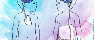
Currently, occlusive (obstructive) hydrocephalus is treated surgically. Surgical intervention consists of draining excess cerebrospinal fluid outside the cerebrospinal fluid system: into the abdominal (abdominal) cavity or into the atrium. Sometimes CSF may be drained into the pleural cavity. In these cavities, cerebrospinal fluid is absorbed and excreted along with waste products of the body.
To drain the CSF, the surgeon implants a drainage system (shunt). The system material is silicone and polypropylene. Both of these materials are well tolerated by the body. All elements of the system are implanted under the skin; there are no external areas.
Shunt System Components
The system consists of two catheters and one one-way valve. The ventricular catheter is located in the ventricle of the brain, and the peripheral (peritoneal or cardiac) catheter is placed in the abdominal cavity or in the right atrium, respectively. Both catheters are connected to a valve that regulates the unidirectional flow of cerebrospinal fluid. The valves are designed to operate in different pressure ranges (high, medium, low and very low). The neurosurgeon, having determined the patient’s intracranial pressure, selects the appropriate valve, depending on the severity of the disease, the patient’s age and clinical nuances.
Almost all valve models have a reservoir that your doctor can use to “bleed” the system to determine if it is working properly. From the reservoir, by inserting a thin needle through the skin, you can take samples of cerebrospinal fluid for laboratory tests or administer medications. Patients and their loved ones are not recommended to test the drainage system by “bleeding” the reservoir. This action can be dangerous unless your doctor has given you specific instructions about it. In patients suffering from non-communicating (obstructive) hydrocephalus, cerebrospinal fluid should be drained from the cerebral ventricle using a ventricular catheter. In patients with communicating hydrocephalus, a CSF drainage system is implanted from the lumbar space of the spine into the abdominal cavity - the lumboperitoneal system.
Features of the intervention
Before the intervention is carried out, the patient must give his consent. In addition, he must have absolute indications for bypass surgery. In this case, the patient must sign the agreement after reading it first.
The fact is that inside the tube there is a special valve that opens only when intracranial pressure increases to a certain level.
The shunting technique is as follows:
- the patient is given general anesthesia;
- the person is covered with sheets so that only those places where the holes will be made remain free;
- all these areas are pre-treated with an antiseptic;
- The shunt itself is implanted. It can be non-cerebral (if the abdominal cavity is used) or ventricular. The second is performed if a cardiac sac is used;
- For the tube, a path is cut to the brain in the subcutaneous tissue.
- Finally, the shunt is placed to the ventricles.
The patient remains in the hospital for several weeks. During the first days he lies in intensive care. Before the patient regains consciousness, doctors insert a tube into the trachea, with the help of which the breathing process will be carried out.
The discomfort in the throat that remains after its removal passes quickly enough.
Surgery and hospitalization
Implantation of the shunt system is carried out by a neurosurgeon under sterile operating room conditions. In preparation for surgery, the hair is shaved to achieve maximum cleanliness. The operation is performed under general anesthesia, usually not for a very long time. The neurosurgeon drills a small hole in the skull bone, then makes a small incision in the dura mater that protects the brain and inserts a ventricular catheter into the lateral ventricle. To implant the rest of the bypass system, another incision is made and a tunnel is created subcutaneously to implant a peritoneal or cardiac catheter. The end of the catheter is carefully inserted either into the abdominal cavity or into the jugular vein leading to the ventricle of the heart. Catheters are connected to the valve using connectors on the valve itself. All parts of the shunt are thus connected into a single drainage system. At the end of the operation, small sterile dressings are applied to each incision.
Immediately after the operation, the patient is taken to the postoperative department. The patient is here from 1 hour to 1 day under close observation, then he is transferred to the ward. Most patients are discharged 7-10 days after clinical rehabilitation.
Necessary examination before surgery
Common preoperative investigations include:
- Complete blood count;
- Coagulation pattern;
- Creatinine and electrolyte levels;
- Liver function tests;
- Screening for methicillin-resistant Staphylococcus aureus;
- Chest X-ray;
- Electrocardiography (ECG);
- Echocardiography or ventriculography (to assess left ventricular function);
- Coronary angiography (to determine the extent and location of coronary artery disease).
If patient demographic characteristics (eg, age or smoking) indicate an increased risk of cancer, gastrointestinal or urinary tract malignancy should be excluded.
When determining whether a patient is indicated for cardiac bypass surgery, the risks are assessed using the Euroscore system. This is a European risk calculator, adopted in many countries around the world (including the USA) to predict mortality in CABG patients. The results can be presented logistically or as a simple score, which can be calculated using the interactive calculator on the Euroscore website.
Participation in care
Children with implanted shunts should be monitored by a neurosurgeon throughout their lives. Most patients suffering from hydrocephalus, after implantation of a shunt system, can lead a normal life, but constant monitoring by loved ones in collaboration with a neurosurgeon is necessary.
The neurosurgeon monitors each patient to prevent drainage failure. The first time after implantation or re-operation - regularly, with a gradual transition to examinations once a year.
Relatives are advised to be able to recognize early signs of complications. Quick and accurate assessment of health problems is very important. Flu symptoms may mask symptoms of a blocked shunt. Quickly identifying them will allow you to plan a repeat operation and avoid an emergency.
Patients and their loved ones should pay close attention to signs and symptoms of complications. The main causes are: blockage, infection and excessive drainage.
Coronary artery bypass surgery for myocardial infarction
Coronary artery bypass grafting may be performed as an emergency procedure in the context of ST-segment elevation myocardial infarction in cases where percutaneous coronary intervention (PCI) has not been possible or when there is a persistent threat of pain and ischemia of a large area of the myocardium despite medical therapy.
Other Class I indications for emergency open heart surgery in the setting of AMI include:
- ventricular septal defect associated with AMI;
- rupture of the papillary muscle;
- free rupture of the heart wall;
- ventricular pseudoaneurysm;
- life-threatening ventricular arrhythmias;
- cardiogenic shock.
Blockage
The main type of complication is blockage of the system. Blockages can occur at any level of drainage. The holes in the ventricular catheter may become clogged with brain tissue or villous plexus tissue. It can also be blocked due to excessive contraction of the ventricular cavity due to too intense drainage of fluid from the ventricular cavity (constricted ventricle syndrome). Intestinal loops or necrotic tissue may adhere to the peritoneal catheter. Shunts inserted into a ventricle of the heart can become blocked by blood clots, pieces of brain matter, or tumor cells.
The shunt system may also be disconnected by disconnection of various elements of the shunt or by changes in the position of the catheter caused by the growth of the child. X-rays are necessary to check the integrity of the shunt.
Partial blockage of the system parts provokes the appearance of symptoms of increased intracranial pressure. Episodic headaches, nausea, apathy and decreased sensory functions may occur. Decreased performance at school or at work is the most common phenomenon in these conditions.
In case of complete blockage, symptoms develop more quickly (headaches, nausea, vomiting, blurred vision, loss of coordination and confusion). The patient falls into a stupor or coma. In such cases, urgent hospitalization is necessary for observation and appropriate treatment.
The surgeon performs a series of tests to locate and determine the extent of drainage blockage. Sometimes it is necessary to remove and replace part or all of the system.
After operation
During bypass surgery, an endotracheal tube will be inserted into the patient's trachea, which is used to maintain breathing while under anesthesia.
It will be removed only after its effect wears off. Therefore, minor pain in the throat is normal. There is also nothing strange or scary in the fact that a patient will end up in the intensive care unit after surgery.
This is a mandatory precaution for the first day or more.
Common consequences of surgery include headache and nausea. Your doctor may prescribe medications. It will be possible to stand on your feet and walk – only around the ward for now – no earlier than on the second day.
A day after surgery, an MRI is performed to make sure everything went well. In total, if there are no complications, the patient will need to spend another 5-6 days in the hospital.
You may need to take the following medications in the first days after discharge:
- Painkillers;
- Anticonvulsants.
Both must be prescribed by a doctor, as well as their dosage.
Possible consequences and complications
What should you not do in the first weeks after bypass surgery?
- Any housework or housework is excluded until the surgeon allows it (usually after the first examination this restriction is lifted).
- Driving is prohibited; it will also be possible to resume it after the doctor checks all the changes and makes a positive decision on this issue.
- Any objects heavier than 2 kg cannot be lifted!
- Alcohol should also be avoided while you are taking prescribed medications.
What is recommended?
- As a physical exercise, the only thing that definitely won’t cause harm and will be useful at the same time is walking. Start your daily walks with 10-15 minutes and gradually increase the time slightly.
- Swimming in ponds is not recommended, but taking a bath and washing your hair can be done without problems. In this case, it is better to use children’s shampoo, and, if possible, avoid touching the area of the bypass surgery. It is enough to treat it with a sponge using gentle movements. Do not rub, do not pour water from the shower, do not plunge headlong into the bath.
- Home regime – 2-4 weeks.
In the first days you may feel very tired, and this is normal. But other sensations may be suspicious. You should tell your doctor about your concerns if:
- Temperature above 38 appeared;
- The prescribed medications caused a rash and itching;
- When walking - unsteadiness, dizziness;
- Drowsiness;
- The scar remaining after the operation is swollen and red;
- Weakness, neck pain;
- Increased headaches and nausea;
- Vomit.
Remember that the occurrence of complications is often associated with the patient’s irresponsible attitude to the doctor’s instructions. The most severe consequences are stroke and convulsive syndrome, much less often - shunt thrombosis. No other complications were recorded.
The risk of seizures is easily reduced by taking medications prescribed after bypass surgery.
Rehabilitation
Patients after bypass surgery are prescribed constant (lifelong, but in courses) medications such as acetylsalicylic acid, clopidogrel, ticlopidine. When wearing glasses, you need a gauze pad on the temple so that it does not press on the donor artery.
For approximately six months after bypass surgery, the patient will undergo several examinations to monitor changes. Based on the results, other rehabilitation options may be prescribed.
Infection
Infection is the second type of complication. It poses a significant risk for any surgical procedure, most often when implanting a foreign body.
It manifests itself in the form of redness or suppuration along the edges of the suture or along the path of the drainage system under the skin. The surgeon fixes his attention on these signs. If left untreated, the wound may erode or open, and in more serious cases, the infection may cause chills and fever. Typically, the drain needs to be removed. Sometimes antibiotic therapy can be administered without removing the system.
Since the shunt is a foreign body, the patient may experience an allergic or inflammatory reaction. Inflammation at one of the drainage sites should be immediately shown to a neurosurgeon.
Contraindications
Coronary artery bypass grafting (CABG) may not be performed on patients even if there are indications for surgery. This may be due to the presence of contraindications. These are diseases and conditions of the body that make surgery too dangerous or unsuccessful.
Main contraindications for cardiac CABG:
- Reduced left ventricular ejection fraction by up to 30%
- Congestive heart failure
- Impaired blood flow in all coronary arteries
- Severe concomitant diseases
In case of circulatory disorders in all coronary arteries, preference is given to laser myocardial revascularization or cell therapy.
Excessive drainage
Excessive drainage of cerebrospinal fluid occurs when the valve is incorrectly selected according to the pressure parameter. If the valve opening pressure is too low, it can cause excessive drainage, causing the cerebral ventricle to compress and deforming the brain tissue. The patient experiences headaches that are most severe when standing.
In addition, nausea, vomiting, drowsiness and nervous system disorders, in particular double vision, appear. School-age children experience a decline in mental abilities.
Complications of cardiac bypass surgery
Complications from coronary artery bypass surgery can occur in both the short and long term.
They are most often associated with anesthesia, cardiopulmonary bypass, sternotomy and the operation itself.
These complications may include the following:
- Myocardial dysfunction
- Cerebrovascular complications
- Acute renal failure
- Cardiac tamponade
- Respiratory tract infections
- Aortic dissection
In the initial postoperative period, there is a decrease in myocardial function due to secondary edema and ischemia-reperfusion injury.
Additional factors (eg, incomplete revascularization and postoperative graft failure) may exacerbate dysfunction. Patients may present with low output syndrome, with 4-9% requiring inotropes or intra-aortic balloon pulsation.
In addition, segmental transmural myocardial infarction occurs in 1-3% of patients, and postoperative arrhythmias occur in approximately 30% of patients after CABG.
Adverse neurological outcomes represent a serious problem in cardiac surgery.
Major neurological deficit and coma occur in 3.1% of cases.
Postoperative renal failure is also a common complication of CABG - 4% of cases. Dialysis is required in 20% of patients with postoperative acute renal failure.
More information about shunt systems for the treatment of hydrocephalus
The result of the operation largely depends on the quality of the shunt system. In recent years, in many hospitals, neurosurgeons have been using shunt systems produced by the largest American company Medtronic. The company's engineers, together with leading US neurosurgeons, have developed a range of valves of various models and sizes (including valves for newborns).
One of the latest achievements in the production of liquor drainage devices is the Delta valve. This is a unique valve, has no analogues from other manufacturers. It was created to avoid such a common complication as excessive CSF drainage. If all other valves pass as much fluid as they are designed for pressure, then the Delta valve allows as much fluid as is needed to remain in the ventricle to maintain intracranial pressure within physiological limits. When implanting the Delta valve, the patient is maintained at normal pressure, regardless of the rate of cerebrospinal fluid production and, most importantly, regardless of the patient’s body position (lying/standing).
The technological features of the materials from which the system valves are made prevent deformation and sticking during pumping; the dome of the valve reservoir is designed to withstand repeated punctures with a thin needle (the holes self-tighten). Catheters are made of high-quality latex-free silicone, so they do not stick together or form loops, which significantly reduces the risk of system blockage.
The valves are equipped with connectors for connection to catheters; their design facilitates connection and reduces the possibility of disconnection and disconnection of the system.
A radiopaque mark is applied along the length of the catheters, this allows you to see the shunt on an x-ray. The same substance is applied to the valve with a dot code indicating the pressure of the valve. There are no metal parts in the shunt system. This is very important when conducting CT and NMR studies, because metal will produce artifacts, and a magnet in NMR may shift the location of the system (if it had metal parts).
All systems are sterile and supplied in double sterile packaging. To reduce the risk of infection, Medtronic has developed a unique BioGlide hydrogel. It is applied to the inner and outer surfaces of catheters, as well as the outer surface of the valve, and does not peel off. Before implantation, the neurosurgeon can treat the system parts with antibiotics, and the hydrogel will hold them for 3 days for postoperative antibiotic therapy inside the patient’s body. This way, the risk of infection is minimized.
Benefits of beating heart surgery
The risk of stroke, intraoperative heart attack, and death was similar and low in patients undergoing both on-pump and off-pump CABG.
One concern that has emerged from recent multicenter studies is that patients undergoing off-pump heart surgery had less complete revascularization, meaning that fewer patients had completely restored blood flow compared with patients with CPB.
The advantage of beating heart surgery is that the recovery period can be significantly reduced.
Which patients are most suitable for beating heart surgery?
Patients at high risk for complications from cardiopulmonary bypass, such as people with vascular disease (blood vessels), previous strokes, or liver disease, may benefit from off-pump revascularization.
How is the blood supply to the heart organized during beating heart surgery?
One of the possible options for providing blood access to the ischemic area is an intracoronary bypass. It is a synthetic tube connecting the aorta to the distal end of the coronary artery
Emotional support
The physical side of hydrocephalus is only part of the problem of this disease. The patient and his relatives need to take into account his emotional factors.
Although surgery should resolve your hydrocephalus to some extent, you may feel fearful, depressed, irritable, or doubtful. If the patient is a child, it should be taken into account that he has the same feelings as an adult. If your child is feeling out of shape or uncomfortable because they need to visit the doctor frequently or have repeated tests, it is best to reassure them with simple explanations. If he knows what awaits him, he will be more willing to cooperate with you. Children, like adults, as a rule, do not like unpleasant surprises. A calm atmosphere among loving loved ones is the best environment for children. It is advisable to explain the phenomenon of hydrocephalus in words that the child can understand.
It is very important to know how the child feels and be able to explain to him what he is experiencing. The needles hurt. It is natural to cry and want to get rid of them. Being admitted to hospital is a new challenge for your child. You must tell him the truth in order to gain and secure his trust. Sincerity is the best way to maintain your child's trust.
Children over ten years of age are generally able to comprehend more complex concepts. They may associate signs and symptoms with their illness. Restrictions associated with the disease are easier for them to bear. Tell your doctor about your feelings and give him the right to guide you. Some people share their feelings with close friends, others need professional help. The health professionals treating you or your child are interested in your well-being and their goal is to do what is best for you and your loved ones.
Patients and the patient's parents should communicate with their doctor quite frequently. It is important to actively participate in this communication so that your doctor can better understand your needs and the needs of your loved ones.
How is the operation performed?
The operation can be performed using a heart-lung machine. In addition, in developed countries, including Germany, off-pump CABG is increasingly being used.
The essence of surgery is to create a bypass path for blood flow. For this purpose, shunts are created - additional vessels that leave the aorta and flow into the coronary arteries - beyond the level of narrowing. A person’s own veins or arteries are used as prostheses. This may be the great saphenous vein or the radial artery.
Usually 3 bypasses are created - for the largest coronary arteries:
- Anterior interventricular
- Envelope
- Right
In 20% of patients, 4-8 anastomoses are installed. More preferable is CABG (coronary artery bypass grafting) on a beating heart.
The advantages are as follows:
- Blood cells are not damaged
- Rehabilitation after CABG is faster
- The operation has a shorter duration
In this case, complications after CABG on the heart are less common. Because there is no risk from those that are directly related to the use of a heart-lung machine. This is pulmonary edema, embolic damage to the blood vessels of the brain or kidneys, hypoxia (oxygen starvation) of internal organs.