Reflex arc
The activity of the nervous system is based on reflexes (Latin reflexus - reflected). Reflex is the body's response to the action of a stimulus.
Any reflex exists on the basis of a reflex arc - a set of nerve elements connected to each other, through which a nerve impulse is sequentially transmitted during the implementation of the reflex. The simplest example is the knee reflex, which is often checked by a neurologist, which allows one to quickly draw a conclusion about the safety of the elements of the reflex arc.
Neurons connect to each other using processes: axons and dendrites, at the end of which there are special contacts - synapses, which we studied in detail in the article about nerve tissue.
Reflex arc device
Reflex arcs can be very simple: consist of two neurons, like the reflex arc of the knee reflex (there is no interneuron), or they can include dozens of different neurons. The reflex arc can be divided into 3 parts:
- Sensitive (afferent, centripetal)
- Intercalary (associative, intermediate)
- Motor (efferent, centrifugal)
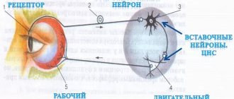
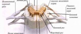
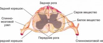
It consists of a receptor (can be located in the skin, internal organs, blood vessels) of a sensory neuron and a sensory fiber coming from this neuron, which penetrates the spinal cord through the dorsal horns.
The body of the sensory neuron is located in the dorsal roots (!) of the spinal cord. Introduced? Now imagine a dendrite running from the tip of your index finger all the way to your spinal cord. That is why it is incorrect to assume that a dendrite is always a “short” process, and an axon is always a “long” one. We discussed this issue in the article about nerve tissue.
Consists of an interneuron and its processes. The interneuron communicates between the sensory and motor parts of the reflex arc. Interneurons can communicate with other parts of the central nervous system.
The bodies of interneurons are located in the dorsal horns of the spinal cord.
Represented by a motor neuron (efferent, executive, motor neuron), from which nerve fibers go to the working organ (effector, executive organ).
Depending on what the effector is - a muscle, a gland - when nerve impulses arrive at it, its work is activated: the muscle begins to contract, the gland begins to secrete secretions.
Motor neurons lie in the anterior horns of the spinal cord, from where their processes emerge.
Let's consider the diagram of a reflex arc, on the basis of which the reflex of withdrawing a hand from a hot object is realized. Try to describe the path that a nerve impulse takes and remember the 3 parts of the reflex arc. Name the location of each neuron.
This may seem obvious, but it must be emphasized that afferent nerve fibers enter the spinal cord through the dorsal roots. Efferent nerve fibers exit the spinal cord through the ventral roots.
Types of reflex arcs
Reflex arcs are divided into somatic and autonomic. With the help of somatic reflex arcs, reflexes are carried out, providing the possibility of voluntary movements (performed by the will of a person). With the help of vegetative ones - coordination of the activity of internal organs, that is, functions that are not amenable to our conscious control (remember the autonomic nervous system).
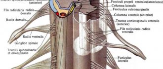
Below you will see diagrams of somatic and autonomic reflex arcs. Under the picture there will be written a significant difference between them, which you must remember, but first try to draw a conclusion yourself by studying the picture.
The difference between the somatic and autonomic reflex arcs is that in the latter, the efferent neuron lies outside the spinal cord - in the autonomic ganglion. These ganglia can be located on the sides of the spine, near internal organs or in their wall.
Also, you most likely noticed that the interneuron of the autonomic arch is localized in a different place - in the lateral horns of the spinal cord (and not in the posterior ones, as in the somatic horn).
Nervous regulation
The reflex arc is the foundation on which the reflex is carried out. In the nervous system, not only processes of excitation occur, but also inhibition, which we will talk about in more detail in the topic devoted to higher nervous activity. Inhibition consists of weakening or delaying excitation that has already arisen.
Thus, coordination and regulation of the processes of excitation and inhibition is the basis for the coordinated work of organs and organ systems that make up a single organism.
Diseases
Paresis (Greek πάρεσις - weakening) is a neurological syndrome caused by damage to the motor (efferent) pathway and weakness in the limb, or in another organ that innervated this nerve pathway. Paresis is manifested by a decrease in muscle strength, movements are preserved in an incomplete volume.
Paralysis (Greek παράλυσις - relaxation) is the complete absence of voluntary movements, due to the same reasons as paresis.
Hypothermia may cause paresis of the facial nerve. The reason for this is tissue inflammation, as a result of which the nerve in a narrow bone canal is compressed by inflamed tissue. Nerve impulses partially or completely stop reaching the facial muscles, making it impossible for the patient to move them.
© Bellevich Yuri Sergeevich 2018-2021
This article was written by Yuri Sergeevich Bellevich and is his intellectual property. Copying, distribution (including by copying to other sites and resources on the Internet) or any other use of information and objects without the prior consent of the copyright holder is punishable by law. To obtain article materials and permission to use them, please contact Yuri Bellevich
.
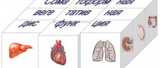
Autonomic nervous system
All functions of the body are divided into somatic (animal) and vegetative (autonomous). Somatic functions include the perception of external stimuli and motor reactions of skeletal muscles. These reactions can be voluntarily evoked, enhanced or inhibited and are under the control of consciousness. Vegetative functions ensure metabolism, thermoregulation, the functioning of the cardiovascular, respiratory, digestive, excretory and other systems, growth and reproduction. Autonomic reactions, as a rule, are not controlled by consciousness.
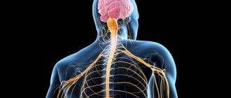
The autonomic nervous system (ANS) is a complex of central and peripheral nervous structures that regulate the activity of internal organs and the necessary functional level of all body systems. More than 80% of diseases are associated with a disorder of this system.
Physiological significance:
1. Maintaining homeostasis - the constancy of the internal environment of the body.
2. Participation in the vegetative support of various forms of mental and physical activity.
Morphological and functional features of the ANS.
General properties of the somatic and autonomic nervous system.
1. Reflex arcs are built according to the same plan - they have afferent, central and efferent links.
2. The reflex arc of somatic and autonomic reflexes may have a common afferent link.
1 - receptor
2 - afferent nerve and afferent neuron
3 - interneuron in the spinal cord
4 - efferent nerve, which leaves the efferent neuron
5 - effector organ
The structure of the reflex arc of somatic and autonomic reflexes
Structure of the ANS.
The ANS consists of central and peripheral sections.
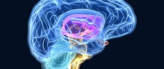
The central section is represented by segmental and suprasegmental centers. Segmental centers are the spinal cord, medulla oblongata and midbrain. Suprasegmental centers - hypothalamus, cerebellum, basal ganglia, cerebral cortex, limbic system. Suprasegmental centers exert their influence only through underlying segmental centers.
The peripheral section includes microganglia of the metasympathetic nervous system, para- and prevertebral ganglia, preganglionic and postganglionic fibers of the ANS.
Central nervous control of autonomic activity
The activity of the autonomic nervous system varies depending on the information it receives from visceral and somatic afferent fibers. Regulation also depends on information coming from higher centers of the brain, in particular from the hypothalamus.
Internal organs are innervated by afferent fibers that respond to mechanical and chemical stimuli. Some visceral afferent fibers reach the spinal cord through the dorsal roots along with somatic afferents. These fibers form synapses at the segmental level and transmit information through second-order ascending fibers within the spinothalamic tract of the spinal cord. They project to the nuclei of the solitary tract, various motor nuclei in the brain stem, the thalamus and hypothalamus. Other visceral afferents, such as those from arterial baroreceptors, reach the brainstem via vagal afferents.
Information from visceral afferents causes certain visceral reflexes, which, like reflexes of the somatic motor system, can be either segmental or can be associated with the participation of neurons in the brain. Examples of autonomic reflexes are the baroreceptor reflex, pulmonary respiratory reflexes, and the micturition reflex.
In response to perceived danger and damage, there is a behavioral warning response that can lead to aggressive or defensive behavior. This is known as the defense response, which originates in the hypothalamus. During a defensive reaction, there are noticeable changes in the activity of the autonomic nerves, in which the normal control of reflexes is altered.
The hypothalamus regulates the homeostatic activity of the autonomic nervous system and is the highest central organ for the regulation of the sympathetic and parasympathetic systems. The activity of the autonomic nervous system and the functions of the endocrine system are under the control of the hypothalamus, which is part of the brain and regulates mainly those
functions that are associated with maintaining homeostasis of the body. If the hypothalamus is destroyed, homeostatic mechanisms do not work. The hypothalamus receives afferents from the retina, sensory organs, somatic organs, and afferents from internal organs. It also receives a lot of information from other parts of the brain, including the limbic system and cerebral cortex, which can influence the functioning of the autonomic nervous system indirectly through changes in the functioning of the hypothalamus. Neurons of the hypothalamus play an important role in thermoregulation, in the regulation of tissue osmolarity and water-salt balance, in the control of food and drink intake, and in reproductive activity.