Physiology of the medulla oblongata, cerebellum and midbrain
The medulla oblongata, like the spinal cord, performs two functions - reflex and conductive. Eight pairs of cranial nerves (V to XII) emerge from the medulla oblongata and the pons and it, like the spinal cord, has a direct sensory and motor connection with the periphery. It receives impulses through sensory fibers - information from the receptors of the scalp, mucous membranes of the eyes, nose, mouth (including taste buds), from the organ of hearing, the vestibular apparatus (organ of balance), from the receptors of the larynx, trachea, lungs, as well as from the interoceptors of the heart. -vascular system and digestive system.
Through the medulla oblongata, many simple and complex reflexes are carried out, covering not individual metameres of the body, but organ systems, for example, the digestive, respiratory, and circulatory systems. The reflex activity of the medulla oblongata can be observed in a bulbar cat, that is, a cat in which the brain stem above the medulla oblongata has been cut. The reflex activity of such a cat is complex and diverse. The following reflexes occur through the medulla oblongata: Protective reflexes: coughing, sneezing, blinking, lacrimation, vomiting. Food reflexes: sucking, swallowing, juice production (secretion) of the digestive glands. Cardiovascular reflexes that regulate the activity of the heart and blood vessels. The medulla oblongata contains an automatically functioning respiratory center that provides ventilation to the lungs. The vestibular nuclei are located in the medulla oblongata.
From the vestibular nuclei of the medulla oblongata begins the descending vestibulospinal tract, which is involved in the implementation of posture reflexes, namely in the redistribution of muscle tone. A bulbar cat can neither stand nor walk, but the medulla oblongata and cervical segments of the spinal cord provide those complex reflexes that are elements of standing and walking. All reflexes associated with the standing function are called positioning reflexes. Thanks to them, the animal, contrary to the forces of gravity, maintains the posture of its body, as a rule, with its crown upward.
The special importance of this part of the central nervous system is determined by the fact that the medulla oblongata contains vital centers - respiratory, cardiovascular, therefore not only removal, but even damage to the medulla oblongata ends in death. In addition to the reflex function, the medulla oblongata performs a conductive function. Conducting pathways pass through the medulla oblongata, connecting the cortex, diencephalon, midbrain, cerebellum and spinal cord with a bilateral connection.
The cerebellum does not have a direct connection with the body's receptors. It is connected in numerous ways to all parts of the central nervous system. Afferent (sensitive) pathways are sent to it, carrying impulses from proprioceptors of muscles, tendons, ligaments, vestibular nuclei of the medulla oblongata, subcortical nuclei and cerebral cortex. In turn, the cerebellum sends impulses to all parts of the central nervous system.
The functions of the cerebellum are studied by irritating it, partially or completely removing it, and studying bioelectrical phenomena.
The Italian physiologist Luciani characterized the consequences of removal of the cerebellum and loss of its function with the famous triad A - astasia, atony and asthenia. Subsequent researchers added another symptom: ataxia. A dog without a cerebellum stands on widely spaced legs, making continuous rocking movements (astasia). She has impaired proper distribution of flexor and extensor muscle tone (atonia). Movements are poorly coordinated, sweeping, disproportionate, abrupt. When walking, the paws are thrown beyond the midline (ataxia), which does not happen in normal animals. Ataxia is explained by the fact that movement control is impaired. Analysis of signals from proprioceptors of muscles and tendons also falls out. The dog cannot get its muzzle into the food bowl. Tilt of the head downwards or to the side causes a strong opposite movement.
The movements are very tiring; the animal, after walking a few steps, lies down and rests. This symptom is called asthenia.
Over time, movement disorders in a cerebellar dog smooth out. She eats independently and her gait is almost normal. Only biased observation reveals some violations (compensation phase).
As shown by E.A. Asratyan, compensation of functions occurs due to the cerebral cortex. If the bark of such a dog is removed, then all the violations are revealed again and are never compensated. The cerebellum is involved in. regulation of movements, making them smooth, precise, proportionate.
As studies by L.A. Orbeli have shown, cerebellar dogs have impaired autonomic functions. Blood constants, vascular tone, the functioning of the digestive tract and other autonomic functions become very unstable and easily shift under the influence of certain reasons (food intake, muscle work, temperature changes, etc.).
When half of the cerebellum is removed, motor dysfunction occurs on the side of the operation. This is explained by the fact that the cerebellar pathways either do not cross at all or cross twice.
The midbrain plays an important role in regulating muscle tone and in the implementation of righting and righting reflexes, which make standing and walking possible.
Fig. Transverse (vertical) section of the midbrain at the level of the superior colliculus.
The role of the midbrain in the regulation of muscle tone is best observed in a cat in which a transverse incision is made between the medulla oblongata and the midbrain. Such a cat has a sharp increase in muscle tone, especially extensors. The head is thrown back, the paws are sharply straightened. The muscles are so strongly contracted that an attempt to bend the limb ends in failure - it immediately straightens. An animal placed on outstretched paws like sticks can stand. This condition is called decerebrate rigidity.
If the incision is made above the midbrain, then decerebrate rigidity does not occur. After about 2 hours, such a cat makes an effort to get up. First she raises her head, then her body, then stands on her paws and can begin to walk. Consequently, the nervous apparatus for regulating muscle tone and the functions of standing and walking are located in the midbrain.
The phenomena of decerebrate rigidity are explained by the fact that the red nuclei and reticular formation are separated from the medulla oblongata and spinal cord by transection. The red nuclei do not have a direct connection with receptors and effectors, but they are connected with all parts of the central nervous system. They are approached by nerve fibers from the cerebellum, basal ganglia, and cerebral cortex. The descending rubrospinal tract begins from the red nuclei, through which impulses are transmitted to the motor neurons of the spinal cord. It is called the extrapyramidal tract. The sensitive nuclei of the midbrain perform a number of important reflex functions. The nuclei located in the superior colliculi are the primary visual centers. They receive impulses from the retina and participate in the orientation reflex, i.e. turning the head towards the light. In this case, a change in the width of the pupil and the curvature of the lens (accommodation) occurs, facilitating clear vision of the object.
The nuclei of the inferior colliculi are the primary auditory centers.
They participate in the orienting reflex to sound - turning the head in the direction of the sound. Sudden sound and light stimulation cause a complex alarm reaction, mobilizing the animal to quickly respond. (Visited 188 times, 1 visits today)
Medulla oblongata, hindbrain, midbrain and diencephalon
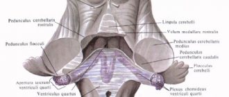
The medulla oblongata is a section of the brain that looks like a truncated cone. The gray matter of the medulla oblongata consists of individual nuclei of the cranial nerves. White matter is the pathways of the spinal cord and brain that extend up into the brain stem and from there into the spinal cord.
The anterior median fissure is located on the anterior surface of the medulla oblongata, flanked by thickened white fibers called pyramids. The pyramids taper downward due to the fact that part of their fibers move to the opposite side, forming an intersection of pyramids that form a lateral pyramidal path. The part of the white fibers that do not intersect form a straight pyramidal path.
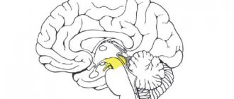
The hindbrain includes the pons and cerebellum. Varoliev bridge or bridge
- a section of the brain, together with the cerebellum, is part of the hindbrain. The pons lies above the medulla oblongata. This is a thickened roller with transverse fibers. The main groove runs through its center, in which the main artery of the brain lies. On both sides of the furrow there are significant elevations formed by pyramidal tracts. The bridge consists of a large number of transverse fibers that form its white matter - nerve fibers. Between the fibers there are many accumulations of gray matter, which forms the nuclei of the bridge. Continuing to the cerebellum, the nerve fibers form its middle peduncles. The cerebellum is located dorsal to the pons and medulla oblongata. The cerebellum has 2 hemispheres and the middle part - the vermis. The surface of the cerebellum is covered by a layer of gray matter (cerebellar cortex) and forms narrow convolutions separated by grooves. With their help, the surface of the cerebellum is divided into lobules. The hemispheres and the cerebellar vermis consist of white matter, it is located medially, and outside is gray matter, which is formed by 3 layers of nerve cells. The white substance when cut resembles a branched tree (the tree of life).
The midbrain consists of the cerebral peduncles and the roof plate (quadrigeminal). The cavity of the midbrain is the cerebral aqueduct. The cerebral peduncles are located between the upper edge of the pons and the diencephalon. Behind the cerebral peduncles there is a quadrigeminal aqueduct between them - it connects the third ventricle with the fourth and is filled with cerebrospinal fluid. The roof plate consists of two upper and two lower hillocks (tubercles), which contain the nuclei of gray matter. The superior colliculi are associated with the visual pathway, and the inferior colliculi with the auditory pathway. From them originates the motor path that goes to the cells of the anterior horns of the spinal cord; the hillocks have the appearance of hemispheres, separated from each other by perpendicular grooves.
The pineal body (epiphysis) is located in the longitudinal groove. A transverse groove separates the pair of superior colliculi from the lower ones. From each mound, a thickening extends from the sides - the handle of the mound. It ends in the geniculate bodies of the diencephalon. A vertical section of the midbrain reveals three sections: the roof, the tegmentum, and the base or peduncles of the brain. The cerebral peduncles are like vertical pillars on which the entire brain rests. The legs diverge towards the brain and form an interpeduncular fossa between themselves; it contains a perforated substance - this is the place of entry of blood vessels into the brain.
The diencephalon is located above the midbrain, under the corpus callosum. The diencephalon is divided into: thalamic brain, hypothalamus, third ventricle.
The thalamus or optic thalamus is a paired, ovoid-shaped formation that consists mainly of gray matter. The medial and superior surfaces are free, and the lateral-inferior surface communicates with other parts of the brain. The thalamus is the subcortical center of all types of sensitivity (pain, temperature, tactile, proprioceptive). The thalamus is the switching point for all sensory pathways coming from extero-, proprio- and interoreceptors.
The hypothalamus or subthalamic region is located below the thalamus. The hypothalamus includes the mammillary bodies, which are the subcortical centers of smell, the pituitary gland, the optic chiasm, the second pair of cranial nerves, the gray tubercle, which is the autonomic center of metabolism and thermoregulation. The hypothalamus contains nuclei that control endocrine and autonomic processes.
The hypothalamus is divided into four parts:
Anterior hypothalamic part
Intermediate hypothalamic part
Posterior hypothalamic part
Dorsolateral hypothalamic part
The third ventricle is the cavity of the diencephalon. It is a narrow slit-like space located in the sagittal plane.
The third ventricle has five walls:
1.The lateral wall is represented by the visual tubercle
2. The lower wall is represented by the subthalamic region and partially by the cerebral peduncles
3. The posterior wall is represented by the posterior commissure and the pineal recess
4. The upper wall is represented by the choroid of the third ventricle
5. The anterior wall is represented by the columns of the arch, the anterior commissure and the terminal plate.