Hospitalization and treatment under the compulsory medical insurance quota. More details after viewing the pictures.
- Aneurysm clipping
A cerebral aneurysm is a local protrusion of the arterial walls. The disease often occurs without symptoms. As the brain aneurysm grows, a significant thinning of the walls of the formation occurs, which can lead to its rupture and the development of hemorrhagic stroke.
Depending on the shape, a cerebral aneurysm can be spindle-shaped or saccular. The exact type of disease can only be determined during a comprehensive diagnosis. Saccular aneurysm is much more common.
Causes of the disease
An aneurysm is a dangerous condition that primarily requires surgical treatment. Otherwise, complications will not be avoided. Experts believe that the disease is a consequence of developmental abnormalities that disrupt the structure of the vascular wall. Often, a cerebral aneurysm is combined with connective tissue dysplasia, polycystic kidney disease, and vascular disorders. The acquired form of the disease can be a consequence of traumatic brain injury. Also, a cerebral aneurysm occurs against the background of atherosclerosis and hypertension.
The vascular wall defect itself occurs gradually. Against the background of degenerative processes, tissue damage or underdevelopment, the area loses its former elasticity. Under the pressure of blood flow, the vascular wall begins to bulge. This is how a brain aneurysm forms. Most often, the formation occurs in the place where the arteries branch and there is the strongest pressure on the vessel wall.
Diagnosis of brain aneurysm
If you suspect a disease, make an appointment at the clinic for diagnosis of a cerebral aneurysm according to the international protocol and start treatment right now. We will arrange for you the assistance of neurosurgeons of the highest class.
Examination for cerebral aneurysms includes MRI, endovascular examination, angiography with contrast or CT angiography, an appointment with a neurosurgeon and preoperative preparation (laboratory tests, ECG, X-ray of the lungs).
What do you get when you seek treatment from us:
- quick and accurate understanding of your problem,
- carrying out all diagnostic measures as soon as possible,
- our consultant is always in touch with you,
- the right strategic decision from a council of top-class specialists, the decision is made not by one doctor, but by the heads of departments and clinics: leading specialists in your problem areas, a specialized surgeon if necessary, a diagnostician - an Israeli general practitioner,
- the best treatment option in the Russian Federation according to international standards, these are the most qualified doctors, and their work according to Western standards, guaranteeing the best result,
- solving all problems at all stages, that is, Dr. Aronov is always “with you”, for everything we will offer the best solution,
- peace of mind, clear steps in treatment - you get all the benefits of Israeli and European medicine in Russia, medical care from an Israeli doctor in Russia.
Symptoms of an aneurysm
Two variants of the course of the disease can be distinguished: apoplexy and tumor-like. The characteristics of the clinical picture depend on the types of aneurysm. The tumor-like form of the disease is accompanied by an active course. In a short period of time, the aneurysm increases in size and compresses important anatomical structures of the brain.
A tumor-like aneurysm is most often localized in the area of the cavernous sinus or chiasm. When the chiasmal region is damaged, visual acuity decreases. Over a long period of time, optic nerve atrophy occurs. If a cerebral aneurysm is located in the cavernous sinus, paresis occurs and damage to the branches of the trigeminal nerve occurs. As the pathology develops, oculomotor disturbances occur and strabismus may appear.
Signs of a ruptured aneurysm
A sharp headache is the first symptom of a ruptured aneurysm. The pain syndrome is initially local in nature. But very quickly the headache spreads and becomes diffuse. In this case, patients note the appearance of nausea and repeated vomiting.
In the future, characteristic meningeal symptoms arise:
- stiffness of the neck muscles;
- Kernig's and Brudzinski's symptoms;
- loss of consciousness.
When a cerebral aneurysm ruptures, subarachnoid hemorrhage occurs. It may be accompanied by focal symptoms, which depend on the location of the formation. The most severe is hemorrhage into the ventricles, which often leads to the death of the patient.
With an aneurysm of the anterior cerebral artery, leg paresis develops, which is often combined with mental disorders. Damage to the middle cerebral artery leads to speech impairment and the development of hemiparesis.
If the pathology is localized in the area of the vertebrobasilar system, during the rupture of the aneurysm, swallowing disorders occur, nystagmus develops in combination with central paresis of the face and trigeminal nerve.
At the first sign of a possible aneurysm, you should immediately seek emergency medical help. It is important to maintain physical rest and avoid overexertion. Specialists will conduct an examination, diagnose, make an accurate diagnosis, and prescribe the necessary medications to reduce the risk of life-threatening complications.
How is it diagnosed?
Since an arterial aneurysm can occur without significant symptoms, it is necessary to pay special attention to the first signs in women and men. A neurologist must diagnose the disease based on the examination, the diagnostic results obtained and the study of cerebrospinal fluid.
During a neurological examination, focal and meningeal symptoms are detected. Based on the location of complaints, it is possible to determine presumably where the pathological focus is located.
The neurologist obtains accurate information about the patient’s condition using the following studies:
- X-ray of the skull. A cerebral aneurysm is characterized by signs of bone tissue destruction.
- Computed tomography, magnetic resonance imaging of the brain.
- Angiography. Basic information about the location, shape and size of the brain aneurysm can be obtained.
- Lumbar puncture. The study reveals blood in the cerebrospinal fluid, which indicates the presence of hemorrhage.
Specialists carry out differential diagnosis of cerebral aneurysm. It is important to make an accurate diagnosis in a short time. This will avoid the adverse effects of a brain aneurysm. It is necessary to differentiate an aneurysm from tumor-like processes, cysts and abscesses.
What you need to know about cerebral aneurysms
An aneurysm is a pathological expansion of the wall of a vessel and filling it with blood.
In the presence of risk factors such as arterial hypertension, congenital vascular disorders, atherosclerosis, previous head injuries, connective tissue pathology, aneurysm rupture is possible, and it can happen suddenly, which is associated with hemorrhage in the brain and serious complications, even death. As a rule, in the absence of various injuries, aneurysms can develop after 50 years, and can be asymptomatic, which is the insidiousness of the disease. Aneurysms can vary in shape, size, or type. They can be derived from one or more chambers, be saccular (or berry-shaped, this is the most common form), fusiform, lateral, arterial or arteriovenous. Small aneurysms reach 11 mm in diameter, and giant aneurysms exceed 25 mm. Why they arise: congenital pathology of the muscular layer of blood vessels may play a role; they can form due to certain hereditary disorders, after head injuries, with tumors, atherosclerosis, due to high blood pressure; smoking and drug addiction are also risk factors. Symptoms usually appear only after the aneurysm ruptures. It usually involves sharp and sudden severe pain in the head. There may also be pain in the eyes, a feeling of numbness or paralysis in the face, and blurred vision. There may be speech impairment, dizziness, hearing loss, nausea, drooping eyelids, etc. Do not hesitate, vascular diseases of the brain are very dangerous and can manifest themselves unexpectedly. Call us or leave a request on the website and sign up for a consultation with a leading specialist and diagnosis of a cerebral aneurysm.
Principles of treatment
Aneurysm treatment is carried out by a neurologist. The operation is performed only by a neurosurgeon. Surgical methods are aimed primarily at preventing aneurysm rupture, which can result in the death of the patient. Conservative therapy for this pathology is used to slow the progression of the disease.
Surgical treatment of a cerebral aneurysm involves two types of main operations: clipping and endovascular occlusion. Surgery prevents the aneurysm from rupturing. A radical method of treatment is the removal of arteriovenous malformation during a microsurgical operation.
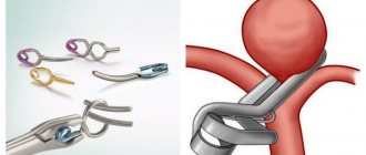
Aneurysm clipping
Aneurysm clipping is an open operation during which the neurosurgeon blocks blood flow in a certain area of the vessel and applies a clip. At the stage of preparation for surgery, specialists prescribe a procedure that allows them to stabilize the patient’s condition and minimize the risk of possible complications. Preventive examination makes it possible to identify possible disorders and diseases that can lead to adverse consequences of surgical intervention.
The operation is performed using modern microsurgical techniques through a small trepanation hole. During surgery, the neurosurgeon prevents rupture of the wall of the malformation.
After isolating the neck of the aneurysm, the specialist places a clip on it. Additional control with Doppler ultrasound allows you to assess the state of blood flow and ensure the effectiveness of the operation.
Treatment of an aneurysm using the clipping method is considered quite complex and should only be performed by an experienced neurosurgeon. A qualified specialist will do everything possible to ensure accurate and technically correct application of the clip. Otherwise, dangerous complications and a long rehabilitation period cannot be avoided.
The clip is applied to the neck of the aneurysm vessel. The accuracy of the neurosurgeon’s actions is confirmed by conducting high-quality diagnostics (Dopplerography) during the procedure.
Endovascular occlusion
During endovascular occlusion of an aneurysm, the neurosurgeon blocks the lumen of the dilated vessel with a special implant. The operation is used when clipping is not possible, for example, when the aneurysm is spindle-shaped. During surgery, a specialist inserts a catheter balloon through the femoral artery using angiographic control. It closes the lumen of the aneurysm. You can also use a microspiral to perform thrombosis. The choice remains with the attending physician. The microspiral in the cavity of the affected vascular area forms blood clots, which clog the lumen of the vessel and shut off the aneurysms from the blood circulation.
A comment
The implementation in Russia of the Federal program for providing medical care to patients with vascular diseases led to the organization of regional vascular centers (RVCs) in almost all regional or republican hospitals. Improvements in non-invasive brain diagnostic techniques have increased the number of patients with cerebral aneurysms before they rupture. Many questions have arisen regarding the management of such patients. The authors of the article provide an overview of world experience on this issue.
In Western European countries, Japan, and Finland, treatment of unruptured aneurysms has long been considered as a way to prevent subarachnoid hemorrhages. In these countries, the number of operations for unruptured aneurysms is constantly increasing. However, to date there are no generally accepted recommendations for the management of patients with unruptured aneurysms.
The introduction of the so-called surgical prevention of SAH in Russia is an extremely urgent task and requires determining the principles of patient selection and choosing a method of treatment for unruptured aneurysms (microsurgery, endovascular neurosurgery).
The article is undoubtedly necessary and important. Subsequently, it is necessary to discuss and prepare a recommendation protocol for the treatment of such patients within the framework of the Association of Neurosurgeons of Russia.
V.A. Lazarev (Moscow)
Rehabilitation after surgical treatment of an aneurysm
The rehabilitation period takes place under the supervision of specialists. Professionals do everything necessary to prevent the occurrence of postoperative complications. The patient remains in the intensive care unit for several days under the constant supervision of specialists. Afterwards, the patient is transferred to a general ward, where strict bed rest must be observed for some time. The return to a normal lifestyle occurs gradually.
If the patient's well-being worsens after surgery, transcranial Doppler ultrasound may be prescribed. Rehabilitation is aimed primarily at preventing vasospasm and increased blood pressure. Specialists also monitor the general condition of the cardiovascular system and, if necessary, prescribe osmodiuretics to eliminate swelling of the brain tissue. Additionally, anti-inflammatory therapy is carried out.
Rehabilitation after surgical treatment of an aneurysm necessarily includes a program to restore impaired body functions and further socialize the patient. The recovery period lasts at least 6 months. With the help of high-quality rehabilitation, it is possible, among other things, to eliminate the adverse consequences of surgical treatment.
Rehabilitation measures include physiotherapy, massage, and the implementation of an individually selected rehabilitation program. After endoscopic clipping, the patient can return to normal life within a few weeks.
Forecast for life
The prognosis of the disease depends on the location of the vascular protrusion and its size. The initial condition of the patient also has a huge impact. The mortality rate in case of rupture of a cerebral aneurysm exceeds 30-50%. Surviving patients sometimes retain the consequences of the condition in the form of movement restrictions, cognitive impairment and a significant decrease in quality of life. Mortality after recurrent hemorrhage exceeds 70%.
Therefore, it is so important to carry out surgical treatment in time, turning to a qualified neurosurgeon. The operation is postponed only if there are certain medical indications.
Successful neurosurgical operations are carried out at the Burdenko Research Institute. Patients with signs of an aneurysm or suspected aneurysm are admitted here. At the Institute of Neurosurgery, it is possible to carry out comprehensive diagnostics using the latest technology and further treatment of identified diseases.