Eye pain is a symptom of a serious illness!
Therefore, do not self-medicate, but consult a doctor. You can make an appointment at a time convenient for you.
Leave your phone number. The clinic administrator will call you back.
Make an appointment
Appointment with an ophthalmologist
from 750 rub.
We all experience eye pain at some point in time. Sometimes it goes away on its own, and sometimes it persists for a long time. Doctors at the Laser Eye Microsurgery Clinic on Maerchak warn that eye pain can be caused by serious diseases. Therefore, if you experience eye pain, consult a doctor.
Causes of eye pain
Eye burn
A burn, regardless of type (chemical, thermal), leads to severe pain, the intensity of which increases over time. The patient cannot open his eyes on his own. Associated symptoms include photophobia, increased lacrimation, eyelid edema, and conjunctival chemosis. In severe burns, hyperemia is replaced by blanching of the eye tissue.
Eye injuries
Eye injury results in severe pain. The patient is unable to open his eyelids without the help of a doctor. The surrounding tissues are swollen and hyperemic. Symptoms are extremely pronounced when the integrity of the cornea is violated. The most common types of traumatic eye injuries are:
- Foreign bodies.
They can be located on the ocular surface or have intraocular localization. In the practice of an ophthalmologist, foreign bodies of the conjunctiva, eyelids and cornea are more often encountered. The location of foreign particles under the upper eyelid injures the cornea when blinking, causing pain. - Corneal erosion.
The appearance of erosive defects is often caused by microtrauma when applying makeup or being hit by a branch. Symptoms are especially noticeable upon awakening and include soreness, flushing, lacrimation, blurred vision and photophobia.
Conjunctivitis
Redness and soreness of the eye are common signs of inflammation of the conjunctiva, regardless of etiology. Therefore, it is very important to pay attention to the specific symptoms of individual forms. Differential diagnosis should primarily be carried out among the following types of conjunctivitis:
- Bacterial.
Pain syndrome is of moderate intensity, most pronounced in the morning. Purulent discharge may appear, which is yellow in color and has a viscous consistency. Patients are unable to open their eyes after sleep. - Allergic.
Inflammation of the conjunctiva is accompanied by itching, pain, and lacrimation. Discomfort increases upon contact with an allergen. In the conjunctival fornix, large follicles are often formed in a “cobblestone” type. - Adenoviral.
This conjunctivitis is characterized by increasing chemosis, hyperemia and pain. The course of the disease is protracted (3 or more weeks). Often the pathological process spreads to the cornea.
Keratitis
The leading symptom of keratitis is severe pain, which is combined with conjunctival hyperemia, swelling of surrounding tissues and photophobia. Common causes of corneal inflammation: contact with infectious patients, failure to comply with personal hygiene rules. The main forms of keratitis:
- Herpetic.
Accompanied by acute pain, blepharospasm, photophobia. The conjunctiva and soft tissues of the eyelids are swollen and hyperemic. Pathognomonic signs are tree-like defects on the surface of the cornea and a decrease in its sensitivity. - Bacterial.
The disease is characterized by an acute onset. The clinical picture includes pain, pericorneal or mixed injection of conjunctival vessels, and photophobia. Mucopurulent discharge is characteristic of keratoconjunctivitis. - Adenoviral.
Patients note decreased visual acuity, pain and swelling of surrounding tissues. Corneal damage is represented by “coin-shaped” subepithelial opacities that persist for a long time (from 2-3 months to 1-1.5 years).
Uveitis
Uveitis is an inflammation of the choroid (uveal tract). Depending on the location of the pathological process, anterior (iridocyclitis) and posterior uveitis are distinguished. Symptoms include pain, decreased visual acuity, and redness of the eye. Anisocoria, lacrimation, and photophobia may develop. With iridocyclitis, patients often report blurred vision.
Ophthalmohypertension
Increased intraocular pressure above 30 mm. rt. Art. may lead to pain in the eye that radiates to the head. Due to diurnal fluctuations in IOP, pain is most pronounced in the morning. An associated manifestation is congestive conjunctival injection. When looking at a light source, rainbow circles appear. A similar clinical picture is observed with angle-closure glaucoma.
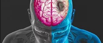
Neurological disorders
Pain syndrome is a common sign of pathology of the central nervous system. In most cases, eye pain is associated with intracranial hypertension, in which patients' vision deteriorates, diplopia, photophobia and blurred vision may occur. At the same time, a decrease in intracranial pressure leads to positional pain, photophobia and double vision. Also, pain in the eye occurs with the following pathologies:
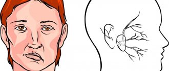
- Trigeminal neuralgia.
Short-term attacks of intense pain or prolonged burning pain on the affected side are typical. The pain is clearly limited to the zone of innervation. The clinic increases with irritation of trigger points. - Supraorbital neuralgia .
The affected area is limited to the supraorbital region, the brow ridge and the lower part of the forehead. The pain is paroxysmal or constant. Lacrimation is determined only on the side of neuralgia. - Optic neuritis.
Patients complain of pain, which increases with eye movements. The clinical picture is represented by deterioration or blurred vision. The anterior part of the eyeball is unchanged. - Ophthalmoplegic migraine.
This is one of the forms of associative migraine, characterized by transient paresis of one or more oculomotor nerves. Pain in the eye spreads to the corresponding half of the head and is combined with diplopia, ptosis, and anisocoria.
Do your eyes always hurt from fatigue?
Sometimes the true causes of eye pain are associated with diseases that are not related to ophthalmology. Pain in the area of the organ of vision can occur due to diseases of the trigeminal nerve, damage to nerve fibers in the area of 1-2 cervical vertebrae. But there are other reasons why your eyes hurt:
- Trochleitis. Pain in the inner corners and above the eyeball often occurs due to inflammatory damage to the oblique eye muscle.
- Spasm of blood vessels in the head. This phenomenon is usually accompanied by pain in the periorbital region and forehead. Sometimes visual disturbances appear in the form of floating dots and light sparks. Vascular spasm often occurs due to changes in weather or overwork.
- Migraine. The condition is manifested by intense headache, increased photosensitivity, dizziness, blurred vision, sore eyes, and the appearance of visual impairment.
- Occipital neuralgia. One of the common types of headaches. Typically, a sharp pain occurs in the neck area, after which it rises higher and radiates to the periorbital area.
- Pathologies of the paranasal sinuses. Inflammation or swelling may be of allergic, fungal, or viral origin. When the sinuses are blocked, a cough, fever, and pain around the eyes appear.
- Cluster headache. One of the most intense headaches. It occurs unexpectedly, and the attacks are prolonged. Severe, sharp pain is usually concentrated near the eyes. It is often accompanied by profuse lacrimation and redness of the conjunctiva.
Sometimes fatigue appears, and the eyes hurt due to reflective pain. If pain occurs in the frontal lobe, it will certainly affect the organs of vision.
Diagnostics
Severe pain in the eye leads to blepharospasm, which makes ophthalmological examination difficult. To alleviate the patient's condition, instillation of analgesics is recommended, after which diagnosis begins. It is important to assess the condition of the eyelids, the shape of the palpebral fissure and the position of the eyes. The following are specific studies:
- Visometry.
Visual acuity is determined at the beginning of the examination of the patient. In the absence of object vision, it is necessary to study light projection. Visometry is performed with and without distance correction. - Non-contact tonometry.
Penetrating eye injuries are often accompanied by hypotension. With iridocyclitis, intraocular pressure increases. It is important to compare the IOP in both eyes and also measure the central corneal thickness. - Biomicroscopy of the eye.
First, a detailed examination of the anterior segment of the eyeball is carried out with mandatory eversion of the upper eyelid. Next, fluorescein staining and examination with a cobalt filter are performed, which allows visualization of small erosive defects. - Ophthalmoscopy.
Fundus examination is carried out after cycloplegia, if intraocular pressure is compensated. The ophthalmologist evaluates the transparency of the optical media and the condition of the retina down to the dentate line. - Ultrasound of the eye.
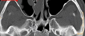
Ultrasound examination is used when there are difficulties in visualizing the structures of the eyeball due to miosis, corneal edema, hyphema or hemophthalmos. The advantage of this method is the ability to detect x-ray negative foreign bodies. - Radiography.
It is performed in case of severe injuries in order to prevent damage to the bone walls of the orbit. The special Komberg-Baltin prosthesis makes it possible to determine the location of intraocular radiopaque foreign bodies.
Eye examination by an ophthalmologist
Prevention
To reduce the likelihood of developing pain in the left eye, it is recommended to adhere to the following rules of prevention:
- increase the duration of walks in the fresh air;
- eliminate the impact of ophthalmological pathologies on the visual apparatus;
- avoid eye strain;
- read and work with small particles in good lighting;
- reduce the amount of time you spend working at the monitor;
- get rid of bad habits;
- take a course of vitamins;
- balance the diet;
- Take medications only under the supervision of your doctor.
Prevention will not completely eliminate the cause of pain in the left eye, but it will significantly reduce the likelihood of the symptom reoccurring.
Treatment
Help before diagnosis
In case of a burn or injury, you must immediately call an ambulance. Before the doctor arrives, in case of a burn, you need to thoroughly rinse your eyes with running water at room temperature. This will minimize exposure to the active substance. For washing at home, it is strictly forbidden to use acids or alkalis for the purpose of neutralization.
In case of injury, it is necessary to ensure functional rest, eliminate visual stress and eye movements. To do this, it is recommended to apply a binocular bandage. Independent removal of a foreign body can lead to expansion of the wound channel and additional trauma. In case of inflammatory diseases, it is important to follow the rules of personal hygiene and avoid uncontrolled use of antibacterial agents.
Conservative therapy
Therapeutic tactics depend on the etiology of eye pain. The prescription of etiotropic and pathogenetic drugs is justified. You should not give preference to physiotherapeutic and folk remedies for the purpose of relieving pain. Conservative treatment may include:
- Analgesics
. Anesthetizing drops are used at the diagnostic stage, as well as before performing subconjunctival injections. In case of severe pain, oral analgesics are indicated, since local agents inhibit the regeneration process. - Antibiotics
. If the pathology is bacterial in nature, instillation of antibacterial agents is indicated based on the result of an antibiotic sensitivity test. Broad-spectrum drugs are used empirically. The average course of treatment is 7-10 days. - Antiseptics
. Prescribed in the presence of small foreign bodies located under the eyelids or in the area of the conjunctival fornix, if there are no signs of inflammation or deep damage to the tissues of the eye. Frequent instillations (every 2-3 hours) are justified for viral conjunctivitis. - Reparants
. These drugs promote corneal regeneration. Tissue repair stimulators are indicated for erosions, post-traumatic defects, and postoperative wounds. The duration of use varies from 10 days to 1 month. - Antihistamines
. For allergic conjunctivitis, pathogenetic therapy is reduced to the prescription of histamine blockers. However, the type of allergen should be determined and etiotropic therapy should be started by an allergist.
Physiological reasons
Physiological disorders provoke pain in the visual apparatus on the left side, which goes away after the impact factors are relieved. The reasons that provoke the development of pain in the area of the left eye are:
- Migraine. Physiological deviation is accompanied by nausea and dizziness. Possible attacks of vomiting. Luminous areas appear before the gaze, and sometimes hyperemia of the eyeball appears.
- Allergic reaction. With a pathological disorder, there is an increase in the production of tear secretion, conjunctival hyperemia and a feeling of scratching. Additional symptoms include the appearance of sinusitis and sneezing.
- Long-term wearing of contact correction products. During the adaptation period to the contact product, soreness, redness and dryness of the mucous membrane develop. When wearing lenses for a long time or the product is unsuitable, a feeling of foreign particles in the eyes develops.
- Temperature increase. In the case of an infectious or viral lesion, hyperthermia develops, which acts as a response to pathogenic microflora. The symptom is localized in any area of the visual apparatus. It stops after the temperature drops to normal values.
- Increased blood pressure levels. Elderly people are especially susceptible to left-sided pain due to the influence of this factor. As pressure increases, fluid accumulates, which provokes the development of complications.
- Fatigue. Emotional shocks and prolonged work on analytical tasks lead to depletion of the visual apparatus. To eliminate pain, you need to increase sleep to 8 hours and take 15-minute breaks from work.
- Disturbances in the patency of the cerebrospinal fluid. In case of deviation, an increase in intracranial pressure is observed. As the physiological disorder progresses, it is recommended to avoid drinks containing caffeine.
- Hematoma. Severe head trauma can lead to pain on one side of the skull. To rule out brain damage, a tomogram is performed.
- Pre-stroke condition. If you have problems in the vascular system, you need to be especially careful about your own health. A sudden headache may indicate the presence of a stroke or mini-stroke.
- Vascular aneurysm. With a physiological deviation, severe pain appears, which is pulsating in nature. The symptom worsens with head movements.
- Inflammation of the membranes of the brain. Inflammatory processes in the head are accompanied by a headache, which increases all the time.
- Neoplasm in the brain. There is increasing discomfort that intensifies. Additional symptoms of tumor development include dizziness and nausea.