What body systems begin to work in a new way?
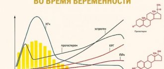
After conceiving a child, global physiological changes occur in a woman’s body. Great changes are observed in the functioning of the endocrine system. The production of hormones of the anterior pituitary gland, which are responsible for the formation and development of the follicle in the ovary, is suppressed. The corpus luteum is formed - a hormone-producing gland that plays a key role in maintaining pregnancy in the early stages.
A temporary gland forms in the right or left ovary after ovulation and produces progesterone. Functions of progesterone:
- prevents the release of new eggs and the development of menstrual bleeding;
- stimulates the growth of the endometrium, prepares the uterine mucosa for the implantation of the fertilized egg,
- ensures implantation of the embryo and its further normal development;
- promotes the formation of a mucus plug in the cervix, which is necessary to protect the embryo from infections;
- reduces the tone of the muscular layer of the uterus, preventing its contraction and miscarriage.
In non-pregnant women, the corpus luteum dissolves two weeks before menstruation. During the period of bearing a child, it exists until 10-12 weeks of gestation, after which the temporary gland ceases to exist and its functions are performed by the placenta.
The functioning of the corpus luteum is ensured by the hormone hCG, which begins to be produced by chorion tissue 6-8 days after the fusion of male and female gametes. HCG enhances the synthesis of sex hormones and corticosteroids produced in the adrenal cortex.
An important hormone during pregnancy is prolactin, which is produced by the pituitary gland. Starting from the 8th week of pregnancy, prolactin production increases. This is necessary to prepare the mammary glands for breastfeeding, as well as for the normal formation of the lung tissue of the embryo.
In the early stages of pregnancy, the functioning of the thyroid gland changes. The organ increases in size and begins to synthesize more hormones that are important for the development of the embryo.
The immune system
During the period of bearing a child, even in an absolutely healthy woman, the body's defenses decrease. This is due to changes in hormonal levels and the body’s adaptation to new conditions.
The likelihood of acute inflammatory processes and exacerbation of chronic diseases increases. Pathologies of the respiratory, digestive, and genitourinary systems may worsen. The situation is often complicated by the fact that many preventive and therapeutic medications are contraindicated during pregnancy.
What can a pregnant woman do to strengthen her immune system?
- Create a useful menu. Fresh vegetables and fruits, dairy products, fish, nuts, green tea - all this strengthens the immune system.
- Lead an active lifestyle and monitor your body weight. Excess weight and inactivity reduce the body's defenses.
- Avoid stress. Emotional experiences suppress the functioning of the immune system.
In addition, during pregnancy it is important to treat any inflammatory processes in a timely manner. All body systems are interconnected. For example, a common cold can cause acute pyelonephritis or other serious illness.
Psycho-emotional changes
Almost all women during pregnancy are subject to sudden emotional changes, because changes in hormonal levels affect the functioning of the central nervous system. The psycho-emotional state of a pregnant woman often worsens during toxicosis. In this regard, a pregnant woman may experience:
- tearfulness;
- irritability;
- dizziness;
- drowsiness.
As a rule, such manifestations are characteristic only of early pregnancy. Good sleep, moderate physical activity, pleasant activities and hobbies will help you cope with emotional stress in the first trimester.
Online consultation with a gynecologist
Online consultation
During the consultation, you will be able to voice your problem, the doctor will clarify the situation, interpret the tests, answer your questions and give the necessary recommendations.
Fetal development by week in the first trimester
The conception of a child occurs during the period of ovulation, after the fertilization of the egg by the sperm. As a rule, ovulation coincides with days 10-16 of the menstrual cycle.
Human intrauterine development is divided into two stages: embryonic and fetal. The first stage is the period from the moment of fertilization of the egg to the tenth week of pregnancy.
During the embryonic stage, important processes occur: crushing and implantation of the embryo into the uterine cavity, the formation of the neural plate (the rudiment of the central nervous system) and its closure into the neural tube, the formation of organs and the placenta.
It is worth distinguishing between embryonic and obstetric stages of pregnancy. The first is counted from the moment of conception, the second - from the beginning of the last menstruation. As a rule, the difference between these periods is two weeks.
It turns out that the formation and ontogenesis of the embryo begins only in the third week of pregnancy. Let's take a closer look at how the fetus develops week by week during the first trimester.
3rd week of gestation
After the sperm penetrates the egg, a zygote is formed - a diploid cell with a set of chromosomes equally obtained from female and male gametes. The duration of formation of a diploid cell is 25-30 hours.
The zygote begins dividing and moving towards the uterus. At the beginning of division, the zygote splits into 2-4 blastomeres (round-shaped cells that form the embryo). The number of blastomeres increases every day.
On the fourth day after conception, the zygote consists of 12-14 blastomeres. The density of embryonic cells increases, and they are closely connected with each other. The embryo enters the uterine cavity.
A blastocyst is formed (an early stage of embryo development), which is implanted into the endometrium of the uterus. During implantation, a pregnant woman may experience uterine contractions and spotting, which is often mistaken for menstruation. However, unlike menstruation, bleeding during implantation is mild and short-lived.
4 week
Implantation accelerates the development of the embryoblast (internal cells of the embryo), resulting in the formation of extra-embryonic organs:
- chorion is a membrane that performs excretory, respiratory and protective functions. After implantation of the embryo, the first chorionic villi begin to appear on its surface. Subsequently, some of the chorion cells destroy the uterine wall and participate in the formation of the placenta;
- amnion is a water membrane that provides the fetus with optimal development conditions and protects it from mechanical stress. Amnion fluid consists of proteins, sugars, mineral salts and other substances;
- the yolk sac, which performs a hematopoietic function. At first, its size exceeds the size of the embryo. After the 12th week of gestation, this temporary organ decreases in size and completely disappears.
The formation of the primary intestine occurs. The organs of the digestive system will subsequently develop from its departments. In the fourth week of gestation, the formation of the liver and pancreas occurs.
After implantation of the embryo into the uterine cavity, placenta rudiments form at the site of attachment of the embryo. The process of its formation will be completed by the 16th week of gestation. The placenta provides the fetus with the necessary oxygen and nutrients, protects against infections, and removes metabolic products.
By the end of the fourth week of gestation, a woman notices that the expected menstruation does not occur. During this period, mood swings, fatigue, and increased sensitivity of the mammary glands may occur.
5 week
By the fifth week, the neural tube appears - the basis of the spinal cord and brain. The heart forms from the bulge in the central part of the fruit. During this same period, hemoglobin begins to form, so the rate of iron consumption by the embryo increases.
The umbilical cord begins to form, which will become the connecting link between the embryo and the placenta. As the fetus grows, the umbilical cord also increases in size.
During this period, a woman may experience the first signs of toxicosis: nausea, vomiting, intolerance to certain foods and odors.
week 6
The embryo’s heart begins to beat, and the thymus gland, which is responsible for immunity, is formed. Formed:
- rudiments of arms and legs;
- cerebral hemispheres;
- eye sockets and ear canals;
- organs of the excretory system.
Vessels are actively growing, blood circulation is established.
week 7
Nerve fibers, esophagus, stomach, and pharynx develop. The embryo develops semblances of hands and primary germ cells. The pancreas begins to produce insulin. The respiratory system is currently represented only by the trachea.
A woman may experience increased signs of toxicosis, so it is important to drink plenty of fluids.
8 week
This period is characterized by the formation of facial features in the embryo, as well as the development of the lower and upper limbs. The retina forms in the eyes, the upper limbs are able to bend at the elbows, and the lower limbs at the knees.
The genitals acquire a characteristic appearance, but this is still not enough to determine the sex of the unborn child.
Week 9
Left and right atria and ventricles appear in the heart. Large blood vessels and endocrine glands develop. The placenta begins to produce hormones. The fingers on the lower and upper extremities are fully formed.
10 week
If the genotype of the embryo contains a gene that initiates the development of the male body, then in the tenth week of gestation the testicles actively develop and they begin to produce the hormone testosterone.
At the same time, the fetus develops the optic nerve, and urine begins to form in the kidneys. The strength of bone tissue increases. The diaphragm forms between the abdominal and thoracic cavities.
11 week
From the 11th week the fetal period of intrauterine development begins. The state of health of most pregnant women stabilizes: the symptoms of toxicosis, irritability, and fatigue disappear.
The fetus has formed the most important organs, the face, and the hemispheres of the brain. Muscle tissue develops. The fetus begins to make movements, but the woman does not feel them yet.
12 week
Taste buds on the tongue are formed. The cerebellum is actively developing - the part of the brain that is responsible for performing targeted movements, their speed and maintaining muscle tone. The rudiments of nails and teeth appear.
The thymus gland is a full-fledged organ in which T-lymphocyte differentiation occurs. The body proportions of the fetus are still incorrect: the head is large, the arms are long, and the legs are short and bent at the knees.
The intestines enlarge and begin to fold into loops. Leukocytes appear in the blood, which protect the body from external and internal pathogenic agents.
The formation of the gonads continues. By the end of the 12-13th week of pregnancy, it becomes possible to determine the sex of the fetus.
Normal fetal movement
The baby in the mother’s belly moves almost constantly. At the 20th week of pregnancy, the fetus makes about 200 movements per day, and between the 28th and 32nd weeks the number of movements reaches 600 per day. Naturally, a pregnant woman does not feel all the movements of the fetus, but only a small part of them. So, after 28 weeks, the frequency of fetal movement, as a woman feels, is usually from 4 to 8 times per hour, with the exception of periods of fetal sleep (3-4 hours in a row).
During the third trimester, a pregnant woman may notice that her baby has certain sleep and wake cycles. Children are usually most active from 19:00 to 4:00 in the morning, and the period of “rest” occurs more often from 4:00 to 9:00 am. Of course, the movements of the fetus depend on the mother’s mood; if the mother is worried or happy, the baby may move more actively, or, conversely, become quiet. The fact is that when a mother is happy, the amount of joy hormones in her body increases significantly - endorphins, which regulate the functioning of the heart and blood vessels, including the blood vessels of the placenta. During times of stress or expressed negative emotions, biologically active substances are also produced - stress hormones; they also affect the functioning of the heart and blood vessels. It is thanks to this biological interaction between the mother and baby that the fetus senses the mother’s condition. When the expectant mother is resting, the baby usually becomes more active; if a pregnant woman is active and busy with some kind of work, the child most often becomes quiet. The movements also change depending on the satiety of the expectant mother. Usually the baby begins to move actively after the mother eats, especially something sweet. At the same time, the level of glucose in the blood increases sharply, which causes the fetus to be more active.
Fetal movements are the language in which the unborn baby speaks to his mother. Naturally, a pregnant woman should listen to movements because in some cases, changes in fetal movements may indicate a violation of its intrauterine state and a not entirely successful course of pregnancy.
If after 20 weeks of pregnancy the expectant mother does not feel the fetus moving, it may be worth consulting a doctor and making sure that everything is fine with the baby.