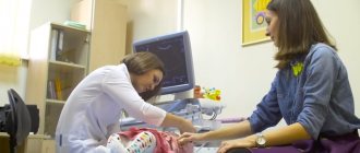
All pregnant women carrying a child must undergo routine ultrasound examinations of the fetus in each of the three trimesters. This is a requirement of the Ministry of Health of the Russian Federation. Fetal ultrasound allows you to fully monitor the condition of the woman and child, while the examination is absolutely safe and most informative.
Appointment with a gynecologist - 1000 rubles. Comprehensive pelvic ultrasound - 1000 rubles. Ultrasound during pregnancy - from 1300 rubles. Appointment based on ultrasound or test results - 500 rubles (optional)
CLICK TO MAKE AN APPOINTMENT or ultrasound tests
Timing of planned ultrasound scans during pregnancy and indications for unscheduled examinations
The content of the article
Routine fetal screenings are carried out within the following framework:
- The first screening is from the 10th to the 14th week.
- The second screening is from the 20th to the 24th week.
- The third screening is from the 30th to the 32nd week.
However, most women undergo the procedure more often. The reasons for non-screening ultrasound are the following:
Up to ten weeks:
- The need to confirm the fact of pregnancy and determine the due date.
- There is experience of miscarriage due to miscarriage, habitual miscarriages, missed pregnancies.
- Pregnancy took place through the use of assisted reproductive technology
- There have been negative experiences with fetal pathologies in the past.
- There was a multiple pregnancy in the family.
- Determination of intrauterine or ectopic pregnancy.
After the first ultrasound, you need to undergo an unscheduled examination if you have:
- Nagging pain in the area of the reproductive organs - lower abdomen.
- Presence of bleeding or unusual discharge.
- The size of the uterus is inappropriate for the given period, the belly is too large.
- Suspicion of leaking amniotic fluid.
- The need to control insufficiency (weakness) of the cervix.
You also need to be examined to confirm or refute the conclusions of doctors from previous ultrasound examinations that showed pathologies.
General indications for all three trimesters:
- Bleeding, discharge.
- Stomach ache.
- A uterine size that is not appropriate for the current week is noticed during an examination by a leading gynecologist - then an ultrasound is necessary to exclude an undeveloped pregnancy
Objectives of ultrasound throughout pregnancy
The main task of fetal ultrasound
- monitoring the growth, development and well-being of the baby. But each trimester has its own characteristics and clarifying tasks.
In the first trimester, the doctor needs to:
- Determine the location of the fertilized egg. If the pregnancy turns out to be ectopic, urgent surgery will be required, otherwise the organ where the embryo is attached will burst over time. In this case, the woman will at least lose her tube, and there are also deaths.
- Specify the deadline. Knowing the due date will allow you to calculate the due date.
- Find out the exact number of fruits. This is important, since multiple pregnancies often occur with pathologies and end in early birth.
- Analyze extracorporeal circulation and calculate the amniotic fluid index.
- Diagnose non-compliance with the norms of fetal development. During this period, you can refuse a pathological pregnancy and have a medical abortion or vacuum aspiration.
In the second trimester, during a fetal ultrasound, the gynecologist will be able to:
- Examine the woman’s uterus to rule out threats of miscarriage.
- Study the features of the umbilical cord, its loops and the presence of entanglements.
- Inspect the amniotic fluid for its quantity and purity.
- An exploration of the maturity and texture of a child's place.
- Diagnose in detail all the formed organs of the fetus, record the number of heartbeats.
- Determine the sex of the child.
- Carry out fetal fetometry in the context of the current period.
- Determine the position of the fetus.
- Study fetal mobility.
In case of serious developmental disorders, a surgical abortion can be performed during this period. By law, such a procedure is available only if indicated.
The third trimester is considered crucial in terms of the quality of childbirth. Using ultrasound, a gynecologist-obstetrician can:
- Monitor placental blood flow and its effect on fetal development.
- Determine the size of the fetus and its correspondence to the given period.
- Study the location and “age” of the placenta
- Record the location of the fetus relative to the uterine outlet
- Consider the presence and number of entanglements of the fetus with the umbilical cord.
- Identify late-onset pathologies and current diseases of the fetus.
- Determine the condition of the amniotic fluid.
- Study the features of the structure and functioning of internal organs, in particular the brain and heart.
- Specify the due date.
The information obtained from ultrasound during this period is important for determining possible options for delivery. If the fetal position is abnormal, the doctor will prescribe a caesarean section. In case of placental abruption, threat of fetal suffocation, etc., artificial early labor can be performed with stimulation of the process.
Changes in the facial part of the skull
Facial assessment should be performed in three planes to evaluate different facial structures as this facilitates the detection of abnormalities (Table 3). According to the ISUOG guidelines, the minimum assessment of the fetal face includes the presence of both orbits, an assessment of the nose/nostrils, the presence of a mouth, and preferably an assessment of the facial profile and lips.
Table 3.
| Plane | Structure | Index | Anomaly |
| Venechnaya | Lips | Integrity violation | Cheiloschisis |
| Mouth | Small or continuous, open | Microstomia or syndromes | |
| Nose | Flattened or one nostril present | Hypoplasia or single nostril syndrome | |
| Palpebral groove | Tilt up or down | ||
| Transverse | Orbits | Small, absent, abnormal interocular diameter | Microphthalmia/anophthalmia, hypotelorism/hypertelorism |
| Medial cyst | Lacrimal sac cyst | ||
| Lens | Echogenic | Cataract | |
| Tooth buds | Clefts, abnormal number | Cleft palate, oligodontia/anodontia | |
| Lower jaw | Small | Micrognathia | |
| Tongue | Missing or double equal sign | Cleft uvula | |
| Ears | Incorrect size, shape, location or unfolded | Ear developmental anomaly | |
| Sagittal | Forehead | Convex | Skeletal dysplasia |
| Flattened | Microcephaly | ||
| impending | Additional nose | ||
| Nose | Flattened | Syndromes | |
| Absent or shortened nasal bone | Aneuploidy | ||
| Upper jaw | Premaxillary protrusion | Bilateral facial cleft | |
| Soft sky | No soft palate or "equals sign" | Cleft soft palate, accessory uvula | |
| philtrum | Long or short | Syndromes | |
| Lower jaw | Small | Micrognathia | |
| Symmetry | Facial asymmetry | ||
| Language | Large, protruding, massively displaced back | Macroglossia, tumor glossoptosis | |
| Ears | Abnormal size, shape or location or unfolded | Ear developmental anomaly |
A cleft is diagnosed when the integrity of the lip is lost on one or both sides on the coronal projection (Fig. 4, 5). Bilateral cleft lip suggests the presence of a premaxillary convexity in sagittal view (Fig. 6). It is difficult to diagnose an incomplete cleft lip (Fig. 7), often only the cleft palate is visible. Using color Doppler, it is possible to see the flow of amniotic fluid that normally passes through the nostrils during breathing, in the case of an abnormality through the palate when it has a cleft.
Figure 4: Second trimester fetus with unilateral cleft lip.
Coronal view of the fetal face shows loss of integrity (arrow) of the upper lip (L).
Figure 5: Second trimester fetus with bilateral cleft lip.
Coronal view of the fetal face shows loss of integrity (arrows) of the upper lip (L) on both sides (1 and 2).
Figure 6: Second trimester fetus with premaxillary bulge.
Sagittal view of the fetal face shows a soft tissue mass (arrow) protruding forward under the nose (N).
Figure 7: Second trimester fetus with a partial unilateral cleft lip. The coronal plane of the fetal face shows partial loss of integrity (arrows) of the upper lip (L).
The facial profile can be assessed using the midsagittal projection. In particular, frontal protuberances (Figure 8), micrognathia (Figure 9) or a flat nose (Figure 10) may be found. From the lateral side, ear abnormalities can be assessed (Fig. 11). Both eyes and their abnormalities can be assessed in the axial plane (Fig. 12-14).
Figure 8: Second trimester fetus with prominent forehead.
The midsagittal view shows the straight convexity (arrow) of the forehead (FH).
Figure 9: Second trimester fetus with micrognathia.
The midsagittal view shows a small and receding (arrow) chin (C).
Figure 10: Second trimester fetus with a flat nose.
Midsagittal view shows a flat (arrow) nose (N).
Figure 11: Second trimester fetus with ear anomaly.
Lateral sagittal view shows a small (arrows) right (R) ear (E) with loss of normal structure.
Figure 12: Second trimester fetus with hypotelorism.
The axial view shows the abnormally reduced distance (arrows) between the two orbits (circle).
Figure 13: Second trimester fetus with bilateral cataracts.
Axial view shows echogenicity (arrows) in the lens (L) of both eyes.
Figure 14: Fetus in the second trimester with a protrusion between the two orbits. Axial view shows a soft tissue mass (arrows) protruding between the two orbits (circles).
It is worth distinguishing between micrognathia (small lower jaw) and retrognathia - displacement of the jaw towards the back. It is differentiated by determining the lower angle of the face in relation to the width of the lower jaw to the width of the upper jaw. If there is a strong family history or if an anomaly is suspected, measurements of fetal structures such as the length of the nasal bones, the length of the ear, the length of the upper jaw, and the diameter of the eye and interocular space may be performed.
Preparation for fetal ultrasound, procedure progress
No special actions are required from the woman to prepare for the ultrasound examination, since all internal organs are displaced by the uterus and do not interfere with the view. But you will have to adhere to several recommendations to make the pregnant woman feel as comfortable as possible.
The doctor will recommend:
- Avoid foods rich in allergens one week before the procedure. Allergies are dangerous not only for the woman - her poor health will certainly affect the behavior of the fetus.
- In a couple of days you need to give up fatty, fried, spicy and salty foods. These products stimulate the secretion of bile and affect the liver - it increases in size.
- For 24 hours, do not consume carbonated water and foods that cause gas formation in the intestines. Air bubbles obstruct vision, distorting echogenicity characteristics.
- Immediately before the ultrasound, control the amount of food and liquid consumed so that the natural urge to empty the intestines and bladder does not constrain the pregnant woman
Failure to follow these recommendations certainly affects the results obtained during the ultrasound, but not so significantly as to refuse diagnosis if you ate a piece of meat or drank a glass of soda the day before.
The procedure is painless. As usual, the woman lies on her back on the couch, and the specialist rubs her stomach, which was lubricated a minute earlier with the gels necessary as conductors. The procedure lasts on average from 10 minutes to half an hour.
Often in the first trimester, a transvaginal ultrasound is performed - a sensor with a condom on it is inserted into the pregnant woman’s vagina. This does not cause pain, the procedure lasts no more than a few minutes. Transvaginal ultrasound may be repeated in the second trimester if a detailed examination of the cervix is necessary.
What can a specialist see on an ultrasound?
First trimester
The first planned ultrasound examination is a mandatory part of screening a pregnant woman and, by law, must be carried out no later than the sixth day of the thirteenth week. Globally, it is aimed at excluding pathologies or risk factors for their occurrence. If you suspect any abnormalities after an ultrasound, you may need an additional examination of the amniotic fluid or a chorionic villus biopsy.
In detail, a study in the first trimester provides the following fetometric data:
- The size and weight of the fetus, which confirms or refutes the expected date. Only three values are taken into account: the distance from the head to the coccyx (KTR), between the parietal bones of the head (BPR), and the volume of the fertilized sac (FV).
- Data on the size and structure of the brain - whether its hemispheres are symmetrical, the presence of the necessary structures.
- Information about the work and structure of the fetal heart.
- Development of the digestive, nervous and cardiovascular systems in accordance with the current week of pregnancy.
- Are the fetal limbs normally developed?
- The presence of chromosomal abnormalities such as Down and Edwards syndrome.
Second trimester
The main purpose of ultrasound from the 20th to the 24th week is to examine the size and organs of the fetus to exclude anomalies and developmental deviations, as well as to monitor the condition of the pregnant woman’s organs to avoid premature birth. The data obtained in many cases helps to maintain pregnancy and improve the vital functions of the fetus using medication.
- Location and structure of the children's place. The specialist can see the presentation, low location (in these cases, measures are required) or normal. Compaction of the structure of the placenta (more than 4.5 cm) indicates hydrops fetalis, diabetes or rhesus conflict. Its thickness serves as an indicator of maturity - if it is less than 2 cm, then premature aging is diagnosed, due to which the fetus does not receive the nutrients it needs. If placental abruption occurs, a woman must be hospitalized urgently.
- The amount and purity of amniotic fluid. Their volume is constantly changing and if oligohydramnios is suspected, it is necessary to check this parameter at unscheduled ultrasounds. The reason for the insufficient amount of water may be leakage or lack of kidneys in the fetus. Polyhydramnios is caused by Rh conflict or infection. The latter is evidenced by the presence of flakes in the waters.
- Vessels, loops and entanglements of the umbilical cord.
- Uterine tone, the presence of myomatous nodes, the condition of scars after previous surgery.
- The cervix is measured to exclude isthmic-cervical insufficiency. Its length during this period should be 35 millimeters, at least 33 for the first pregnancy and 22 for repeated ones. The opening of the cervix is also controlled.
Fetometrics of the fetus:
- Dimensions: length of the femur and ulna, ridge, diameter of the head, chest, abdomen,
- weight in grams and height in millimeters. Based on these data, we can conclude that the physical development of the fetus corresponds to the current period.
- The study of facial bones is aimed at excluding Down syndrome pathologies such as cleft lip or cleft palate.
Internal organs of the fetus:
- The head section is examined for double contour, abnormal shapes, and integrity of the cranial bones.
- The brain is assessed by the size of the ventricles and the presence of tumors.
- The lungs are examined for maturity.
- The structure of the heart, blood vessels, and whether the heart rate corresponds to the norms are checked.
- The presence of abnormalities and defects in the kidneys, digestive tract and diaphragm is examined.
Third trimester
Routine ultrasound screening of the last trimester of pregnancy is carried out from the 30th to the 34th week. Its global goal is to determine the readiness of the fetus and the pregnant woman’s body for childbirth. The results of the study are the most voluminous, which is why the procedure lasts about 30 minutes.
Female reproductive organs:
- Uterus - its walls are examined for tone and thickness, the presence of fibroids and the condition of scars in case of previous operations.
- The length, patency, shape and maturity of the cervix are studied, especially in the context of previously diagnosed isthmic-cervical insufficiency. It must be closed, otherwise there is a risk of premature birth.
- Ovarian health is examined.
- Thickness and degree of maturity of the placenta - its walls must be at least 20 mm and no more than 45. During this period, it must mature to the first degree.
- The umbilical cord is examined for attachment to the placenta, the presence of loops and entanglements, and anechoic formations.
- The volume of amniotic fluid should be no more than one and a half liters.
Fetometrics of the fetus and its correspondence to the normal development of this period:
- Child's height and weight.
- Biparietal, fronto-occipital dimensions, head circumference.
- Size of the tibia, femur, humerus, forearm.
- Abdominal girth.
- Maturity of the lungs - during this period it should be the first.
- Nucleus of ossification of the lower part of the thigh (Beclara).
Internal organs of the fetus:
- Cistern, lateral ventricles and cerebellum of the brain.
- Facial bones are examined to exclude cleft lip, cleft palate and other pathologies. The lips, palate, nasal bone, eye sockets, and nasolabial triangle are examined.
- The structure of the spine is examined for hernias.
- The structure of the heart is studied.
- The structure and compliance with the normal sizes of the organs of the digestive and excretory systems are considered.
- The sex of the child determined in the second trimester is confirmed.
Possible fetal diseases:
- Diabetic fetopathy: polyhydramnios, large fetus, enlarged liver size, universal or intense swelling of the subcutaneous tissue.
- Nonimmune hydrops fetalis.
- Hemolytic disease: swelling of the placenta, due to which there is a double contour of the head, back and hip of the fetus, enlarged liver, hydrocele in boys and others.
Common soft ultrasound markers of fetal malformations
In the chapter on prenatal diagnosis, it was mentioned that often during ultrasound they find “soft” or “small” signs that can be associated with congenital malformations or be a manifestation of the norm. They are found in 5% of all pregnancies. But often such findings cause a stressful situation for the woman, since she does not receive enough information regarding the pregnancy prognosis or the information is false.
Common soft ultrasound signs include an enlarged neck crease and a short nasal bone in the first trimester, which were discussed in the chapter on prenatal genetic screening. In the second trimester, ventriculomegaly, hyperechoic intestine, dilation of the renal pelvis, intracardiac hyperecogenic focus, short femur and others are common.
Ventriculomegaly
Enlargement of the ventricles of the brain is called ventriculomegaly. Very often this brain condition is mistakenly confused with hydrocephalus. Ventriculomegaly (most often we are talking about the lateral (lateral) dilated ventricles) occurs as an isolated symptom in 30-60% of cases, in the rest it is associated with the presence of other developmental defects. This brain condition is found in 0.3-1.5 per 1000 births.
There are several causes of ventriculomegaly. Ventricular dilatation can occur as a result of increased intracranial pressure, disturbances in the exchange of cranial and cerebrospinal fluid, and due to damage to brain tissue. In 5% of cases, the development of ventriculomegaly is hereditary (transmitted through the X chromosome), and can also be a manifestation of complex hereditary syndromes. Primary infection with a number of infections (toxoplasmosis, syphilis, CMV infection, rubella) can cause ventriculomegaly. Most often the cause is unknown.
Until 16-18 weeks of pregnancy, dilation of the ventricles of the brain almost does not occur, so ventriculomegaly is detected most often in the second half of pregnancy. As the fetus grows, it is important to consider the gestational age and know it before diagnosis. The average diameter of the lateral ventricles is 7.6 mm +/- 0.6 mm from 14 to 38 weeks. If the diameter is more than 1 cm (10 mm), they speak of ventriculomegaly.
There are mild or borderline (10-15 mm), moderate or moderate (more than 15 mm, but with preserved cerebellar cortex - more than 3 mm), and severe or acute (ventricles larger than 15 mm and cerebellar cortex less than 3 mm) ventriculomegaly.
Isolated mild and moderate ventriculomegaly does not worsen the outcome of pregnancy, however, in 2-3% of cases, chromosomal abnormalities will be detected, most often Down syndrome. In association with other developmental defects, the pregnancy prognosis is poor, accompanied by stillbirth, the birth of crippled or mentally retarded children.
If ventriculomegaly is detected, it is necessary to conduct a detailed anatomical ultrasound, fetal karyotyping and genetic counseling. The management of pregnancy and delivery will depend on the results of the examination - from observation without intervention to termination of pregnancy.
Hyperechoic bowel
Increased echogenicity of the fetal intestine during ultrasound occurs in 0.1-8% of pregnancies in the second and third trimesters. Some doctors suggest using an echogenicity scale to determine the degree of “normal” and abnormal. The first degree is characterized by insignificant (weak) echogenicity, the second degree is considered moderate, and the third degree is characterized by increased echogenicity, or hyperechogenicity.
The causes of hyperechogenicity may vary and pregnancy risk factors, fetal condition and the presence of other abnormalities must be taken into account. Most often, intestinal hyperechogenicity is associated with infection of the fetus, chromosomal and genetic abnormalities, and fetal hypoxia.
In isolated form, intestinal hyperechogenicity does not affect the prognosis of pregnancy. In the presence of other abnormalities, such an ultrasound sign is combined with intrauterine growth retardation and intrauterine fetal death in almost 25% of cases. In 3-25% of cases, hyperechoic intestine occurs in Down syndrome, and in 3-5% of cases in cystic fibrosis.
If hypercontinuous bowel is detected, the same examination is performed as if other mild ultrasound signs associated with chromosomal abnormalities are detected.
Dilatation of the renal pelvis
Enlargement of the renal pelvis (pyelectasia), unilateral or bilateral, can be associated with many diagnoses, including congenital defects and inherited chromosomal and genetic syndromes. It is found in 2% of pregnancies.
Since the size of the fetus, including its kidneys, increases with the progress of pregnancy, there are still no clear criteria for diagnosing pyelectasis. The most difficult question is what minimal dilatation of the renal pelvis is considered normal. Most doctors consider pyelectasis to be dilation of the renal pelvis greater than 4 mm before 32 weeks and greater than 7 mm after 36 weeks. Enlargement of the renal pelvis greater than 10 mm is considered severe pyelectasis or hydronephrosis. Pyeelectasia occurs twice as often in male fetuses as in female fetuses.
The most common causes of severe pyelectasis and hydronephrosis are blockages (obstructions) of the ureter, when the flow of urine from the kidney is impaired. Less common is lower urinary tract obstruction, most often in boys, due to the presence of a urethral valve (lower urethral obstruction syndrome - LUTO). Pyeelectasia occurs with malformations of the renal system and lower intestine.
Unilateral pyelectasis, if the expansion does not exceed 8 mm, resolves spontaneously in most cases in the third trimester or after childbirth. If the dilation is greater, children often require examination and surgical correction in early childhood.
Bilateral dilatation of the renal pelvis requires a serious approach in the search for anomalies in the development of the urinary system, as well as in the diagnosis of chromosomal and other abnormalities of the fetus. In severe cases, to preserve the kidneys, a puncture of the kidney or bladder and drainage of accumulated urine, as well as shunting (insertion of a probe) to drain urine, are performed. Often after birth, such children require surgical treatment in the first days of life.
Intracardiac hyperechoic focus
An intracardiac hyperechoic focus is a small structural area at the base of the papillary muscles and is often described as microcalcification or micromineralization of the area. Most often, such changes are found in the left ventricle, but sometimes a hyperechoic focus occurs in the right or both ventricles.
An isolated hyperechoic focus occurs in 0.5-20% of pregnancies. On average, up to 7% of pregnancies are accompanied by the presence of such changes. It is believed that in 17-20% of cases such a diagnosis may be erroneous due to technical errors of the ultrasound machine.
Although some doctors argue that such an ultrasound marker can be associated with chromosomal abnormalities, in isolated form, that is, in the absence of other abnormalities, an intracardiac hyperechoic focus is not a sign of the presence of possible chromosomal abnormalities.
Evidence that an intracardiac hyperechoic focus is associated with Down syndrome is conflicting.
Short femur
Measuring fetal thigh length is carried out from the second half of pregnancy. A short femur or short femur is said to occur when the hip dimensions fall outside the 5th to 10th percentile. In small fetuses and those who are stunted, this sign may be a manifestation of the norm.
In 50% of cases where such a deviation is found, there are other developmental abnormalities, and in 20% of cases, deviations in fetal growth are found without the presence of anomalies. A femur that is too short may be associated with skeletal dysplasia (15% of cases) or chromosomal abnormalities, including Down syndrome.
Isolated short femur is not associated with chromosomal abnormalities.
When a short femur is detected, it is always important to clarify the gestational age, exclude fetal growth restriction, the presence of other developmental abnormalities and chromosomal abnormalities.
Share link:
- Click to share on WhatsApp (Opens in new window)
- Click to share on Telegram (Opens in new window)
- Click here to share content on Facebook. (Opens in a new window)
- Click to share on Twitter (Opens in new window)
- Click to share on Skype (Opens in new window)
- Send this to a friend (Opens in new window)
- Click to print (Opens in new window)
By
Detailed explanation of fetal ultrasound results, norms and pathologies
Since the goals and subjects of medical interest in ultrasound examinations of different trimesters differ, the abbreviations and abbreviations in the reports are different.
First trimester
Estimated date of birth (EDD) - in fact, this is the same as the exact date of pregnancy, only from a different angle. Determined by data on the menstrual cycle and the size of the fetus
- The cervical-neck space (CN) is the main criterion for ultrasound examination in the first trimester. The norm is from 1.5 mm at the 10th week to 2.7 mm at the 13th. If the indicator is increased, the pregnant woman needs to undergo further examination.
- The coccygeal-parietal size (CPR) is one of the main parameters for assessing the size of the fetus. Normally, it should range from 41 mm at week 10 to 73 mm at week 12
- Heart rate - (h/s, heart rate) normally for the first trimester should be 146-179 beats per minute, and this figure is lower the longer the pregnancy period
- Nasal bone – starting from the 12th week should be at least 3 mm, otherwise Down syndrome is suspected
- Biparietal size (BSD) should be from 14 mm at 10 weeks to 26 and higher at 13.
- The yolk sac is studied as an indicator of gestation-appropriate development. It grows in the first weeks and then decreases. Should be round in shape and 4-6 mm in diameter. If the shape is different, and the internal length of the incision is less than 2 mm at 8-12 and more than 6 mm after 10 weeks, a frozen pregnancy is stated.
NSG cyst - Protocol
The brain structures are formed correctly and asymmetrically. The furrows and convolutions are pronounced. In the projection of the lobe, an anechoic formation is visualized - mm, communicating with the cavity of the lateral ventricle.
The lateral ventricles are asymmetrically dilated. Anterior horns of the lateral ventricles: on the right - 3 mm; on the left - 3 mm (norm up to 5 mm). Body of the lateral ventricle – on the right – 3 mm; on the left - 3 mm (norm up to 5 mm). Temporal horns are not detectable (normal). Atrium - filled with choroid plexuses (normal up to 12-16 mm). Occipital horns are not detected (normal).
Periventricular tissues and areas of the basal ganglia have increased echogenicity.
CONCLUSION: cyst in projection
Norms of fetal ultrasound indicators in the first trimester in the summary table
| Term | BPR | Yolk sac | Nasal bone | Heart rate | KTR | VP |
| 10 weeks | 14 mm | 4-6 mm, but not less than 2 | Yes, but not measured during this period | From 161 to 179 beats/min | From 41 mm | 1.5 to 2.2 mm |
| 11 weeks | 17 mm | 4-6 mm, begins to decrease | Yes, but not measured during this period | From 153 to 177 beats/min | 1.6 to 2.4 mm | |
| 12 weeks | Not less than 20 mm | Disappears | Not less than 3 mm | From 150 to 174 beats/min | up to 73 mm | 1.6 to 2.5 mm |
| 13 weeks | Approximately 26mm | Disappears | Not less than 3 mm | From 141 to 171 beats/min | 1.7 to 2.7 mm |
Second trimester
The ultrasound report from the 20th to the 24th week contains the following abbreviations:
- BPR – biparietal size of the fetal head;
- LZR – fronto-occipital size;
- BPR/LZR – cephalic index;
- AB – abdominal circumference;
- RS – heart size;
- DG – tibia length;
- DP – shoulder length;
- DB – thigh length;
- OG – head circumference;
- s/b or heart rate - heartbeat;
- IUGR – intrauterine growth restriction;
- DgRK – chest diameter;
- AFI - amniotic fluid index - an indicator of the amount of amniotic fluid;
- ICI - isthmic-cervical insufficiency - the cervix with this pathology is not long enough for full gestation.
Classification of fetal anomalies
Developmental pathologies include:
- abnormalities in the development of the neural tube - absence of the cerebral hemispheres, hydrocephalus, cerebral herniation, microcephaly;
- spinal defects - myelomeningocele, spina bifida, cystic hygroma;
- heart pathologies - abnormal position of the heart, underdevelopment of the organ, ventricular septal defect;
- anomalies of the gastrointestinal tract - atresia of the cardia, duodenum, jejunum;
- malformations of the anterior abdominal wall - omphalocele, gastroschisis, ascites in the fetus;
- congenital kidney anomalies - underdevelopment, urinary tract obstruction, polycystic disease;
- pathologies of the amount of amniotic fluid - oligohydramnios, polyhydramnios;
- intrauterine fetal death.
Neural tube malformations
- Anencephaly is a pathological condition characterized by the congenital absence of the cranial vault and cerebral hemispheres. This is the most common pathology of the central nervous system in the fetus. It is diagnosed at 11-12 weeks. As a rule, the defect is combined with hydramnios and other disorders. The level of alpha-fetoprotein in the mother's blood increases.
- Signs of hydrocephalus in the form of expansion of the anterior and posterior horns of the lateral ventricles will be visible at 18 weeks of pregnancy.
- Microcephaly - a pathological reduction of the fetal head - occurs when the biparietal size decreases by more than three standard deviations. To rule out intrauterine growth retardation, the doctor will compare the size of the head with the size of the body of the unborn child. In isolated form, the defect is rare, and in borderline cases, the diagnosis is quite difficult to make. In this regard, the woman is recommended to undergo an ultrasound again in a few weeks. The data obtained are interpreted quite carefully, especially if the fetus grows and develops normally.
- Encephalomeningocele usually appears as a rounded protrusion in the area of the calvarial bones. Inside the protrusion is fluid or brain matter. Most often, anomalies are localized at the back of the head, but sometimes anterior localization of the defect occurs. To minimize the likelihood of diagnostic error, if an encephalomeningocele is suspected, the woman will be advised to undergo a repeat ultrasound, possibly from a different doctor.
Anomalies of the spine
Most often, fetal pathologies in the form of anomalies in the development of the spine are localized in the cervical and lumbar region. Since the spine is clearly visualized from the 15th week of pregnancy, its defects are diagnosed at 18-20 weeks, during the second screening ultrasound.
- Myelomeningocele on ultrasound is visualized as a sac containing fluid and elements of the spinal cord. In rare cases, an open defect is diagnosed, in which only protrusion of soft tissue through the defect is detected. Such anomalies are particularly difficult to detect.
- Spina bifida in the fetus is diagnosed using expert-class, high-resolution ultrasound machines. Normally, the posterior ossification centers of the spine are visualized as two hyperechoic, almost parallel structures. In spina bifida they are displaced laterally.
- Cystic hygroma is a pathology of the development of the lymphatic system. In this case, an ultrasound reveals a cyst with septa located in the cervical spine. Sometimes internal partitions are visible, resembling the spokes of a wheel. The skull, brain and spinal cord, unlike other anomalies, develop normally. Sometimes cystic hygroma is combined with a generalized anomaly of the lymphatic system. In most such cases, the fetus dies.
Congenital heart defects
Diagnosis of congenital heart defects requires special equipment. The doctor should be able to perform Doppler ultrasound. If congenital heart defects are suspected, an expert opinion is required. In difficult cases, clinicians are warned about possible complications so that they are ready to provide specialized care at the birth of the child.
Anomalies of the development of the gastrointestinal tract
The most common pathology of the development of the digestive tract is duodenal atresia. In this case, on an ultrasound, the doctor sees round structures similar to cysts in the upper part of the fetal abdomen. If the cyst is located on the left, it is an enlarged stomach, on the right - the duodenum. This is the so-called “double bubble” sign. Very often the pathology is combined with polyhydramnios, anomalies of the heart, kidneys, and central nervous system.
Diagnosis of small bowel atresia is difficult. In the upper part of the fetal abdomen, the doctor sees cystic structures - overstretched loops of the small intestine. As a rule, fetal pathology is detected in the middle of pregnancy or at a later stage. With high atresia, polyhydramnios is usually diagnosed. Colonic atresia is almost impossible to diagnose using ultrasound.
Pathology of development of the anterior abdominal wall
The most commonly diagnosed defect in this group is an anterior abdominal wall defect - omphalocele. In the hernial sac, formed by the amniotic membrane and the parietal peritoneum, intestinal loops, part of the liver, stomach and spleen are found.
Other pathology is mainly localized in the right periumbilical region (gastroschisis) and is usually isolated. Only intestinal loops that are not covered by the amniotic membrane prolapse through this defect.
Ascites in the fetus
Free fluid is visualized on ultrasound as an anechoic zone surrounding the internal organs of the unborn child. If ascites is suspected, the doctor must carefully examine the fetus and evaluate its anatomy to exclude combined defects. The urinary system must be carefully examined, as ascitic fluid may be urine. If the unborn child has thickened skin or fluid is contained in at least two natural cavities, they speak of hydrops fetalis.
Its reasons are:
- Rhesus conflict;
- congenital heart defects;
- arrhythmias;
- obstruction of blood or lymphatic vessels.
To find out the exact cause of dropsy, the doctor will recommend an additional expert ultrasound to the woman.
Abnormalities of the urinary system
Some congenital defects of the urinary system are incompatible with life. If such a pathology is detected in the early stages, the doctor may advise the woman to terminate the pregnancy. If the anomaly was diagnosed late, the doctor may change the pregnancy management tactics.
Diagnosing agenesis, or absence of kidneys, is difficult due to the significant enlargement of the adrenal glands. In the last weeks of pregnancy, these glands can take on a bean-shaped shape, which makes diagnosis even more difficult. The bladder is small or absent altogether. To make an accurate diagnosis, you need to examine the fetus in several planes. Measuring the kidneys during an ultrasound helps to identify hypoplasia - underdevelopment of the organ.
Obstruction, hydronephrosis is manifested by dilation of the renal pelvis. However, it must be borne in mind that the expansion of the renal pelvis may be transient. Such dilations are most often bilateral and disappear after some time. If hydronephrosis is suspected, an ultrasound scan should be repeated after two to three weeks.
Pathological bilateral obstruction of the urinary system is usually combined with oligohydramnios and has a poor prognosis. If the obstruction is unilateral, the amount of amniotic fluid remains within normal limits.
With a multicystic kidney, ultrasound will reveal several cysts of varying diameters. They are located diffusely, less often in one part of the organ. Renal parenchyma can be detected between the cysts, although it is not clearly visualized. Autosomal recessive polycystic kidney disease is diagnosed in the third trimester of pregnancy. In such situations, there is a family history and oligohydramnios. On ultrasound, the kidneys are enlarged in size and their echogenicity is sharply increased.
Amniotic fluid
The most common causes of polyhydramnios are:
- high obstruction of the digestive tract;
- neural tube pathology;
- hydrops fetalis;
- defects of the anterior abdominal wall;
- skeletal dysplasia of the chest;
- development of several embryos simultaneously;
- diabetes mellitus in the mother.
The causes of oligohydramnios are:
- bilateral renal anomaly;
- damage to the amniotic membranes with leakage of water;
- intrauterine growth retardation;
- post-term pregnancy;
- antenatal fetal death;
- intrauterine developmental delay;
- post-term pregnancy;
- intrauterine embryonic death.
Oligohydramnios with bilateral renal anomalies is a poor prognostic sign.
Norms of fetal ultrasound indicators in the second trimester in the summary table
| Term | BPR | LZR | coolant | DP | DG | OG | Heart rate | DgRK | DB |
| 20 weeks | From 43 to 53 mm | From 56 to 68 mm | From 124 to 164 mm | From 26 to 34 mm | From 26 to 34 mm | From 154 to 186 mm | 120-160 beats/min | About 48mm | From 29 to 37 mm |
| 21 weeks | From 46 to 56 mm | From 60 to 72 mm | From 137 to 177 mm | From 29 to 37 mm | From 29 to 37 mm | From 166 to 200 mm | 120-160 beats/min | About 50mm | From 32 to 40 mm |
| 22 weeks | From 48 to 60 mm | From 64 to 76 mm | From 148 to 190 mm | From 31 to 39 mm | From 31 to 39 mm | From 178 to 212 mm | 120-160 beats/min | About 53mm | From 35 to 43 mm |
| 23 weeks | From 52 to 64 mm | From 67 to 81 mm | From 160 to 201 mm | From 34 to 42 mm | From 34 to 42 mm | From 190 to 224 mm | 120-160 beats/min | About 56mm | From 37 to 45 mm |
| 24 weeks | From 56 to 67 mm | From 71 to 85 mm | From 172 to 224 mm | From 36 to 44 mm | From 36 to 44 mm | From 201 to 237 mm | 120-160 beats/min | About 60mm | From 40 to 48 mm |
Third trimester
The last planned ultrasound examination is the most extensive and consists of measuring and studying the following fetal data:
- BPR – biparietal head size;
- LZR – fronto-occipital size;
- OG – head circumference;
- AB – abdominal circumference;
- DG – length of the tibia;
- DB – length of the femur;
- DP – length of the humerus;
- Height – 430 -470 mm;
- Weights – 1400 – 2400 grams;
- Beclair's nuclei - with normal development less than 5 mm;
- Thickness of placenta;
- The maturity of a child's place;
- Placenta placement, its presentation and the presence of abruption;
- AFL - the amount of amniotic fluid - 1-1.5 l is considered the norm;
- The length of the neck is considered normal to be from 30 to 35 mm.
Fetal fetometry: table of body measurements of the unborn baby
Fetal ultrasound The table of fetal fetometry indicators is the norms of various sizes of an intrauterine developing child, determined by ultrasound . It was compiled on the basis of measurements taken in a large sample and subsequent statistical analysis of the results obtained. It is this table that is the basis for determining the duration of pregnancy.
In addition, by comparing the sizes obtained for a particular child with the norms, the symmetry of its development and compliance with the calculated gestational age are assessed. The content of the article
- How to prepare for research
- How is the procedure for measuring baby parameters carried out?
- Which table will help you find out what age your baby is at?
- What parameters are assessed from week 13
- What is "norm"
How to prepare for research
The measurement of the developing baby in utero is carried out during an ultrasound. This study can be performed either with a transvaginal sensor or through the anterior abdominal wall.
If the procedure will be performed through the vagina, there is no need to prepare for fetal fetometry. If you are planning a transabdominal ultrasound, then in the early stages of pregnancy you will need to drink about half a liter of water an hour before the examination and not go to the toilet after that.
This is necessary so that through a full bladder, like through a window, you can look into the uterine cavity. After 12 weeks, this is no longer required, since the necessary “window” is created by amniotic fluid.
How is the procedure for measuring baby parameters carried out?
A woman comes to the ultrasound room, where she undergoes a regular ultrasound examination - through the wall of the abdomen (then the abdomen is lubricated with a special gel) or through the vagina (a condom is put on the sensor).
Next, the doctor performing the ultrasound also performs fetometry: measures the main indicators of circumferences, diameters and lengths. Then these indicators are compared with standard fetal fetometry tables by week.
Which table will help you find out what age your baby is at?
The fetal fetometry data table consists of quite a few indicators. From 5 to 13 weeks, only three fetal sizes are assessed by ultrasound:
- coccygeal-parietal size (CRL, KTP) is the maximum distance that can be measured from the head to the coccyx
- biparietal distance (BPD) can be measured from one parietal bone to the other, that is, we can say that this is the diameter of the head
- the fertilized egg (FE) is the volume of the cavity in which the baby develops; later the placenta and water around it will appear there.
| Term | PY, mm | BPD, mm | CRL, mm |
| 5 | 5 | 1-3 | |
| 6 | 13 | 4-6 | |
| 7 | 21 | 3 | 8-10 |
| 8 | 29 | 7 | 14-16 |
| 9 | 36 | 10 | 22-23 |
| 10 | 44 | 14 | 31-32 |
| 11 | 51 | 17 | 41-43 |
| 12 | 57 | 20-22 | 42-73 |
| 13 | 63 | 24-26 | 51-87 |
This is what the table of fetal sizes looks like according to ultrasound, with which comparison is made to determine whether it corresponds to the norm.
What parameters are assessed from week 13
The size of the child according to ultrasound is estimated based on:
- OG – head circumference (girth)
- BDP – biparietal size (BPD), that is, the distance between the most distant points of the parietal bones
- DB – femur lengthwise
- LZR – fronto-occipital size, that is, the distance between the most distant points of the forehead and the back of the head
- OB – circumference (girth) of the abdomen
- SDA - abdominal diameter in the anteroposterior direction, that is, sagittal size
- PJ is the diameter of the abdomen in diameter
- OG (or DrGK) – circumference (girth) of the chest
- DP – humerus length
- There are also lengths of the tibia, fibula, radius, ulna, and foot. Each of them has its own norm.
You can evaluate pregnancy using fetometry by comparing the indicators indicated in the transcript of your personal ultrasound with the standard parameters in the available tables.
Fetal ultrasound
It is especially important to compare the BDP or coccygeal-parietal distance - these 2 criteria are fundamental for determining the gestational age using ultrasound.
| Duration in weeks | LZD | BPD | OG | coolant | SJ | PJ | DB | DP | Dl.M.Bertz | L.B.berts | DgrK |
| 13 | 32 | 96 | 67 | 21 | 22 | 11 | 10 | 8 | 9 | 24 | |
| 14 | 35 | 26 | 106 | 78 | 24 | 25 | 15 | 13 | 11 | 12 | 26 |
| 15 | 39 | 32 | 118 | 89 | 28 | 29 | 18 | 17 | 14 | 15 | 28 |
| 16 | 43 | 35 | 130 | 100 | 31 | 32 | 21 | 20 | 17 | 18 | 34 |
| 17 | 47 | 39 | 143 | 111 | 35 | 36 | 24 | 23 | 20 | 21 | 38 |
| 18 | 52 | 42 | 155 | 122 | 38 | 39 | 27 | 26 | 2 | 24 | 41 |
| 19 | 56 | 44 | 168 | 132 | 42 | 43 | 30 | 28 | 25 | 26 | 44 |
Fetal fetometry at 20 weeks presents the following indicators (data in mm)
In the photo: embryo on ultrasound at 6-7 weeks of pregnancy
- BPR: 47-49
- OG:181
- DGrK: 48
- LZR: 60
- Coolant: 143
- PJ: 46
- SJ: 45
- thigh length: 33
- normal length of tibia: 29
- fibula length: 28
- shoulder length: 31
- length of forearm bones (radius/ulna) = 25/28
- foot of a 20-week fetus – 33.
Fetal fetometry at 22 weeks is represented by the following indicators in millimeters
- BPR: 54-56
- OG: 206
- DGrK: 53
- Coolant: 165
- PJ: 53
- SJ: 52
- hip: 39
- fibula/tibia (tibia): 33/34
- shoulder: 36
- Radius/ulna = 30/33
Fetometric parameters at 23 weeks (indicators in millimeters)
In the photo: embryo on ultrasound at 6-7 weeks of pregnancy
- BPR: 57-59
- OG: 218
- DGrK: 56
- Coolant: 175
- PJ: 55
- SJ: 56
- hip: 41
- shin = 35/36
- shoulder: 38
- forearm: 32/35
- height: 31 cm
- 580-600 g.
Fetal fetometry upon reaching 31 weeks (figures are in mm)
- BPR: 80-82
- OG: 301
- DGrK: 81
- Coolant: 258
- PJ: 82
- SJ: 81
- hip: 60
- shin = 51/52 mm
- shoulder: 54
- forearm bones = 44/50
- height: 42 cm
- body weight: 1590-1690.
Fetometry norms at 32 weeks on average look like this (mm)
- BPR: 83-85
- OG: 309
- DGrK: 83
- Coolant: 266
- PJ: 85
- SJ: 84
- hip: 62
- shin = 52/54
- shoulder: 55
- forearm bones = 46/52 mm
- height: 43
- weight: 1790-1890.
Fetal fetometry at 33 weeks can be presented in the form of the following table
| BPR | OG | coolant | SJ | PJ | Hip | M. Berts/B. Berts | Shoulder | Beam/Elbow | Weight – 2030-2130 gHeight – 44-45 cm |
| 87 | 316 | 276 | 87 | 88 | 64 | 54/56 | 57 | 47/53 |
Fetometric data at 34 weeks in millimeters
- maximum distance between parietal bones (BPD): 89
- head circumference (OG): 323
- chest diameter (DGrK): 88
- abdominal circumference (AC): 285
- PJ: 91
- SJ: 90
- hip: 66
- shin = 56/57 mm
- shoulder: 59
- forearm bones = 48/55
- height: 46-47 cm
- body weight: 2270-2390.
Fetometry at 36 weeks (mm)
- BPR: 92
- OG: 335
- DGrK: 94
- Coolant: 303
- PJ: 97
- SJ: 96
- hip: 70
- shin bones: 58/60
- shoulder: 61
- forearm bones = 50/57
- height: 49 cm
- body weight: 2750-2860.
Fetometry at 37 weeks
- BPR: 94
- OG: 339
- DGrK: 97
- Coolant: 311
- PJ: 100
- SJ: 98
- hip: 71
- shin: 60/62
- shoulder: 63
- forearm bones = 51/58
- height: 50 cm
- weight: 2970-3090.
The main parameters that are important for the correct assessment of the baby in the first two trimesters are abdominal circumference, biparietal size and thigh length.
After this period, just before childbirth, the chest circumference, abdominal circumference and length of the femur become more important.
What is "norm"
In the photo: 3D ultrasound of a child
In the photo: 3D ultrasound of a child
It is considered normal if your child’s data corresponds to the size of the fetus according to ultrasound weeks, and if it lags behind or exceeds the period calculated by your gynecologist, then by less than 14 days.
That is, this will not yet be considered either a delay or an advance in development. Evaluate yourself, find out what weight you and your husband were born with: maybe just one of you is short, and the child inherited this trait.
It is also normal if the indicators are not on the same line, but have a slight scatter, which occurs because the child grows spasmodically.
If, for example, everything “fits” into the norm, except for the length of the femur and tibia, this does not indicate pathology. Perhaps the child's parents or older relatives do not have long legs.
A situation is considered dangerous when, not for the first time, it is noticeable from fetometry that one of the circles is more or less than 2 lines in comparison with other indicators. This should be considered a health hazard.
For example, if the head circumference increases, this may indicate intrauterine hydrocephalus; if it corresponds less and less to the deadline, this may indicate another brain pathology. In this case, measurement of the baby’s size by ultrasound is supplemented by Doppler measurements, CTG and mandatory dynamic monitoring.
However, making a diagnosis of “Intrauterine growth retardation” or “Hypotrophy” is the responsibility of a joint assessment of ultrasound fetometry parameters by two doctors: an obstetrician-gynecologist and a medical geneticist.
Only these specialists can assess your genetic predisposition, and also, in case of a disappointing diagnosis, will be able to find the causes of this pathology. So, these can include chromosomal abnormalities, bad habits, the age of one or two parents, and an infection acquired in utero by a developing child.
Considering all of the above, it is necessary for an obstetrician-gynecologist to decipher the indicators of this study: the ultrasound doctor only provides him with data in which he indicates what and how different it is from the norm.
Thus, what the table of fetal fetometry indicators represents is averaged data. They are used to determine whether the gestational age calculated by the obstetrician corresponds to that obtained from these data. Using the same parameters, they look at whether the baby’s growth is symmetrical (when everything is 1-2 lines lower or higher), or, conversely, asymmetrically. This allows you to identify some developmental defects in time.
ATTENTION! The information on the site is for reference or popular information only. Correct treatment and prescription of medications can only be carried out by a qualified specialist, taking into account the diagnosis and medical history.
You can always make an ultrasound appointment with an ultrasound doctor in Sochi at the medical ]VegaMed[/anchor], ultrasound prices start from 400 rubles, you can visit an ultrasound doctor on the same day as the clinic has free rooms for receiving patients.
You can make an appointment by calling +7 (862) 295-00-95 We also invite you to follow us on Instagram You can use WhatsAPP Make an appointment without a phone call to the medical center Click here to sign up
Norms of fetal ultrasound indicators in the third trimester in the summary table
| Duration, weeks | BPR | LZR | OG | coolant | DG | DB | DP | Height | Weight | Placenta thickness | Placenta maturity |
| 30 | From 71 to 85 mm | From 89 to 105 mm | From 265 to 305 mm | From 238 to 290 mm | From 49 to 57 mm | From 52 to 62 mm | From 49 to 57 mm | 270 mm | 1400 g | 13.6 to 28.6 mm | 1st degree |
| 31 | From 73 to 87 mm | From 93 to 109 mm | From 273 to 315 mm | From 247 to 301 mm | From 51 to 59 mm | From 54 to 64 mm | 50 to 60 mm | Up to 390 mm | 1500-1600 g | 17.4 to 29.7 mm | 1st degree |
| 32 | From 75 to 89 mm | From 95 to 113 mm | From 283 to 325 mm | From 258 to 314 mm | From 52 to 60 mm | From 56 to 66 mm | From 52 to 62 mm | Up to 400 mm | Up to 1700 g | From 18.1 to 30.7 mm | 1st degree |
| 33 | From 77 to 91 mm | From 98 to 116 mm | From 289 to 333 mm | From 267 to 325 mm | From 54 to 62 mm | From 58 to 68 mm | From 53 to 63 mm | Up to 430 mm | Up to 1800 g | 18.8 to 31.8 mm | 1st degree |
| 34 | From 79 to 93 mm | From 101 to 119 mm | From 295 to 339 mm | From 276 to 336 mm | From 55 to 63 mm | 60 to 70 mm | From 55 to 65 mm | 430-450 mm | Up to 2400 g | 19.6 to 32.9 mm | 1-2 degree |
Pathologies visible on fetal ultrasound by trimester
In the first trimester, pathologies that arise due to chromosomal abnormalities are detected - frozen pregnancy and lack of embryo growth. In addition to them, signs of Down, Patau, Edwards, Cornelia de Lange, Smith-Opitz syndromes, as well as triploidy and omphalocele are clearly visible.
In the second trimester, in addition to the syndromes of the first trimester, Shershevsky-Turner, Klinefelter, hypertensive-hydrocephalic syndromes, as well as types of polysomy, polyploidy, deformations or absence of internal organs, heart defects, facial bone defects, hypertensive-hydrocephalic, multiple births, poly- or oligohydramnios, umbilical cord pathologies, fetal presentation, intrauterine growth retardation.
In the third trimester, infections of the fetus, defects of the brain, heart muscle, internal organs, intrauterine growth retardation, cleft lip, cleft palate, and fetal presentation may be detected.
The earlier fetal pathologies are detected, the better. If chromosomal abnormalities are diagnosed, the pregnant woman will likely be offered termination of pregnancy in the first and second trimester. Acquired pathologies are most often treated with medication during pregnancy and, if necessary, after the birth of the child.
Experts offer solutions to the problem, and future parents choose them, taking into account all the risks. Each situation is individual and there are no uniform schemes of action. But even if it happened that the pregnancy turned out to be problematic, you need to understand that the level of development of medicine and plastic surgery makes it possible to correct most abnormalities.
Third planned ultrasound at 30-32 weeks of pregnancy
Ultrasound examination of the fetus during pregnancy is a mandatory examination method in a complex of diagnostic measures. This technique is used 3 times during the entire period of pregnancy (in the 1st, 2nd, and 3rd trimesters). Ultrasound diagnostics makes it possible to assess the development and condition of the fetus at various stages of pregnancy, identify pathological conditions, including genetic diseases, and, if necessary, take a set of measures to maintain pregnancy. These studies also provide important information that allows the specialist to select the most appropriate method of labor management.
The third trimester of pregnancy begins at 28 weeks. An ultrasound examination is usually performed at 30, 31 or 32 weeks. It is during this period of time that the information is most revealing and allows for the necessary assessment of the condition of the fetus.
Why is an ultrasound performed at 32 weeks of pregnancy?
It is carried out with the aim of definitively determining the presentation, position, position and type of position of the fetus. They also perform a number of measurements - fetometry (determine the child’s weight, body size, head size, height, etc.) - and compare the indicators with the average normal values. All received data is carefully analyzed, and if necessary, specific measures are prescribed. The issue of delivery tactics is being resolved.
Ultrasound at 32 weeks
allows you to diagnose and analyze the functioning of the fetal heart. The heart rate and rhythm are assessed.
These results will give an idea of the current state of the child, whether he is developing correctly and whether his development is on time.
Where to get an expert fetal ultrasound in St. Petersburg
You can undergo such an examination only in modern clinics with high-tech equipment. Such a specialized medical center in St. Petersburg is located on Zanevsky Prospekt, 10. The latest device with Doppler, 3D, 4D, etc. functions is installed here. The examination is carried out by doctors of the highest category with extensive experience in maternity hospitals, antenatal clinics and gynecological clinics. The cost of the examination depends on the period and starts from 1000 rubles.
If you find an error, please select a piece of text and press Ctrl+Enter