The main task of the human body is to maintain a constant internal environment (i.e. homeostasis), as well as adaptation (i.e. adaptation) to the environment. These tasks are performed by 3 body systems: 1. The immune system, which is responsible for protecting against foreign genetic information. 2. The humoral (endocrine) system is responsible for the slow regulation of the activity of individual organs and tissues of the body. 3. The nervous system, which appeared later than the previous two; is responsible for the rapid and precise regulation of the activity of individual organs and tissues.
Content
- Functions of the nervous system
- NS departments
- Reflex as the basic principle of the nervous system
Neurophysiology views the nervous system as part of a living system that specializes in the transmission, analysis and synthesis of information, and neuropsychology as the material substrate of complex forms of mental activity, formed on the basis of the integration of various parts of the brain into functional systems.
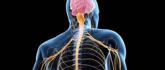
The nervous system (NS) is a set of anatomically and functionally interconnected nervous structures that provide regulation and coordination of the activities of the human body and its interaction with the environment.
The structural unit of the NS is a cell with a process ( neuron , or neurocyte). The nervous system is a collection of neurons that communicate with each other through synapses.
Functions of the nervous system
The nervous system occupies a special position among other body systems. It ensures the relationship of the organism with the environment . Receptors respond to any signals from the external and internal environment, converting them into streams of nerve impulses that enter the central nervous system. Based on the analysis of the flow of nerve impulses encoding information, the brain forms an adequate response.
Together with the endocrine glands, the nervous system regulates the functioning of all organs . This regulation is carried out due to the fact that the spinal cord and brain are connected by nerves to all organs through bilateral connections. The central nervous system receives signals from the organs about their functional state, and the nervous system, in turn, sends signals to the organs, correcting their functions and ensuring all vital processes - movement, nutrition, excretion, etc. The nervous system ensures coordination of the activities of cells, tissues, organs , organ systems. In this case, the body functions as a single whole.
The nervous system is the material basis of mental processes : attention, memory, speech, thinking, etc., with the help of which a person not only learns about the environment, but can also actively change it.
Thus, several functions of the nervous system can be distinguished: 1. Connects the body with the environment (perception and transmission).
2. Provides interaction between tissues of organs and body systems and their regulation.
NS departments
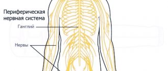
Central nervous system and peripheral nervous system
According to topographical principles, the nervous system is divided into central and peripheral. The central nervous system (CNS) includes those parts that are contained in the cranial cavity and the spinal canal, i.e. brain and spinal cord. The spinal cord is a tube with a small canal in the middle, surrounded by neurons and their processes. The brain is an extension of the spinal cord. The topographic boundary with the spinal cord is a plane passing through the lower edge of the foramen magnum. The average brain weight is 1400 g with individual variations from 1100 to 2000 g.
The peripheral nervous system (PNS) includes all the nerve structures located outside of it. These are nodes and bundles of fibers that connect the central nervous system with sensory organs and various effectors (muscles, glands, etc.), i.e. ganglia and nerves. The peripheral nervous system connects the spinal cord and brain with receptors and effectors. It consists of 12 pairs of cranial and 31-33 pairs of spinal (spinal) nerves.
Somatic nervous system and autonomic nervous system
According to the functional classification, the nervous system is divided into somatic nervous system and autonomic nervous system. Both somatic and autonomic nervous systems include central and peripheral parts.
The somatic nervous system includes the parts of the nervous system that regulate the functioning of skeletal muscles. Responsible for connecting the body with the external environment, providing sensitivity and movement, causing contraction of skeletal muscles. It regulates primarily the functions of voluntary movement. Its neurons are located in the anterior horns of the spinal cord, and their axons through the anterior roots of the spinal cord are directed to the skeletal (striated) muscles.
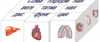
The autonomic (autonomic) nervous system is a collection of nerves and ganglia through which the heart, blood vessels, internal organs, glands, etc. are regulated. Internal organs receive double innervation (supply of organs and tissues with nerves, which ensures their connection with the central nervous system) - from the sympathetic and parasympathetic divisions of the autonomic nervous system. These two departments have excitatory and inhibitory influences, determining the level of activity of the organs.
The vegetative nervous system provides metabolism, respiration, and excretion. Influencing the metabolic activity in various organs and tissues in accordance with the changing conditions of their functioning and the external environment, it carries out an adaptive-professional function.
The autonomic nervous system innervates smooth muscles; it is also called the visceral nervous system. The division of the peripheral nervous system into somatic and autonomic is quite arbitrary, since in the central nervous system there is a significant overlap of projections of both, and somatic and autonomic reactions are equal components of any behavioral reaction.
In the autonomic nervous system, there are 2 divisions that are functional antagonists : sympathetic and parasympathetic . They differ in the localization of centers in the brain and peripheral nodes, as well as the nature of the effect on internal organs.
Differences between sympathetic and parasympathetic nervous system
1. The fibers of the sympathetic nervous system emerge from the thoracic and lumbar spinal cord, where the first sympathetic neuron lies. They then converge on the sympathetic ganglia located along the spine, where the second sympathetic neuron is located. The fibers of the parasympathetic nervous system begin in the spinal cord above or below the place where the sympathetic nerves exit the cranial and sacral regions, and then converge in ganglia located not along the spinal column, but close to the innervated organ.
2. The peculiarities of the location of the ganglia of these two systems suggest a difference in the effect they have. The action of the sympathetic nervous system is more diffuse, while the parasympathetic nervous system is more specific, since it is associated only with changes in the organ next to which the ganglion is located.
3. These systems also differ in the mediators involved in synaptic transmission. The main transmitter for the sympathetic nervous system is adrenaline, and for the parasympathetic nervous system it is acetylcholine.
4. The results of the activity of these two systems are largely opposite. While the main function of the sympathetic nervous system is to mobilize the body for fight or flight, the parasympathetic nervous system primarily ensures the maintenance of homeostasis.
5. Activation of the sympathetic nervous system underlies the behavior of a person eager to fight. The stimulation of the parasympathetic nervous system ensures digestion in a person lying on the couch after a hearty lunch.
The sympathetic nervous system excites, and the parasympathetic nervous system inhibits the activity of the heart, the first weakens the motor activity of the intestines, the second strengthens it. At the same time, they can act at the same time: together they increase the motor activity of the salivary and gastric glands, although the composition of the secreted juice varies depending on the share of participation of each system.
- 1. Basic principles of the functioning of the central nervous system. Structure, functions, methods of studying the central nervous system
- 2. Neuron. Structural features, meaning, types
- 3. Reflex arc, its components, types, functions
- 4. Functional systems of the body
- 5. Coordination activities of the central nervous system
- 6. Types of inhibition, interaction of excitation and inhibition processes in the central nervous system. Experience of I. M. Sechenov
- 7. Methods for studying the central nervous system
LECTURE No. 6. Physiology of the central nervous system
1. Basic principles of the functioning of the central nervous system. Structure, functions, methods of studying the central nervous system
The basic principle of the functioning of the central nervous system is the process of regulation, control of physiological functions, which are aimed at maintaining the constancy of the properties and composition of the internal environment of the body. The central nervous system ensures optimal relationships between the body and the environment, stability, integrity, and the optimal level of vital activity of the body.
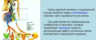
There are two main types of regulation: humoral and nervous.
The humoral control process involves changing the physiological activity of the body under the influence of chemicals that are delivered by body fluids. The source of information transfer is chemicals - utilizons, metabolic products (carbon dioxide, glucose, fatty acids), informons, hormones of the endocrine glands, local or tissue hormones.
The nervous process of regulation involves controlling changes in physiological functions along nerve fibers using excitation potential under the influence of information transfer.
Characteristics:
1) is a later product of evolution;
2) provides quick regulation;
3) has an exact target of impact;
4) implements an economical method of regulation;
5) ensures high reliability of information transmission.
In the body, the nervous and humoral mechanisms work as a single system of neurohumoral control. This is a combined form, where two control mechanisms are used simultaneously; they are interconnected and interdependent.
The nervous system is a collection of nerve cells, or neurons.
According to localization they distinguish:
1) central section – brain and spinal cord;
2) peripheral - processes of nerve cells of the brain and spinal cord.
According to functional features they are distinguished:
1) somatic department, regulating motor activity;
2) vegetative, regulating the activity of internal organs, endocrine glands, blood vessels, trophic innervation of muscles and the central nervous system itself.
Functions of the nervous system:
1) integrative-coordination function. Provides the functions of various organs and physiological systems, coordinates their activities with each other;
2) ensuring close connections between the human body and the environment at the biological and social levels;
3) regulation of the level of metabolic processes in various organs and tissues, as well as in oneself;
4) ensuring mental activity by the higher departments of the central nervous system.
2. Neuron. Structural features, meaning, types
The structural and functional unit of nervous tissue is a nerve cell - neuron
.
A neuron is a specialized cell that is capable of receiving, encoding, transmitting and storing information, establishing contacts with other neurons, and organizing the body’s response to irritation.
Functionally, a neuron is divided into:
1) the receptive part (dendrites and membrane of the soma of the neuron);
2) integrative part (soma with axon hillock);
3) transmitting part (axon hillock with axon).
Perceiving part.
Dendrites
– the main receptive field of the neuron. The dendrite membrane is capable of responding to mediators. A neuron has several branching dendrites. This is explained by the fact that a neuron as an information formation must have a large number of inputs. Through specialized contacts, information flows from one neuron to another. These contacts are called "spines".
The neuron soma membrane is 6 nm thick and consists of two layers of lipid molecules. The hydrophilic ends of these molecules face the water phase: one layer of molecules faces inward, the other outward. The hydrophilic ends are turned towards each other - inside the membrane. The lipid bilayer of the membrane contains proteins that perform several functions:
1) pump proteins - move ions and molecules in the cell against a concentration gradient;
2) proteins embedded in the channels provide selective membrane permeability;
3) receptor proteins recognize the necessary molecules and fix them on the membrane;
4) enzymes facilitate the occurrence of a chemical reaction on the surface of the neuron.
In some cases, the same protein can serve as both a receptor, an enzyme, and a pump.
Integrative part.
Axon hillock
– the point where the axon exits the neuron.
The neuron soma (neuron body) performs, along with an informational and trophic function, relative to its processes and synapses. The soma ensures the growth of dendrites and axons. The neuron soma is enclosed in a multilayer membrane, which ensures the formation and propagation of electrotonic potential to the axon hillock.
Transmitting part.
Axon
- an outgrowth of the cytoplasm, adapted to carry information that is collected by dendrites and processed in the neuron. The axon of a dendritic cell has a constant diameter and is covered with a myelin sheath, which is formed from glia; the axon has branched endings containing mitochondria and secretory formations.
Functions of neurons:
1) generalization of the nerve impulse;
2) receiving, storing and transmitting information;
3) the ability to summarize excitatory and inhibitory signals (integrative function).
Types of neurons:
1) by localization:
a) central (brain and spinal cord);
b) peripheral (cerebral ganglia, cranial nerves);
2) depending on the function:
a) afferent (sensitive), carrying information from receptors to the central nervous system;
b) intercalary (connector), in the elementary case providing communication between afferent and efferent neurons;
c) efferent:
– motor – anterior horns of the spinal cord;
– secretory – lateral horns of the spinal cord;
3) depending on the functions:
a) stimulating;
b) inhibitory;
4) depending on the biochemical characteristics, on the nature of the mediator;
5) depending on the quality of the stimulus that is perceived by the neuron:
a) monomodal;
b) multimodal.
3. Reflex arc, its components, types, functions
The activity of the body is a natural reflex reaction to a stimulus. Reflex
– the body’s reaction to irritation of receptors, which is carried out with the participation of the central nervous system. The structural basis of the reflex is the reflex arc.
Reflex arc
- a series-connected chain of nerve cells that ensures the implementation of a reaction, a response to irritation.
The reflex arc consists of six components: receptors, afferent (sensitive) path, reflex center, efferent (motor, secretory) path, effector (working organ), feedback.
Reflex arcs can be of two types:
1) simple - monosynaptic reflex arcs (reflex arc of the tendon reflex), consisting of 2 neurons (receptor (afferent) and effector), there is 1 synapse between them;
2) complex – polysynaptic reflex arcs. They consist of 3 neurons (there may be more) - a receptor, one or more intercalary and an effector.
The idea of the reflex arc as an expedient response of the body dictates the need to supplement the reflex arc with another link - a feedback loop. This component establishes a connection between the realized result of the reflex reaction and the nerve center that issues executive commands. With the help of this component, the open reflex arc is transformed into a closed one.
Features of a simple monosynaptic reflex arc:
1) geographically close receptor and effector;
2) reflex arc two-neuron, monosynaptic;
3) nerve fibers of group A? (70-120 m/s);
4) short reflex time;
5) muscles contracting according to the type of single muscle contraction.
Features of a complex monosynaptic reflex arc:
1) territorially separated receptor and effector;
2) three-neuron receptor arch (there may be more neurons);
3) the presence of nerve fibers of groups C and B;
4) muscle contraction according to the tetanus type.
Features of the autonomic reflex:
1) the interneuron is located in the lateral horns;
2) the preganglionic nerve pathway begins from the lateral horns, after the ganglion - the postganglionic;
3) the efferent path of the autonomic nervous arch reflex is interrupted by the autonomic ganglion, in which the efferent neuron lies.
The difference between the sympathetic nervous arch and the parasympathetic: the sympathetic nervous arch has a short preganglionic pathway, since the autonomic ganglion lies closer to the spinal cord, and the postganglionic pathway is long.
In the parasympathetic arc, the opposite is true: the preganglionic pathway is long, since the ganglion lies close to the organ or in the organ itself, and the postganglionic pathway is short.
4. Functional systems of the body
Functional system
– temporary functional unification of the nerve centers of various organs and systems of the body to achieve a final beneficial result.
The beneficial result is a self-forming factor of the nervous system. The result of an action is a vital adaptive indicator that is necessary for the normal functioning of the body.
There are several groups of final useful results:
1) metabolic – a consequence of metabolic processes at the molecular level that create substances and end products necessary for life;
2) homeostatic – constancy of indicators of the state and composition of the body’s media;
3) behavioral – the result of biological needs (sexual, food, drinking);
4) social – satisfaction of social and spiritual needs.
The functional system includes various organs and systems, each of which takes an active part in achieving a useful result.
The functional system, according to P.K. Anokhin, includes five main components:
1) a useful adaptive result - that for which a functional system is created;
2) control apparatus (result acceptor) – a group of nerve cells in which a model of the future result is formed;
3) reverse afferentation (supplies information from the receptor to the central link of the functional system) - secondary afferent nerve impulses that go to the acceptor of the result of the action to evaluate the final result;
4) control apparatus (central link) – functional association of nerve centers with the endocrine system;
5) executive components (reaction apparatus) - these are the organs and physiological systems of the body (vegetative, endocrine, somatic). Consists of four components:
a) internal organs;
b) endocrine glands;
c) skeletal muscles;
d) behavioral reactions.
Properties of a functional system:
1) dynamism. The functional system may include additional organs and systems, which depends on the complexity of the current situation;
2) the ability to self-regulate. When the controlled value or the final useful result deviates from the optimal value, a series of reactions of a spontaneous complex occur, which returns the indicators to the optimal level. Self-regulation occurs in the presence of feedback.
Several functional systems operate simultaneously in the body. They are in continuous interaction, which is subject to certain principles:
1) the principle of the system of genesis. Selective maturation and evolution of functional systems occur (functional circulatory, respiratory, nutritional systems mature and develop earlier than others);
2) the principle of multiply connected interaction. There is a generalization of the activities of various functional systems aimed at achieving a multicomponent result (homeostasis parameters);
3) the principle of hierarchy. Functional systems are arranged in a certain row in accordance with their significance (functional system of tissue integrity, functional nutrition system, functional reproduction system, etc.);
4) the principle of sequential dynamic interaction. There is a clear sequence of changing the activities of one functional system to another.
5. Coordination activities of the central nervous system
Coordination activity (CA) of the CNS is the coordinated work of CNS neurons, based on the interaction of neurons with each other.
CD functions:
1) ensures clear performance of certain functions and reflexes;
2) ensures the consistent inclusion of various nerve centers in the work to ensure complex forms of activity;
3) ensures the coordinated work of various nerve centers (during the act of swallowing, the breath is held at the moment of swallowing; when the swallowing center is excited, the breathing center is inhibited).
Basic principles of CNS CD and their neural mechanisms.
1. The principle of irradiation (propagation). When small groups of neurons are excited, the excitation spreads to a significant number of neurons. Irradiation is explained:
1) the presence of branched endings of axons and dendrites, due to branching, impulses spread to a large number of neurons;
2) the presence of interneurons in the central nervous system, which ensure the transmission of impulses from cell to cell. Irradiation has boundaries, which are provided by the inhibitory neuron.
2. The principle of convergence. When a large number of neurons are excited, the excitation can converge to one group of nerve cells.
3. The principle of reciprocity - coordinated work of nerve centers, especially in opposite reflexes (flexion, extension, etc.).
4. The principle of dominance. Dominant
– the dominant focus of excitation in the central nervous system at the moment. This is the center of persistent, unwavering, non-spreading excitation. It has certain properties: it suppresses the activity of other nerve centers, has increased excitability, attracts nerve impulses from other foci, sums up nerve impulses. Foci of dominance are of two types: exogenous (caused by environmental factors) and endogenous (caused by internal environmental factors). The dominant underlies the formation of a conditioned reflex.
5. Feedback principle. Feedback is a flow of impulses into the nervous system that informs the central nervous system about how the response is carried out, whether it is sufficient or not. There are two types of feedback:
1) positive feedback, causing an increase in the response from the nervous system. Underlies the vicious circle that leads to the development of diseases;
2) negative feedback, reducing the activity of CNS neurons and response. Underlies self-regulation.
6. The principle of subordination. In the central nervous system there is a certain subordination of departments to each other, the highest department being the cerebral cortex.
7. The principle of interaction between the processes of excitation and inhibition. The central nervous system coordinates the processes of excitation and inhibition:
both processes are capable of convergence; the process of excitation and, to a lesser extent, inhibition are capable of irradiation. Inhibition and excitation are connected by inductive relationships. The process of excitation induces inhibition, and vice versa. There are two types of induction:
1) consistent. The process of excitation and inhibition alternates in time;
2) mutual. There are two processes at the same time - excitation and inhibition. Mutual induction is carried out through positive and negative mutual induction: if inhibition occurs in a group of neurons, then foci of excitation arise around it (positive mutual induction), and vice versa.
According to I.P. Pavlov’s definition, excitation and inhibition are two sides of the same process. The coordination activity of the central nervous system ensures clear interaction between individual nerve cells and individual groups of nerve cells. There are three levels of integration.
The first level is ensured due to the fact that impulses from different neurons can converge on the body of one neuron, resulting in either summation or a decrease in excitation.
The second level provides interactions between individual groups of cells.
The third level is provided by cells of the cerebral cortex, which contribute to a more advanced level of adaptation of the activity of the central nervous system to the needs of the body.
6. Types of inhibition, interaction of excitation and inhibition processes in the central nervous system. Experience of I. M. Sechenov
Braking
– an active process that occurs when stimuli act on tissue, manifests itself in the suppression of other excitation, there is no functional function of the tissue.
Inhibition can develop only in the form of a local response.
There are two types of braking:
1) primary. For its occurrence, the presence of special inhibitory neurons is necessary. Inhibition occurs primarily without prior excitation under the influence of an inhibitory transmitter. There are two types of primary inhibition:
a) presynaptic in the axo-axonal synapse;
b) postsynaptic in the axodendritic synapse.
2) secondary. It does not require special inhibitory structures, occurs as a result of changes in the functional activity of ordinary excitable structures, and is always associated with the process of excitation. Types of secondary braking:
a) transcendental, which occurs when there is a large flow of information entering the cell. The flow of information lies beyond the functionality of the neuron;
b) pessimal, which occurs with a high frequency of irritation;
c) parabiotic, which occurs during strong and long-term irritation;
d) inhibition following excitation, resulting from a decrease in the functional state of neurons after excitation;
e) inhibition according to the principle of negative induction;
e) inhibition of conditioned reflexes.
The processes of excitation and inhibition are closely related to each other, occur simultaneously and are different manifestations of a single process. Foci of excitation and inhibition are mobile, cover larger or smaller areas of neuronal populations and can be more or less pronounced. Excitation is certainly replaced by inhibition, and vice versa, that is, there is an inductive relationship between inhibition and excitation.
Inhibition underlies the coordination of movements and protects central neurons from overexcitation. Inhibition in the central nervous system can occur when nerve impulses of varying strength from several stimuli simultaneously enter the spinal cord. Stronger stimulation inhibits reflexes that should have occurred in response to weaker ones.
In 1862, I.M. Sechenov discovered the phenomenon of central inhibition. He proved in his experiment that irritation with a sodium chloride crystal of the visual thalamus of a frog (the cerebral hemispheres have been removed) causes inhibition of spinal cord reflexes. After the stimulus was removed, the reflex activity of the spinal cord was restored. The result of this experiment allowed I.M. Secheny to conclude that in the central nervous system, along with the process of excitation, a process of inhibition develops, which is capable of inhibiting the reflex acts of the body. N. E. Vvedensky suggested that the phenomenon of inhibition is based on the principle of negative induction: a more excitable area in the central nervous system inhibits the activity of less excitable areas.
Modern interpretation of the experiment of I.M. Sechenov (I.M. Sechenov irritated the reticular formation of the brainstem): excitation of the reticular formation increases the activity of inhibitory neurons of the spinal cord - Renshaw cells, which leads to inhibition of ?-motoneurons of the spinal cord and inhibits the reflex activity of the spinal cord.
7. Methods for studying the central nervous system
There are two large groups of methods for studying the central nervous system:
1) experimental method, which is carried out on animals;
2) a clinical method that is applicable to humans.
Among the experimental methods
classical physiology includes methods aimed at activating or suppressing the nerve formation being studied. These include:
1) method of transverse section of the central nervous system at various levels;
2) method of extirpation (removal of various parts, denervation of the organ);
3) method of irritation by activation (adequate irritation - irritation with an electrical impulse similar to a nervous one; inadequate irritation - irritation with chemical compounds, graded irritation with electric current) or suppression (blocking the transmission of excitation under the influence of cold, chemical agents, direct current);
4) observation (one of the oldest methods of studying the functioning of the central nervous system that has not lost its significance. It can be used independently, and is often used in combination with other methods).
Experimental methods are often combined with each other when conducting experiments.
Clinical method
aimed at studying the physiological state of the central nervous system in humans. It includes the following methods:
1) observation;
2) method of recording and analyzing electrical potentials of the brain (electro-, pneumo-, magnetoencephalography);
3) radioisotope method (investigates neurohumoral regulatory systems);
4) conditioned reflex method (studies the functions of the cerebral cortex in the mechanism of learning and the development of adaptive behavior);
5) questionnaire method (assesses the integrative functions of the cerebral cortex);
6) modeling method (mathematical modeling, physical modeling, etc.). A model is an artificially created mechanism that has a certain functional similarity with the mechanism of the human body being studied;
7) cybernetic method (studies control and communication processes in the nervous system). Aimed at studying organization (systemic properties of the nervous system at various levels), management (selection and implementation of influences necessary to ensure the functioning of an organ or system), information activity (the ability to perceive and process information - an impulse in order to adapt the body to environmental changes).
Table of contents
Reflex as the basic principle of the nervous system
I. M. Sechenov in 1863 in his work “Reflexes of the Brain” he developed the idea that the reflex is the basic principle of the functioning of not only the spinal cord, but also the brain.
A reflex is the body’s response to irritation with the participation of the central nervous system. Reflexes are divided into: 1) unconditioned reflexes: innate (hereditary) reactions of the body to stimuli carried out with the participation of the spinal cord or brain stem; 2) conditioned reflexes: temporary reactions of the body acquired on the basis of unconditioned reflexes, carried out with the obligatory participation of the cerebral cortex, which form the basis of higher nervous activity.
Each reflex has its own reflex arc - this is the path along which excitation passes from the receptor to the effector (executive organ).
The reflex arc is represented by a chain of neurons that provide the perception of irritation, the transformation of the energy of irritation into a nerve impulse, the conduction of a nerve impulse to the nerve centers, the processing of incoming information and the implementation of a response.
Any reflex arc consists of 5 components
1. A receptor is a specialized cell designed to perceive a stimulus (sound, light, chemical, etc.). 2. Afferent pathway, which is represented by afferent neurons. 3. Part of the central nervous system, represented by the spinal cord or brain; 4. The efferent pathway consists of the axons of efferent neurons extending beyond the CNS. 5. An effector is a working organ (muscle, gland, etc.).
The simplest reflex arc includes 2 neurons and is called monosynaptic (based on the number of synapses). A more complex one is represented by 3 neurons and is called three-neuron or disynaptic. However, most reflex arcs include a large number of interneurons and are called polysynaptic.
Reflex arcs can pass through the spinal cord only (for example, withdrawing a hand when touching a hot object) or through the brain only (for example, closing the eyelids when a stream of air is directed at the face), or through both the spinal cord and the brain.
Reflex arcs are closed into reflex rings using feedback connections. The concept of feedback and its functional role was indicated by Bell in 1826. He wrote that two-way connections are established between the muscle and the central nervous system. With the help of feedback, signals about the functional state of the effector are sent to the central nervous system.
The morphological basis of feedback is the receptors located in the effector and the afferent neurons associated with them. Thanks to feedback afferent connections, fine regulation of the effector’s work and an adequate response of the body to environmental changes are carried out.
Reflex
This is the body's response to irritation from the external or internal environment.
The main form of activity of the nervous system is a reflex (from the English reflection - reflection).
An example of a reflex is withdrawing a hand from a hot object. The nerve ending senses high temperature and transmits a signal about it to the central nervous system. A response impulse arises in the central nervous system, going to the muscles of the arm.
Rice. 3. Reflex arc diagram.
The sequence: sensory nerve - CNS - motor nerve is called a reflex arc.