General information
Motor disorders are a symptom that accompanies various neurological diseases. They are associated with damage to the motor departments of both the central and peripheral nervous systems, which leads to partial or complete impairment of movements. Paresis is a limitation of movement and a decrease in muscle strength. A more severe form is paralysis (or plegia), in which voluntary movements are completely absent.
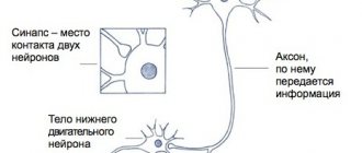
We can say that motor excitation is transmitted along a “two-story” cortical-muscular pathway - from the brain to the muscles (pyramidal pathway). The top floor is the brain, from where the impulse is transmitted to the spinal cord. If this motor pathway is disrupted, central paralysis or paresis develops. Central movement disorders are characterized by weakness in the arms or legs, increased reflexes, and the gradual development of spasticity.
The lower level of the pathway includes the spinal cord, from which the impulse is transmitted to the nerves in the periphery, which provide muscle movement. If this motor pathway is disrupted, peripheral paralysis or paresis develops. Peripheral paresis and paralysis are sluggish due to developing muscle hypotonia and occur with muscle weakness and atrophy. Thus, motor disorders can be caused by lesions not only of the brain, but also of the spinal cord, as well as peripheral nerves, which for some reason stop transmitting impulses from the brain to the muscles. Central paralysis is characterized by muscle spasticity - muscle tension that occurs involuntarily. Spasticity is characteristic of cerebral palsy, multiple sclerosis , encephalomyelitis , traumatic brain injury , and stroke . Code for spastic paraparesis according to ICD 10 - G82.1
Detailed description of the study
Hereditary spastic paraplegia (HSP), or Strumpel's disease, includes a group of rare hereditary diseases that are characterized by:
- The development of lower spastic paraparesis - a decrease in strength in the muscles of the lower extremities, up to its complete absence;
- Progressive deterioration in walking;
- Hyperflexion is a pathological strengthening of reflexes.
Basically, the disease is inherited in an autosomal dominant manner, i.e. develops in offspring in approximately 50% of cases. Cases of autosomal recessive inheritance (the disease manifests itself only when a “defective” gene is inherited from both parents) and X-linked inheritance, when the “defective” gene is located on the X chromosome, have also been described.
The most important clinical manifestation of Strumpel's disease is lower spastic paraparesis, as a result of which the muscles stop receiving nerve impulses and gradually lose muscle strength. Other neurological symptoms may also develop, for example, oculomotor, cognitive impairment, etc.
To date, 32 chromosomal regions are known, disturbances in which can lead to the development of pathology.
10 loci (regions) have been described for autosomal dominant Strumpel disease, 14 loci for the autosomal recessive form, and 3 for the X-linked form.
The SPG4 gene is localized in the chromosomal region 2p22.3 and encodes the protein spastin. More than 500 mutations have been identified in the SPG4 gene. This gene is involved in the development of approximately 40% of cases of all autosomal dominant spastic paraplegia.
The spastin protein is a member of the AAA family of proteins, a special class of ATPases with multiple types of cellular activities. These proteins are involved in a number of cellular processes, such as the cell cycle, intracellular transport, proteolysis (the process of protein breakdown). Spastin is involved in the processes of formation and destruction of special protein complexes, which are important for maintaining the integrity of neurons and transmission of nerve impulses.
Gene mutations change the structure of the spastin protein, as a result of which neurons are damaged and cannot fully conduct nerve impulses.
The average age of onset of clinical manifestations of the disease is 30-40 years, although the disease can also be diagnosed in a child.
The severity of symptoms and how quickly they progress can vary greatly. The disease may progress slowly from birth and stabilize by adolescence. In this case, it is possible to maintain the ability to move independently. In other patients the course is continuous.
The presence of characteristic clinical manifestations of the pathology, as well as a family history, suggests Strumpel’s spastic paraplegia. Genetic testing is used to confirm a preliminary diagnosis.
Pathogenesis
If we consider acquired motor disorders in trauma, then at the moment of exposure to traumatic force, direct damage to brain tissue develops. Compression, contusion of the parenchyma and circulatory disorders occur in it. The outcome of these pathological changes is necrosis , developing in the brain or spinal cord. At the same time, the mechanism of secondary damage is triggered - ischemia and inflammation. The necrotic focus gradually turns into a connective tissue scar, which is surrounded by post-traumatic cysts.
The consequences of brain damage are interruption of pathways at different levels, associated with necrosis and atrophy . With a spinal cord injury, control of the muscles innervated by segments below the level of injury is lost. The possibility of functional restoration depends on the restoration of conduction along the spinal cord. If the conductivity of the spinal cord is completely disrupted when it is interrupted, automation develops in those sections that are located below the interruption. In case of partial disruption of conductivity, there is the possibility of slow restoration of functions after elimination of edema , inflammation and circulatory disorders due to the remaining fibers.
The pathogenesis of cerebral palsy is different. Birth trauma is thought to result in asphyxia and brain damage. However, as a cause of cerebral palsy, birth trauma matters only in 5-10%. There are other and more significant mechanisms for the development of the disease: impaired brain formation, intrauterine damage to neurons, diseases that occur in the first week of life. Brain damage (ischemia or trauma) before the child’s age of three, which also causes motor disorders, cannot be ruled out.
Classification
The following types of movement disorders are distinguished depending on their prevalence:
- Monoparesis (monoplegia) is muscle weakness or loss of movement in one limb and on one side.
- Paraparesis (paraplegia) is muscle weakness or loss of movement in two limbs (or both arms or both legs). Depending on this, in neurology there is paraparesis of the lower extremities or paraplegia of the lower extremities and, accordingly, paraparesis or paraplegia of the upper extremities (synonyms lower paraparesis/plegia and upper paraparesis/plegia).
- Hemiparesis (hemiplegia) is muscle weakness or loss of movement in the upper and lower limbs, but on one side. That is, hemiparesis and hemiplegia can be right-sided or left-sided.
- Diplegia is damage to both halves of the body.
- Triparesis (triplegia) is a disorder of movement of three limbs.
- Tetraparesis (tetraplegia) – in case of movement disorders in four limbs.
According to the level of damage to the nervous system, movement disorders are divided into:
- Peripheral.
- Central.
- Mixed.
According to the degree of severity, movement disorders are:
- Mild degree.
- Moderate.
- Deep.
- Complete paralysis.
According to muscle condition:
- Spastic paralysis (paresis) is a pathological muscle tension with weakness of movements that develops in the arm or leg.
- Flaccid paralysis (paresis) is a pronounced relaxation of the muscles, but their shortening does not occur. When peripheral motor neurons are damaged, peripheral paralysis develops - they are called flaccid paralysis.
The spastic form of paralysis with increased muscle tone indicates damage to the central motor neurons. Spasticity occurs in many diseases of the central nervous system: stroke , encephalitis , Strumpel's disease , traumatic brain injury , cerebral palsy, multiple sclerosis , spinal injury , brain tumors and , amyotrophic lateral sclerosis . Acute damage to central neurons first manifests as flaccid paralysis, and then increased muscle tone. Thus, with strokes, motor impairments of varying degrees develop and, along with a decrease in strength, muscle tone changes towards spasticity. Post-stroke spasticity develops in 68% of patients, affecting the arm and leg.
With spinal injury , multiple sclerosis and spinal stroke, lower spastic paraparesis appears. In multiple sclerosis and cerebral palsy, spasticity is accompanied by muscle pain and the development of contractures.
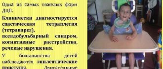
Spastic diplegia is a common form of cerebral palsy, the clinical picture of which is tetraparesis - all limbs are affected, but the legs are predominant. Movement disorders can vary from awkward movements in the limbs to severe paresis.
Spastic diplegia is called Little's disease . Identified in premature infants with intraventricular hemorrhage or leukomalacia . In mild cases of the disease, symptoms appear by six months, and in more severe cases, tetraparesis is noticeable from birth. With this pathology, children begin to sit up late, and when walking they make “dancing” movements, their legs cross and there is emphasis (on their toes). In children, bone growth is disrupted, spinal curvatures and joint contractures develop very early. This disease causes speech delay and speech impairment. There may be pathology of the cranial nerves, which manifests itself in strabismus and atrophy of the optic nerves , as well as hearing impairment.
Tetraplegia is associated with more severe damage to the cerebral hemispheres, brainstem, or cervical spinal cord. May be associated with congenital (cerebral palsy) or acquired diseases. Acquired forms develop after severe traumatic brain injury, hemorrhage, as a result of tumors of the brain or spinal cord, encephalitis, or complications of neurosurgical operations. Flaccid tetraparesis occurs with muscle weakness in the arms and legs. spastic tetraparesis develops (double hemiparesis or double hemiplegia), which in children is considered a form of cerebral palsy. Patients have constantly increased muscle tone and cannot relax them. Without treatment, complete loss of movement and motor skills occurs. Increased spasticity is accompanied by muscle pain. Spasticity is also noted in the neck and tongue, so such children cannot speak (the tongue does not obey them). There is also mental retardation.
Strumpel's disease
Strumpel's spastic paraplegia is a hereditary disease that is caused by degenerative changes in the motor neurons of the spinal cord. Motor neurons enter the pyramidal tract, which begins in the cerebral cortex and ends with mononeurons in the spinal cord. Wikipedia clarifies that a feature of the disease is progressive spasticity and gait disturbance. For an unknown reason, the transport of proteins and lipids, which are necessary to maintain the structure of the cell, is disrupted in motor neuron cells. This disease is not a form of cerebral palsy, although it manifests itself as spastic diplegia .
The initial signs of Strumpel's disease appear at 10-15 years of age, but there are cases of the disease developing from early childhood. Characteristically symmetrical lesions of the legs. Patients are concerned about fatigue and stiffness in the legs, which appear after long walking or intense exercise. The leading cause is muscle spasticity and increased tendon reflexes. An increase in muscle tone causes the symptom of “crossed legs”, protective reflexes and bending of the legs at night appear. The later the disease appears, the more pronounced the spasticity and increased reflexes. Tendon contractures also develop (more so in the ankle joints). As the disease progresses, in the advanced stage, patients have difficulty walking, stumble and fall. There are no trophic disorders in the muscles (no atrophy), and there are also no dysfunctions of the pelvic organs.
Strumpel's disease
Increased muscle tone that occurs in the lower extremities often causes the development of a neurological disease, medically called Strumpel's disease. In most cases, the nature of the occurrence of this disease lies in genetic inheritance and can manifest itself in a person at any age. Despite the fact that Strumpel's disease, or, as it is also called, spastic paraplegia, has been studied for more than a hundred years, the causes of the disease remain not fully understood.
The gene that causes the development of Strumpel's disease was discovered relatively recently, and therefore everything that is in one way or another connected with it is now being actively studied. Neurologists divide the disease into several forms and types that have characteristic symptoms. A simple form of Strumpel's disease is accompanied by paralysis of the legs ; there are no other pathologies. A complicated form of the disease is characterized by paralysis, accompanied by optic nerve atrophy, epileptic seizures, partial or complete hearing , abnormal development of the heart muscle and pathologies in the development of the feet . A characteristic feature of the disease as a whole is the slow onset of symptoms.
Symptoms of Strumpel's disease
Among the main symptoms, neurologists identify difficulty developing walking skills in early childhood; increased leg , cramps, difficulty walking and frequent falls in adolescence; characteristic manifestations of muscle weakness. Neurologists note that increased muscle tone in the legs develops from childhood and persists throughout life. Severe forms of the disease are manifested by a pronounced decrease in intellectual abilities, epileptic seizures, hearing and retinopathy. Delay in seeking help from a medical institution is observed only due to weak symptoms in the initial stages of the development of Strumpel’s disease and the rather slow course of the pathology.
Appointment with a neurologist at Premium Clinics
An appointment with a neurologist includes an examination of the patient, familiarization with the history of his illnesses and a series of tests to determine muscle tone. If the development of Strumpel's disease is suspected, the doctor prescribes a series of instrumental diagnostic procedures. Magnetic resonance and computed tomography can detect disorders in the spinal cord Genetic analysis is of great importance in diagnosing Strumpel's disease. In the diagnostic process, it is imperative to differentiate the disease from a number of diseases with similar symptoms, for example, a spinal cord .
We have to admit the fact that today there are no effective methods for treating this disease. Conservative therapy can reduce symptoms. Drug therapy is accompanied by therapeutic exercises and physiotherapy .
Causes
Most movement disorders are caused by degenerative diseases, either congenital or acquired.
Paraparesis in adults and its causes
If congenital diseases ( adrenoleukodystorophy , which occurs with myeloneuropathy , Strumpel's disease ) and cerebral palsy are excluded, then most often paraparesis of the lower extremities is caused in adults by the following pathologies and conditions:
- Compression lesions (prolapsed intervertebral hernia , bone spines, thickened spinal ligaments that narrow the spinal canal and compress the spinal cord).
- Hepatic encephalopathy , in which spastic paraparesis is associated with the toxic effects of endotoxins.
- Injury to the spine and spinal cord, which leads to loss of movement and dysfunction of the pelvic organs. Spinal cord injuries very often lead to significant motor impairment and disability in patients.
- Tumors of the spinal cord.
- Infectious diseases ( encephalitis , Lyme disease , AIDS, neurosyphilis ).
- Autoimmune diseases ( systemic lupus erythematosus ).
- Epidural abscess.
- Vascular pathology of the spinal cord (epidural and subdural hemorrhages, blockage of the anterior spinal artery).
- Poisoning (medicines and alcohol).
Causes of central paresis:
- Traumatic brain injuries.
- Intervertebral hernia.
- Cerebral palsy.
- Strokes.
- Spinal cord injuries.
- Tumors (brain and spinal cord).
- Amyotrophic lateral sclerosis.
- Multiple sclerosis.
Strumpell's familial spastic paralysis
The diagnosis usually does not cause difficulties if there is a familial nature of the disease and a typical clinical picture.
In atypical sporadic cases, the disease should be distinguished from the spinal form of multiple sclerosis, amyotrophic lateral sclerosis, spinal cord tumors and other pathological processes of various etiologies that cause compression of the spinal cord, as well as funicular myelosis, neurosyphilis and other forms of cerebellar-pyramidal degeneration. The spinal form of multiple sclerosis, along with lower spastic paraparesis, is characterized by a relapsing course, instability and temporary reversibility of individual symptoms, dysfunction of the pelvic organs, loss or asymmetry of abdominal reflexes and asymmetry of symptoms of the lesion in general, changes in immunological parameters of the blood and cerebrospinal fluid. Data on the hereditary nature of the disease are of decisive importance. Unlike amyotrophic lateral sclerosis, Strumpel's disease begins at a young age, there are no signs of peripheral motor neuron damage (fascicular twitching, atrophy of small muscles of the hand, characteristic EMG changes), or bulbar disorders. When differentiating from extramedullary tumors and spinal cord compression syndrome of other etiologies, segmental sensitivity disorders, asymmetry of limb damage, the presence of a block of the subarachnoid space and protein-cell dissociation in the cerebrospinal fluid during lumbar puncture, characteristic of tumors, are important. With neurosyphilis, in contrast to Strumpel's disease, there is a history of skin manifestations. The leading symptoms in the clinical picture are the symptoms of damage to the posterior cords of the spinal cord, characteristic pupillary disorders, changes in the blood and cerebrospinal fluid are determined.
Differential diagnosis of familial spastic paraplegia with other degenerative lesions of the spinal cord can sometimes be difficult. It helps to identify symptoms of damage to other parts of the nervous system (cerebellar, ophthalmic, etc.).
The course of the disease is slowly progressive; a more malignant course is noted when it occurs at an early age. With the late development of the disease, hypertension and hyperreflexia predominate over motor disorders.
Symptoms
Let's look at common motor dysfunctions.
Spastic diplegia
The most common form of cerebral palsy. The clinical picture shows spastic tetraparesis, that is, the function of the arms and legs is impaired, but the lower extremities are more affected. Patients have increased tone not only of the muscles of the arms and legs, but also of the body, as well as the tongue. Small children cannot sit or walk. The majority have pseudobulbar syndrome - articulation (articulate pronunciation), phonation (sound production), swallowing and chewing suffer. This form may be complicated by convulsive syndrome. Pathological attitudes are noted and a spastic gait with a cross is formed. Due to muscle tension, the patient has difficulty bending his legs at the knee joints and does not lift his legs off the floor.
Patients develop early deformities of the arms and legs, contractures , and curvature of the spine . High muscle tone causes pathological attitudes - crossing the legs and bending the knee joints. Characteristic poses develop: the triple flexion pose and the ballerina pose.
Children's intelligence is normal or reduced, and there may be hearing impairment. The degree of increased tone varies from mild to severe. Depending on this, some patients can walk independently, others with support, and others need to move in a wheelchair.
Spastic hemiparesis
With this form, the movements of the arms and legs on any one side of the body deteriorate. In the clinic it can manifest itself as left- or right-sided hemiparesis . At the same time, its expression is greater in the hand. This form is characterized by the fact that spasticity is more pronounced in the leg extensors and arm flexors. Therefore, the affected arm is bent at the elbow and brought to the body, the hand and fingers are also bent (the expression “the hand asks”) is used.
If we talk about cerebral palsy, then from birth the baby has a difference in movements in the arm and leg on the affected and healthy half. Limbs with paresis lag behind healthy ones in growth—the difference in their length is noticeable. The standing posture becomes incorrect and the foot is placed “on its toes”. Shortening the limb leads to poor posture and pelvic distortion.
When a child begins to walk confidently, he develops a hemiparetic gait. When walking, he “circles” a circle with the affected leg: it is extended at the knee joint and performs circular movements outward, and the torso deviates in the opposite direction. This bent position of the arm and leg, which makes circular movements when walking, is called Wernicke-Mann pose .
Hemiparetic gait occurs with lesions of the brain and spinal cord: strokes , abscesses , encephalitis , brain trauma , tumors , toxic injuries and hereditary degenerative processes. In patients, the muscles on the affected side are dense to the touch and their relief is sharply contoured. If several passive movements are performed in a limb in a row, then muscle tension decreases. When examining a patient, a “jackknife” symptom is often encountered - with passive movement in the elbow or knee joint, the patient feels muscle resistance, and then it decreases.
Inferior flaccid paraparesis
This form of motor dysfunction is associated with damage to peripheral neurons and is characterized by:
- decreased or complete loss of muscle tone;
- amyotrophy;
- degeneration of nerves;
- decrease or disappearance of reflexes;
- muscle degeneration.
Acute flaccid paresis was previously associated with polio , but after vaccines were created more than 50 years ago and thanks to mass immunization, the territory of the Russian Federation has been considered a polio-free zone since 2002. This movement disorder can be associated with many other causes and occurs as a symptom in many diseases:
- infectious lesions of peripheral nerves ( myelitis , polyneuropathy , mononeuropathy );
- traumatic neuropathies ;
- encephalomyelitis;
- spinal cord tumors in the thoracic spine;
- spinal cord injury in the cauda equina region;
- demyelinating diseases of the central nervous system;
- transient circulatory disorders of the spinal cord;
- botulism;
- hereditary metabolic diseases (HBD);
- heavy metal poisoning;
- Guillain-Barré syndrome (acute idiopathic polyneuritis - an autoimmune disease);
- toxic effect of drugs ( vincristine , isoniazid );
- the effect of muscle relaxants ;
- myasthenia gravis;
- botulism.
Patients initially develop weakness in the leg muscles, which progresses, and the patient cannot stand or move independently. Movement with great physical stress is possible only with the help of strangers or crutches. Patients cannot maintain an upright posture if their knee joints are not fixed. Attempts to maintain a vertical position lead to bending of the legs. All this leads to the fact that the patient has to stay in bed or in a wheelchair most of the time.
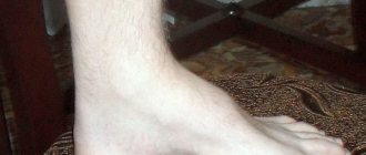
They change their position in bed due to tension in the muscles of the arms and shoulder girdle. Passive movements are performed by patients without muscle tension, but active movements are performed with difficulty, and their amplitude is very limited due to severe muscle weakness. Flaccid paralysis occurs with profound disturbances in muscle movement and trophism. The muscle tone is sharply reduced, the muscles are flabby and atrophic, so the foot acquires the pes equinus (horse foot) position - the foot hangs and is bent towards the sole, which makes walking very difficult.
In acute cases ( encephalomyelitis , acute flaccid paresis ), these phenomena appear within 7-10 days, and in chronic flaccid diseases, muscle weakness and paralysis develop gradually.
Ankylosing spondylitis (Strumpel-Marie-Bechterew disease)
Ankylosing spondylitis (Strumpel-Marie-Bechterew's disease) is a chronic inflammatory ankylosing disease of the joints of the axial skeleton (intervertebral, costovertebral, sacroiliac), belonging to the group of seronegative spondyloarthritis. The disease occurs with a frequency of 2:1000 of the population, and men are affected 3-4 times more often than women.
Cause
disease is unknown. Hereditary predisposition is of great importance, the genetic marker of which is the HLA B27 antigen, which is found in 90-95% of patients, in 20-30% of their first-degree relatives, and only in 7-8% of the population. It is believed that the sensitivity gene for ankylosing spondylitis is linked to the HLA B27 gene.
Pathogenesis
There are two theories of pathogenesis that explain the important role of HLA B27 in the development of ankylosing spondylitis. According to the receptor
theory,
the HLA B27 antigen is a receptor for an etiological damaging factor (for example, a bacterial antigen, virus, arthritogenic peptide, etc.). The resulting complex leads to the production of cytotoxic T lymphocytes, which can then damage cells or tissue areas where B27 antigen molecules are located.
According to the theory of molecular mimicry
a bacterial antigen or some other damaging agent in complex with another HLA molecule may have properties similar to HLA B27 and be recognized by cytotoxic T lymphocytes as HLA B27 or reduce the immune response to the disease-causing peptide (the phenomenon of immune tolerance).
As a result, an immunoinflammatory process develops. More often it begins with damage to the sacroiliac joints, then intervertebral, costovertebral, and less commonly peripheral joints are involved. First, infiltration by lymphocytes and macrophages occurs, then an active fibroplastic process develops with the formation of fibrous scar tissue, which undergoes calcification and ossification.
The main pathomorphological manifestations of ankylosing spondylitis are inflammatory enthesopathies (inflammation of the places of attachment to the bone of tendons, ligaments, the fibrous part of the intervertebral discs, joint capsules), inflammation of the bones forming the joint (osteitis), as well as synovitis.
Subsequently, fibrous and bony ankylosis of the joints of the axial skeleton develops, less often - of the peripheral joints, and ossification of the ligamentous apparatus of the spine occurs early.
Clinical picture
The disease usually begins before the age of 40 and is more often observed in men.
Early stage symptoms
- Pain in the lumbar spine and in the area of the sacroiliac joints is of a constant nature, intensifying mainly in the second half of the night and in the morning, decreasing with movement and in the afternoon (inflammatory “character” of pain).
- Pain in the gluteal region due to damage to the sacroiliac joints, radiating to the back of the thigh, occurring either on the right or on the left.
- A feeling of stiffness and stiffness in the lumbar spine, usually intensifying in the morning and decreasing after exercise or a hot shower.
- Pain in the chest area (when the costovertebral joints are involved in the pathological process) such as intercostal neuralgia or myositis of the intercostal muscles, aggravated by coughing, sneezing, or taking a deep breath.
- Stiffness and tension in the rectus dorsi muscles.
- Flattening of the lumbar lordosis (especially noticeable when the patient bends forward).
- Clinical and radiological symptoms of bilateral sacroiliitis; To identify pain in the sacroiliac joints, indicating the presence of an inflammatory process in them, the following tests are used:
- Makarov test (sacrum tapping);
- Kushelevsky test-1 (pressure on the upper anterior iliac spines with the patient in the supine position);
- Kushelevsky-II test (pressure on the wing of the ilium with the patient in the lateral position);
- Kushelevsky-III test (with the patient in the supine position, simultaneous pressure is applied to the inner surface of the knee joint bent at an angle of 90° and abducted and the upper anterior spine of the opposite wing of the ilium).
- Enthesopathies are manifested by pain in the area of attachment of fibrous structures to bones, in particular, the iliac crests, greater trochanters of the femurs, spinous processes of the vertebrae, sternocostal joints, and ischial tuberosities. As bursitis develops, swelling appears.
- Eye damage in the form of anterior uveitis (iritis, iridocyclitis), usually bilateral, is characterized by an acute onset, lasts 1-2 months, and can take on a protracted recurrent nature; eye pathology is observed in 25-30% of patients.
Late stage symptoms
- Pain in various parts of the spine.
- Poor posture, straightening of the physiological curves of the spine (“plank back”) or “petitioner’s pose,” pronounced kyphosis of the thoracic spine, downward bending and bending of the legs at the knee joints, which compensates for the forward movement of the center of gravity.
- Atrophy of the rectus dorsi muscles.
- A sharp decrease in chest excursions; in many patients, breathing is carried out only through movements of the diaphragm.
- Limitation of mobility in the spine in 3-4 planes: sagittal (flexion, extension), frontal (lateral bending), vertical (rotation).
- Ankylosis of the sacroiliac and intervertebral joints.
- Damage to the “root” (shoulder and hip) or peripheral joints. The hip joints are more often affected in patients whose disease began in childhood or adolescence. Peripheral joints are involved in 10-15% of patients, with predominantly large and medium-sized joints of the lower extremities affected, such as mono- or oligoarthritis. Arthritis of the sternoclavicular (usually asymmetrical), acromioclavicular, mandibular and synchondrosis of the manubrium of the sternum may be observed. Peripheral arthritis can go away without a trace in 20-25% of patients.
- Damage to the cardiovascular system - aortitis, aortic valve insufficiency, pericarditis, myocarditis with varying degrees of atrioventricular conduction impairment, and the incidence of aortitis and atrioventricular conduction impairment is significantly higher with a longer duration of the disease and the presence of peripheral arthritis.
- Lung damage in the form of slowly progressive fibrosis of the apical segments of the lungs.
- Kidney damage is rare, in 5% of cases in the form of amyloidosis or IgA nephropathy, and in contrast to idiopathic IgA nephritis, hematuria is rarely expressed.
Clinical options
- Central form
- damage only to the joints of the spine and sacroiliac joint.
- Rhizomelic form
- damage to both the spine and the shoulder and hip joints.
- Peripheral form
- in some cases, disease of the joints of the extremities precedes damage to the spine, in others - on the contrary, in others - the joints and spine are simultaneously affected. The knee joints are most often affected.
- Option similar to rheumatoid arthritis -
damage to the joints of the hands and feet, morning stiffness. There are no clinical signs of spinal lesions.
- Septic option
is characterized by fever that occurs acutely at the onset of the disease (up to 38-39 ° C), heavy sweat, arthralgia, myalgia, and weight loss.
Tests and diagnostics
First of all, paresis and plegia are detected during a clinical examination. They are characterized by:
- decreased range of motion and muscle strength;
- changes in muscle tone;
- muscle wasting or atrophy;
- increased or decreased normal reflexes;
- fibrillation of the muscles of paretic limbs;
- the presence of pathological reflexes.
To clarify the diagnosis, patients are prescribed the following examinations:
- Echoencephalography of the brain - examination of the brain using ultrasound.
- Electroencephalography is a study of the electrical activity of the brain.
- X-ray contrast and radionuclide studies. With their help, the condition of the brain ventricles and subarachnoid space is assessed.
- Doppler ultrasound is a variant of the study of cerebral vessels.
- Electromyography. Allows you to identify changes in spinal cord motor neurons characteristic of neuropathies.
- Biopsy of the affected muscles with examination of the biopsy material.
- Cerebrospinal fluid puncture.
- CT scan.
- MRI of the brain.
Diet
Patients who do not have impaired swallowing can take regular food and sit on a common table. If a child can chew and swallow independently, his diet should consist of vegetables, fruits, eggs, nuts, meat, fish, cereals, but prepared food must be crushed (pureeed) to make it easy to swallow.
It is necessary to ensure that the diet contains a sufficient amount of protein products (meat, fish, cottage cheese, cheese and other dairy products, eggs) and complex carbohydrates, since rehabilitation treatment requires an increased supply of protein and energy. Fats must be present in the diet - butter and cold-pressed vegetable oils (olive, sesame, flaxseed).
Severe swallowing disorders require special nutrition adapted to the child’s characteristics, since problems with eating gradually lead to a deficiency of essential substances and energy. In such children, protein deficiency and energy deficiency progress. If swallowing continues, the child is additionally given formula through a tube (sipping method). Modern enteral nutrition is a mixture with an optimal composition of nutrients, vitamins, macro- and microelements.
There are dry mixtures ( Nutrien standard ), which need to be prepared by diluting with water, or ready-made mixtures - Nutrizon standard , Nutrien standard , Nutrizon energy and others. Children 3-5 years old with nutritional deficiencies are prescribed formulas for premature infants up to one year old. In severe cases, patients need special nutrition - enteral tube or parenteral.
Prevention
Considering that the main cause of paresis in children is cerebral palsy, its prevention includes:
- improving the health of adolescents and women of childbearing age;
- treatment of sexually transmitted infections when planning pregnancy;
- regular examinations and timely detection of pathology in pregnant women;
- timely diagnosis of coagulopathies in expectant mothers;
- prevention of premature birth;
- adequate obstetric care;
- proper resuscitation, adequate care and use of hypothermia in newborns, timely treatment of hyperbilirubinemia .
In adults, the following are important:
- prevention of injuries (traumatic brain injuries and spinal injuries);
- timely treatment of hypertension , which, if left untreated, is complicated by cerebral hemorrhage;
- exclusion of alcoholism and poisoning by alcohol substitutes.
Prof. Dr. Jörg Wissel, MD, FRCP
Neurological rehabilitation
Head of the Department of Neurological Rehabilitation and Physiotherapy, Department of Neurology and Rehabilitation Center
Specialization
- Neurological rehabilitation after stroke, traumatic brain injury and spinal cord injury
- Rehabilitation therapy for Parkinson's disease and dystonia
- Botulinum therapy
- Treatment of spasticity
- Neurological rehabilitation using intrathecal baclofen therapy and deep brain stimulation
- Author of more than 90 scientific publications, member of several expert commissions
Show doctor's personal profile
Consequences and complications
With paralysis and paresis, complications arise from the musculoskeletal system:
- joint contractures;
- pathological attitudes;
- arthrosis;
- formation of vicious poses;
- muscle atrophy.
Joint contractures are secondary and their severity depends on the severity of the disease and the age of the patient. At first, contractures develop reflexively (from muscle contraction against the background of prolonged excitation), and then become permanent due to degenerative changes in the muscles, ligaments and tendons. The combination of various contractures forms a vicious posture in patients. Motor installations are manifested by a forced position of the limb due to excessively high muscle tone. An example of a propulsion system is the equinus position of the foot, which is formed due to the traction of the gastrocnemius and soleus muscles. With Little's disease, the patient often falls and therefore receives injuries (hematomas, fractures).
Forecast
Functional recovery depends on the severity of the damage to the nervous system. In this regard, compensation of neurological disorders, partial compensation, or the formation of persistent syndromes are ultimately possible.
A quarter of patients move independently, while the rest cannot do without walkers or wheelchairs. With good treatment, children with paraplegia or hemiplegia survive to adulthood and live almost normal lives. The effectiveness of rehabilitation for spastic diplegia depends on how severe the motor impairment is and how much intelligence is preserved. If active rehabilitation has been carried out since early childhood, a person can acquire a profession. If mental abilities are preserved, then they are well adapted in social life. In the presence of epileptic seizures, the prognosis worsens.
With tetraplegia, accompanied by impaired swallowing and the need for feeding through a gastrostomy tube, life expectancy is sharply reduced. Hereditary diseases affecting the pyramidal system ( Strumpel's disease ) progress slowly and have a favorable prognosis for life. However, the ability of patients to work is limited at the onset of the disease, and is lost as it progresses.
Treatment
Strumpel's disease, the treatment of which can only be symptomatic, is a complex disease from a psychological point of view. Patients need to be provided with comprehensive moral support, and courses of symptomatic treatment should be carried out at least twice a year.
Therapy for the disease is aimed primarily at relieving muscle hypertonicity, using muscle relaxants (mydocalm, baclosan, sirdalud and others). Orthopedic methods of influence (wearing orthoses and other devices), neuroprotective drugs (B vitamins) are also used. Patients are prescribed physiotherapeutic procedures, paraffin baths, relaxing massage, and metabolic drugs.
With constant and complete treatment, the progression of the disease is very slow and a person may not significantly lose quality of life for a long time.
List of sources
- Studenikin V. M., Shamansurov Sh. Sh. Neonatal neurology (collective monograph). M.: Medforum, 2014. 120–135.
- Semenova K. A. Restorative treatment of children with perinatal damage to the nervous system and cerebral palsy. M.: Codex, 2007.
- Nemkova S. A., Zavadenko N. N., Medvedev M. I. Modern principles of early diagnosis and complex treatment of perinatal lesions of the central nervous system and cerebral palsy. Toolkit. M., 2013. 76 p.
- Martynova, G.P. Acute flaccid paralysis in children. Diagnostic algorithms and management tactics - Methodological recommendations for pediatricians, infectious disease specialists and neurologists / Krasnoyarsk, 2013. - 48 p.