Classification of nerve fibers
| The amplitude of electrical impulses removed from the entire nerve trunk depends on the strength of the applied stimulation. A small action potential corresponds to stimulation of weak strength, but as stimulation increases, the amplitude of the peak increases, reaches a maximum value and then remains constant, despite a further increase in the strength of the stimulus (Fig. 156). Rice. 156. Responses of the sciatic nerve of the frog to electrical stimuli of increasing strength (according to J. Buresh). The abduction of action potentials is biphasic. At the beginning of each recording there is an irritation artifact. The current increases gradually from 1 to 6. Time stamp 1 ms. This is explained by the fact that the action potential removed from the entire nerve trunk is an algebraic sum of the potentials of its individual nerve fibers . In each fiber, the amplitude of the action potential does not depend on the strength of stimulation in accordance with the “all or nothing” law. The stimulation thresholds of individual fibers differ from each other. With weak stimulus strength, excitation occurs in the most excitable superficial nerve fibers. Increasing the stimulus leads to an increase in the number of excited fibers, so the total peak increases until all fibers are involved in the response. This picture can be observed if the discharge electrodes are placed on the nerve near the stimulating electrodes. As the distance between these two pairs of electrodes increases, the total action potential begins to be divided into several individual oscillations, which become most clearly expressed when the output electrodes move 10-15 cm from the site of stimulation (Fig. 157). |
| This phenomenon was first studied in detail by Erlanger and Gasser (1937). They showed that the reason for the division of the total action potential into components is the unequal speed of excitation along different fibers, as a result of which nerve impulses arrive at the output electrodes through these fibers non-simultaneously. Rice. 157. Complex of the compound action potential of the mixed nerve trunk of the frog (according to Erlanger and Gasser). |
| A detailed analysis showed that there is an approximately proportional relationship between the speed of impulse conduction and the diameter of the nerve fiber: nerve fibers conduct faster the thicker they are. Currently, it is customary to divide nerve fibers according to the speed of excitation, the duration of various phases of the action potential and structure into three main types, designated by the letters A, B and C (Fig. 158). Rice. 158. Schematic representation of all components of the action potential of a mixed nerve (according to Erlanger and Gasser). |
Classification of nerve fibers
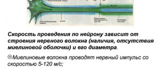
Type A nerve fibers are divided into four subgroups: α, β, γ and δ. They are covered with a myelin sheath. The thickest of them - the so-called alpha fibers (Aα) - in warm-blooded animals and humans have a diameter of 12-22 microns and are characterized by a significant speed of excitation: 70-120 m/sec. Such fibers conduct excitation from the motor nerve centers of the spinal cord to the skeletal muscles (“motor” fibers) and from muscle receptors to the corresponding nerve centers. The action potential of Aα fibers in warm-blooded animals lasts 0.5 ms. After its completion, a trace negative potential develops, which lasts 15-20 ms and turns into a trace positive potential lasting about 40-60 ms.
Three other groups of type A fibers: beta (Aβ)-, gamma (Aγ)- and delta fibers have a smaller diameter - from 2 to 12 microns, a lower conduction velocity and a longer action potential. These are predominantly sensory fibers that conduct excitation from various receptors (tactile, some pain, temperature and receptors of internal organs) into the central nervous system. The only exceptions are gamma fibers, a significant part of which conduct excitation in a centrifugal direction - from the cells of the spinal cord to the so-called intrafusal muscle fibers of which sensitive nerve endings - muscle spindles - are located.
Type B fibers include mpelin, predominantly preganglionic fibers of the autonomic nervous system. The speed of excitation in these fibers in warm-blooded animals is 3-14 m/sec. The duration of the action potential of type B fibers is approximately 2 times longer than the duration of the action potential of type A fibers. On average, it is 1.2 ms. A distinctive feature of these fibers is that they do not exhibit a trace negative potential: the repolarization phase directly transforms into a trace positive potential, which has a long duration - 100-300 ms.
Type C fibers include soft nerve fibers of very small diameter (about 0.5 µm). The speed of excitation in these fibers is 2-6 m/sec. Most C fibers are postganglionic fibers of the sympathetic nervous system.
C-fibers also include those nerve fibers that are involved in conducting excitation from pain receptors and some receptors of cold, heat and pressure. Action potentials of C-fibers are characterized by the longest duration (2 ms in warm-blooded animals). They have a long trace negative potential (50-80 ms), accompanied by an even longer trace positivity (300-1000 ms).
All this data is summarized in the table.
Properties of various nerve fibers of warm-blooded animals
| Fiber type | Fiber diameter (μ) | Conduction speed (m/sec) | Action potential duration (ms) | Duration of negative trace potential (ms) | Duration of positive trace potential (ms) | Function |
| A (α) | 12-22 | 70-120 | 0,4-0,5 | 12-20 | 40-60 | Motor fibers of skeletal muscles, afferent fibers of muscle receptors |
| A(β) | 8-12 | 40-70 | 0,4-0,6 | — | — | Afferent fibers from touch receptors |
| A (γ) | 4-8 | 15-40 | 0,5-0,7 | — | — | Afferent fibers from touch and pressure receptors, efferent fibers to muscle spindles |
| A (Δ) | 1-4 | 5-15 | 0,6-1,0 | — | — | Afferent fibers from some receptors of heat, pressure, pain |
| IN | 1-3 | 3-14 | 1-2 | Absent | 100-300 | Preganglionic autonomic fibers |
| WITH | 0,5-1,0 | 0,5-2 | 2,0 | 50-80 | 300-1000 | Preganglionic autonomic fibers, afferent fibers from some pain, pressure, and heat receptors |
Classification of nerve fibers according to Erlanger-Gasser
In 1939, American [Mf54] physiologists Joseph Erlanger [B55] and Herbert S. Gasser [B56] [B57] recorded action currents from the entire nerve trunk of the frog sciatic nerve at different distances from the stimulating electrode (Fig. 210041905).[B58]
It was found that the recorded total potential has a number of peaks, which were designated by the letters of the Latin alphabet A, B, C (Fig. , I). Peak A had additional peaks labeled with the Greek letters α , β , γ , δ (Fig. ,II). [B59] In 1944, the work of J. Erlanger and G.S. Gasser was awarded the Nobel Prize [B60].
Nerves in vertebrates consist of three main groups of fibers
(A, B and C), differing in the degree of myelization, fiber diameter, duration of the AP peak (AP development rate), electrical excitability, its compensation and conduction speed (all these indicators in the series A - B - C fall).
Group A
includes the thickest well-myelinated motor and sensory fibers;
group B -
weakly myelinated, preganglionic fibers of the autonomic nervous system;
group C -
unmyelinated, postganglionic (sympathetic) fibers.
In group A, in the row a, b, g, d, these indicators also fall. The ratios of the properties of these groups of fibers are shown in table. .
It should be noted that the indicated ratios of the thresholds of electrical stimulation of groups of fibers do not reflect the exact ratio of the electrical excitability of their membranes. The relatively high thresholds of thin fibers when they are irritated in the nerve trunk are determined mainly by the fact that thin fibers have a higher input resistance compared to thick ones. They contain such a small part of the irritating current that at the threshold strength for Aa
-fibers, it is completely insufficient to create any significant depolarization on the membrane of thinner fibers.
For the same reason (high RI
), the PD of thin fibers removed from the trunk (extracellularly) appears significantly smaller than the PD of thick fibers.
Rice. . Components of the mixed nerve action potential.
I – at a relatively slow recording speed.
II – at a relatively high recording speed.
Explanation in the text. The abscissa axis is time, the ordinate axis is the amplitude of the compound potential in mV.
Total electrical activity of the nerve
is created by its fibers, each of which generates its own PD, standard in amplitude and time parameters, spreading in both directions from the point to which the stimulation is applied. The total electrical signal of the nerve depends on the number of active fibers, the synchrony of their activity, the method of abduction and other circumstances.
Let us consider the case of the so-called single-phase derivation,
in which one output electrode (active) is located on a normal section of the trunk, and the other (indifferent) is located on a damaged one, where the fibers are completely depolarized (Fig. 1.19).
Let artificial stimulation be used and the stimulating electrode (cathode) be located sufficiently close (l =
3 mm) to the active output electrode, and the nerve is placed in a non-conducting medium (oil or air). Here, a strong single stimulation leads to synchronous excitation of all fibers, while the active output electrode records the total action potential of the nerve, which is similar in shape to the action potential of an individual A (alpha) fiber, but is slightly more prolonged in time. This nerve action, however, does not follow the all-or-none rule. With threshold stimulation it is negligibly small, with increasing stimulus strength it gradually increases, reaching a maximum equal to 5-10 mV under normal conditions, and 50-100 mV under sucrose bridge conditions.
With a further increase in stimulus strength, this AP slightly lengthens in time. All changes in the amplitude and duration of the AP nerve peak with increasing stimulus are determined by an increase in the number of active fibers, the connection to low-threshold and fast A (alpha) fibers of higher threshold slow beta, gamma, delta fibers of group A, then B and, finally, C-groups.
| Fiber groups (according to Erlanger and Gasser) | Diameter, microns | Conduction speed, m/s |
| Aa | 13 — 22 | 70 — 120 |
| Ab | 8 -13 | 40 — 70 |
| Ag | 4 — 8 | 15 – 40 |
| Ad | 1 – 4 | 5 – 15 |
| B | 1 — 3 | 3 – 14 |
| C | 0,5 – 1,0 | 0,5 — 2 |
Table . Classification of nerve fibers according to J. Erlanger and H. Gasser
| Fiber groups (according to Erlanger and Gasser) | Diameter, microns | Electrical stimulation thresholds (relative to Aa) | PD1 peak duration | Negative Trace Potential (NTP) | Positive trace potential [B61] | Conduction speed, m/s | |
| Duration, ms | SP amplitude, % to AP amplitude | Duration, ms | SP amplitude, % to AP amplitude | ||||
| Aa | 13 — 22 | 1,0 | 0,4 | 15 – 20 | 40 — 60 | 0,2 | 70 — 120 |
| Ab | 8 -13 | 40 — 70 | |||||
| Ag | 4 — 8 | 15 – 40 | |||||
| Ad | 1 – 4 | 5 – 15 | |||||
| B | 1 — 3 | 11,7 | 1,2 | no OSB | 100 – 300 | 3 – 14 | |
| C | 0,5 – 1,0 | 100,0 | 2,0 | 50 – 60 | 300 — 1000 | 0,5 — 2 |
1Absolute refractory phases have approximately the same value
Physiology of nerve fibers
CONTENT
Introduction…………………………………………………………………………………..3
Classification of nerve fibers…………………………………………….4
Classification of nerve fibers by diameter and conduction speed………9
Physiological properties and functional significance of nerve fibers………………………………………………………………………………………13
Features of the structure and types of nerve fibers………….……………….…14
The mechanism of excitation along the nerve fiber……………..……14
Laws of conduction of excitation along the nerve fiber………..………………….16
Conclusion………………………………………………………………………………….17
References……………………………………………………………18
Introduction
A special place in physiology is given to excitable tissues. Not all tissues in the body are able to respond equally quickly to stimuli. Only some of them, in the process of evolution, have developed this property - a quick response to the action of a stimulus.
An irritant is understood as any change in the conditions of the external and internal environment, if it occurs suddenly, has sufficient strength, is maintained for a certain time, and causes reversible changes in the structure and activity of living tissues and cells. The process of exposure of living structures to a stimulus is called irritation.
There are three groups of irritants: physical, physico-chemical and chemical. The nerve impulse is especially distinguished as an irritant.
According to their physiological significance, all stimuli are divided into adequate and inadequate. Adequate are irritants that act on the body and its structures in natural conditions, and the structures of the body are adapted to the perception of this irritant. Inadequate are irritants that naturally do not affect the body, and the structures of the body are not adapted to perceive them. Therefore, such irritants most often cause disruption of body function.
Tissues and cells of the body that are specially adapted to carry out rapid responses to the action of a stimulus are called excitable tissues. These include nervous, glandular and muscle tissue.
Excitable tissues have a number of specific properties: excitability and conductivity.
Excitability is the ability of excitable tissue to respond by changing its structure and activity to the action of a stimulus, i.e. respond to a special biological reaction called arousal.
Excitation is a response of excitable tissue to the action of a pathogen, manifested in a combination of physical, physicochemical, chemical, metabolic processes and changes in activity. Excitation is a wave-like process that manifests itself in different excitable tissues in a specific way: in muscle - by contraction, in glandular - by the formation and release of secretions, in nervous - by the emergence and conduction of a nerve impulse.
The development of excitation is accompanied by a short-term disappearance of excitability. Then it quickly recovers.
An obligatory and general sign of excitation of excitable tissues is the occurrence of a biological current of action, i.e. bioelectric phenomena.
Conductivity is the property of excitable tissue to actively conduct an excitation wave. For example, the motor nerve of a cat conducts excitation at a speed of 1200 cm/s.
Classification of nerve fibers
| The amplitude of electrical impulses removed from the entire nerve trunk depends on the strength of the applied stimulation. A small action potential corresponds to stimulation of weak strength; as stimulation increases, the amplitude of the peak increases, reaches a maximum value and then remains constant, despite a further increase in the strength of the stimulus (Fig. 1). Fig. 1 Responses of the frog sciatic nerve to electrical stimuli of increasing strength (according to J. Buresh). The abduction of action potentials is biphasic. At the beginning of each recording there is an irritation artifact. The current increases gradually from 1 to 6. Time stamp 1 ms. This is explained by the fact that the action potential removed from the entire nerve trunk is the algebraic sum of the potentials of its individual nerve fibers. In each fiber, the amplitude of the action potential does not depend on the strength of stimulation in accordance with the “all or nothing” law. The stimulation thresholds of individual fibers differ from each other. With weak stimulus strength, excitation occurs in the most excitable superficial nerve fibers. Increasing the stimulus leads to an increase in the number of excited fibers, so the total peak increases until all fibers are involved in the response. This picture can be observed if the discharge electrodes are placed on the nerve near the stimulating electrodes. As the distance between these two pairs of electrodes increases, the total action potential begins to be divided into several individual oscillations, which become most clearly expressed when the output electrodes move 10-15 cm from the site of stimulation (Fig. 2). |
| This phenomenon was first studied in detail by Erlanger and Gasser (1937). They showed that the reason for the division of the total action potential into components is the unequal speed of excitation along different fibers, as a result of which nerve impulses arrive at the output electrodes through these fibers non-simultaneously. Fig.2. Complex of the compound action potential of the mixed nerve trunk of the frog (according to Erlanger and Gasser). |
| A detailed analysis showed that there is an approximately proportional relationship between the speed of impulse conduction and the diameter of the nerve fiber: nerve fibers conduct faster the thicker they are. Currently, it is customary to divide nerve fibers according to the speed of excitation, the duration of various phases of the action potential and structure into three main types, designated by the letters A, B and C (Fig. 3). Rice. 3. Schematic representation of all components of the action potential of a mixed nerve (according to Erlanger and Gasser). |
Type A nerve fibers are divided into four subgroups: α, β, γ and δ. They are covered with a myelin sheath. The thickest of them - the so-called alpha fibers (Aα) - in warm-blooded animals and humans have a diameter of 12-22 microns and are characterized by a significant speed of excitation: 70-120 m/sec. Such fibers conduct excitation from the motor nerve centers of the spinal cord to the skeletal muscles (“motor” fibers) and from muscle receptors to the corresponding nerve centers. The action potential of Aα fibers in warm-blooded animals lasts 0.5 ms. After its completion, a trace negative potential develops, which lasts 15-20 ms and turns into a trace positive potential lasting about 40-60 ms.
Three other groups of type A fibers: beta (Aβ)-, gamma (Aγ)- and delta fibers have a smaller diameter - from 2 to 12 microns, a lower conduction velocity and a longer action potential. These are predominantly sensory fibers that conduct excitation from various receptors (tactile, some pain, temperature and receptors of internal organs) into the central nervous system. The only exceptions are gamma fibers, a significant part of which conduct excitation in a centrifugal direction - from the cells of the spinal cord to the so-called intrafusal muscle fibers of which sensitive nerve endings - muscle spindles - are located.
Type B fibers are myelinated, predominantly preganglionic fibers of the autonomic nervous system. The speed of excitation in these fibers in warm-blooded animals is 3-14 m/sec. The duration of the action potential of type B fibers is approximately 2 times longer than the duration of the action potential of type A fibers. On average, it is 1.2 ms. A distinctive feature of these fibers is that they do not exhibit a trace negative potential: the repolarization phase directly transforms into a trace positive potential, which has a long duration - 100-300 ms.
Type C fibers include soft nerve fibers of very small diameter (about 0.5 µm). The speed of excitation in these fibers is 2-6 m/sec. Most C fibers are postganglionic fibers of the sympathetic nervous system.
C-fibers also include those nerve fibers that are involved in conducting excitation from pain receptors and some receptors of cold, heat and pressure.
Action potentials of C-fibers are characterized by the longest duration (2 ms in warm-blooded animals). They have a long trace negative potential (50-80 ms), accompanied by an even longer trace positivity (300-1000 ms)
All these data are summarized in table.1.
Properties of various nerve fibers of warm-blooded animals
| Fiber type | Fiber diameter (μ) | Conduction speed (m/sec) | Action potential duration (ms) | Duration of negative trace potential (ms) | Duration of positive trace potential (ms) | Function |
| A (α) | 12-22 | 70-120 | 0,4-0,5 | 12-20 | 40-60 | Motor fibers of skeletal muscles, afferent fibers of muscle receptors |
| A(β) | 8-12 | 40-70 | 0,4-0,6 | — | — | Afferent fibers from touch receptors |
| A (γ) | 4-8 | 15-40 | 0,5-0,7 | — | — | Afferent fibers from touch and pressure receptors, efferent fibers to muscle spindles |
| A (Δ) | 1-4 | 5-15 | 0,6-1,0 | — | — | Afferent fibers from some receptors of heat, pressure, pain |
| IN | 1-3 | 3-14 | 1-2 | Absent | 100-300 | Preganglionic autonomic fibers |
| WITH | 0,5-1,0 | 0,5-2 | 2,0 | 50-80 | 300-1000 | Preganglionic autonomic fibers, afferent fibers from some pain, pressure, and heat receptors |
Classification of nerve fibers by diameter and conduction speed
− classification of motor and sensory nerve fibers according to Erlanger-Gasser;
− classification of sensory nerve fibers according to Lloyd-Hunt;
Types of nerve fibers and their functions
When recording the electrical activity of the nerve trunk, Joseph Erlanger and Herbert Gasser in 1937 discovered the composite nature of the current action of the mixed nerve.
Based on the data obtained (diameter, conduction velocity, function), a classification of nerve fibers was developed (Table 1, summary Table 3), according to which all nerve fibers are divided into groups A, B and C (Latin letters) with further gradations (α, β, etc.).
Erlanger-Gasser classification of nerve fibers - classification of motor and sensory nerve fibers:
A, B, C - groups of fibers;
α, β, γ, δ - subgroups of group A fibers.
Type A fibers are thick myelinated nerve fibers.
The thickest of them, Aα, have a diameter of 12–22 μm and an excitation speed of 70–120 m/s. These fibers conduct excitation from the spinal motor nerve centers (motor centers of the spinal cord) to the skeletal muscles (motor fibers) and from muscle receptors to the corresponding nerve centers.
Other groups of type A fibers (β, γ, δ) have a smaller diameter - from 8 to
1 µm and lower excitation speed - from 5 to 70 m/s. The fibers of these groups predominantly conduct excitation from various receptors (tactile, temperature, pain; receptors of internal organs or visceroreceptors) in the central nervous system, with the exception of γ-fibers, a significant part of which conduct excitation from the spinal cord to intrafusal muscle fibers.
Table 1
Classification of nerve fibers according to Erlanger-Gasser
| Type fibers | Function (optional) | Average diameter, µm | Average conduction speed, m/s |
| Aα | Primary afferents of muscle spindles, motor fibers of skeletal muscles | 15 | 100 (70 – 120) |
| Aβ | Cutaneous afferents of touch and pressure | 8 | 50 (30 – 70) |
| Aγ | Motor fibers of muscle spindles | 5 | 20 (15 – 30) |
| Aδ | Cutaneous afferents of temperature and pain | <3 | 15 (12 – 30) |
| IN | Sympathetic preganglionic fibers | 3 | 7 (3 – 15) |
| WITH | Cutaneous pain afferents Sympathetic postganglionic fibers | 1 (unmyelinated) | 1 (0,5 – 2) |