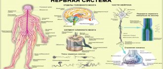
The peripheral nervous system connects the central nervous system to the organs and limbs. Unlike the central nervous system, the peripheral nervous system is not protected by bones and has no physiological barrier separating it from the circulatory system. Therefore, the peripheral nervous system may be susceptible to mechanical damage and is more easily affected by toxins.
Diseases of peripheral nerves are neuropathies. They are characterized by damage to the axons and myelin sheath of the nerves. Diseases of the nerve trunks and plexuses rank second in prevalence among the population after diseases of the spinal roots (“radiculitis”). Therefore, issues of prevention, early diagnosis and subsequent treatment of neuropathies remain pressing problems.
Types
- Demyelinating. The conduction of excitation through neurons is disrupted. Occurs in cases of lead poisoning, diphtheria, polyradiculoneuropathy, and diabetic polyneuropathy. The patient's health is restored within a few weeks if treatment is started in a timely manner.
- Axonopathies. In this case, the damage concerns the axons (the processes of nerve cells) - these are severe disorders of nerve function. As a result, muscle atrophy occurs. The cause of these disorders is the abuse of alcohol and other toxic substances.
Neuropathies of mixed origin are most common. Full recovery and restoration of nerve function depends in part on the severity of the damage.
If the recovery process is absent 3 months after the onset of the disease, then the prognosis is most often unfavorable.
Types of neuropathy
- Mononeuropathies. One nerve or a specific part of the nerve plexus is injured. The causes of damage can be trauma, compression of any level of the nerve trunk. Also, mononeuropathies are observed in diabetes mellitus, atherosclerosis, vascular damage, etc. Hypothermia and herpetic infections are not the least important factors in the functioning of one nerve.
- Multifocal neuropathies are a syndrome of partial damage to individual nerve trunks or their complete damage. Such neuropathies occur slowly and sequentially (from several days to several years). Causes: arthritis, vasculitis and a number of systemic connective tissue diseases.
- Polyneuropathy. Lesions of peripheral nerves are multiple. Moreover, the process is widespread and symmetrical. It occurs both acutely and chronically. It happens that the spinal roots are also affected.
The causes of diseases of the peripheral nerve trunks and plexuses can be:
- injuries received
- decrease in normal immune system function
- hereditary diseases
- intoxication with various substances
- infections
- lack of vitamins
- habitual intoxication (drug addiction and alcoholism)
- allergies
Peripheral nerve conduction studies and electromyography
Peripheral nerve conduction studies provide a simple and reliable way to determine the condition of peripheral nerves. The impulse caused by electrical nerve stimulation is directed along the motor, sensory and mixed nerves, and the characteristics of the impulse are assessed by recording potentials from the muscles or directly from the nerve.
A motor unit consists of a single lower motor neuron and all the muscle fibers it innervates. Motor nerve conduction studies are used to assess the integrity of the motor unit. This provides the investigator with information about the functioning and structural integrity of the motor neuron, nerve, neuromuscular junction, and muscle. It allows you to establish the localization, prevalence, duration and pathophysiological features of damage to the peripheral nervous system (PNS). It can also provide insight into prognosis, treatment effectiveness, and the extent of motor unit recovery. In motor conduction studies, recording electrodes are placed on the skin over the muscle and tendon, and stimulating electrodes are placed on the skin along the nerve being tested. The response of a muscle to electrical stimulation can be measured by recording the net muscle action potential (SMAP), which is the sum of the electrical potentials of all muscle fibers that respond to nerve stimulation. The time it takes for an electrical impulse to reach the muscle (latency) can be determined. The speed at which an impulse travels along a nerve is determined by stimulating the nerve at various locations and determining the distance the stimulus travels.
Motor nerve conduction studies can be used for the following purposes::
- To obtain objective evidence of motor unit damage.
- To identify and pinpoint the location of compression, ischemia, and focal nerve injuries that may manifest as blockade of impulse conduction, slowing of impulse conduction at the site of injury, or abnormal conduction proximal or distal to the injury.
- To determine the extent of nerve damage in patients who show signs of single nerve damage (eg, mononeuropathies).
- For the differential diagnosis of peripheral neuropathies, myopathies and lower motor neuron diseases (eg, amyotrophic lateral sclerosis) in patients with limb weakness.
- To diagnose the disease before it enters the stage of advanced clinical manifestations (for example, with familial neuropathies).
Testing for neuromuscular junction disorders may include rhythmic stimulation of the motor nerves. As the neuromuscular junction becomes fatigued when the SPDM is recorded and compared with a later SPDM, there may be a drop in potential amplitude as fewer and fewer fibers are able to respond to stimulation over time, even when the nerve is stimulated at an intensity that the nerve is normally able to withstand. for a long time.
Sensory nerve conduction studies are carried out by recording action potentials during electrical stimulation of the cutaneous nerve. Selective sensory nerve studies can be performed by stimulating nerves that have only a sensory component (eg, the sural nerve) or, alternatively, by selectively stimulating the sensory component of a mixed nerve. The latter can be done by anatomical isolation of the sensory component (for example, stimulating the fingers and recording over the mixed nerve at the wrist or elbow) or stimulating the mixed nerve and recording over the fingers, in the region of which sensory axons are predominantly located.
Sensory nerve conduction studies may be of value in the following situations::
- For systemic diseases that occur with damage to sensory nerves - to determine the type of sensory nerves involved in the pathological process (for example, thin fibers that conduct pain and temperature sensations, or thick fibers responsible for proprioceptive sensitivity); to determine which part of the peripheral nerve is most affected - the axon or the myelin sheath; to obtain objective evidence of sensory nerve damage.
- For focal neuropathies - to determine the location of damage or blockage, especially with isolated damage to sensory nerves.
- To confirm or quantify impairments when sensory disturbances occur in peripheral neuropathy before motor changes or before the onset of objective clinical signs.
- To establish the location of the injury (proximal or distal) in relation to the dorsal root ganglion (for example, in the differential diagnosis of brachial plexus injury and root injury).
Electromyography (EMG) is usually performed in conjunction with nerve conduction studies to provide additional information. A needle electrode is inserted into the muscle being tested and action potentials generated by groups of muscle fibers (motor unit action potentials, or MUAPs) are recorded. The muscles are examined at rest, in a state of weak contraction and in a state of strong contraction. Normally, muscle activity is not recorded at rest. With active neuropathy, with severe or inflammatory myopathies, spontaneous action potentials from single muscle fibers (fibrillation potentials) can be recorded. In some neurogenic processes (this is especially typical for motor neuron disease), spontaneous contractions of groups of muscle fibers (fasciculation potentials) can be observed. Characteristic changes in PDDE can be observed in pathologies of nerves and muscles. In diseases of peripheral nerves, the amplitude, duration and degree of polyphase of the PDDE are often increased, and recovery is difficult, while in myopathies, the amplitude and duration of the PDDE can be reduced, the polyphase is increased, and recovery is accelerated. Single muscle fiber action potentials can be examined using a more technically sophisticated technique, single muscle fiber electromyography.
In general, electromyography and nerve conduction studies are used to evaluate and clarify the diagnosis in patients with motor neuron disease (for example, amyotrophic lateral sclerosis), pathological processes involving plexuses or nerve roots, compression neuropathies, peripheral polyneuropathies, neuromuscular diseases synapse (for example, myasthenia gravis), as well as with muscle diseases. Because the test requires the insertion of needle electrodes into the muscles and the application of electrical shocks, it is associated with certain inconveniences for the patient. If safety precautions are followed, the study does not pose any danger; The patient's tendency to bleed may limit the EMG.
EMG and determination of the velocity of propagation of excitation (SPT) along the nerve fiber in various diseases
1. EMG and SRV studies are important in the evaluation and electrophysiological diagnosis of motor neuron diseases (eg, amyotrophic lateral sclerosis). In general, peripheral nerve conduction studies give normal results, with the possible exception of some decrease in MUAP amplitudes (since the disease is exclusively of a motor nature, the results of sensitivity studies do not reveal pathology). Needle EMG can detect signs of diffuse anterior horn cell damage, including abnormal spontaneous activity (fibrillations and fasciculations), abnormal parameters (increased amplitude, widening, polyphasicity), and delayed recovery of the MUAP. Often, EMG data indicate an active pathological process even in the absence of clinical manifestations of the disease or minimal manifestations. Using needle EMG, you can also obtain information about the prognosis of the disease; EMG can help diagnose other anterior horn cell diseases such as post-polio syndrome and spinal muscular atrophy.
2. The term radiculopathies combines various symptoms and signs resulting from transient or permanent damage to the nerve as it exits the spinal cord at the level of the intervertebral foramina. Conductivity studies are usually normal. EMG reveals signs of neurogenic changes (for example, fibrillation and changes in the MUAP) in muscles innervated by a specific root, while muscles innervated by roots not involved in the pathological process are intact. The nature of neurological changes depends on the severity of the process, the duration of the disease and the degree of recovery (reinnervation).
In clinical practice, EMG may be useful in the following situations:
- EMG is used to confirm root injury and determine the level of injury. It should be noted that pathological changes according to EMG results were observed in only approximately 90% of patients with cervical or lumbosacral radiculopathy detected during surgery. Thus, normal EMG results do not exclude the presence of radiculopathy.
- EMG allows us to clarify the involvement of specific roots.
- EMG is used to detect active denervation (determined by the presence of fibrillar potentials).
- Using EMG, you can determine the time that has passed since the onset of radiculopathy (acute, subacute, chronic or long-term).
- EMG can provide some information about the severity of radiculopathy.
- EMG may reveal other pathology that may explain the symptoms the patient is experiencing.
- EMG can help determine whether changes detected by MRI or myelography have any physiological significance.
- Using EMG and ENMG, it is possible to diagnose brachial and lumbosacral plexopathies and compression neuropathies, as well as determine the level of damage in these diseases.
3. EMG and ENMG are often prescribed for peripheral polyneuropathies. Electrophysiological characteristics of neuropathies are used as additional information to clarify the nature of the disease, which makes it possible to narrow the range of differential diagnostic searches. EMG/ENMG results allow us to assess the degree of involvement of motor and sensory nerves; determine whether the lesion is primarily the result of damage to the myelin sheath or axon; indicate whether the damage is focal or diffuse; determine whether the process is spreading distally or proximally; provide information about the severity and duration of the pathological process. An increase in distal sensory and motor latencies, a slowdown in conduction velocity, pathology of sensory responses and PDDE, and “neurogenic” EMG changes may be observed. Pathological research results confirm the presence of neuropathies, but it should be noted that in neuropathies with damage to thin sensory fibers (conducting pain and temperature sensitivity), the research results are often normal. Using EMG/ENMG, it is possible to differentiate generalized sensorimotor peripheral polyneuropathy from multiple mononeuropathies in areas of frequent compression (for example, neuropathies of the median and ulnar nerves in the wrist).
According to electrophysiological characteristics, peripheral polyneuropathies can be divided into the following categories::
- Demyelinating mixed sensorimotor neuropathies, including some hereditary neuropathies,
- Segmental demyelinating sensorimotor polyneuropathies, including inflammatory neuropathies (eg, Guillain-Barré syndrome) and neuropathies associated with gammopathies, hypothyroidism, malignancy or lymphoma, AIDS, Lyme disease and exposure to certain toxins.
- Axonal motor-sensory polyneuropathies, including porphyria, some hereditary neuropathies, lymphomatous neuropathies and some toxic neuropathies.
- Axonal sensory neuropathies or neuropathies, including primary amyloidosis, Sjögren's syndrome, paraneoplastic neuropathies, and drug-induced neuropathies and vitamin B12 deficiency.
- Mixed axonal sensorimotor polyneuropathies, including neuropathy due to uremia and diabetes mellitus.
- Axonal sensorimotor polyneuropathies, including neuropathies caused by certain nutritional deficiencies, alcohol intake, associated with sarcoidosis, connective tissue diseases, exposure to toxins, heavy metals and drugs.
4. Diseases of the neuromuscular junction can be diagnosed using rhythmic stimulation. Rhythmic motor nerve stimulation is used mainly to diagnose myasthenia gravis. This pathology is characterized by a progressive decrease in the amplitude of the response to the first few irritating stimuli, obtained when stimulated at a frequency of 3 stimuli per second. The nature of the disease can be clarified by changes in the response to stimulation after a short muscle contraction. In some patients with myasthenia gravis, if stimulation results are normal, the diagnosis can be established using single muscle fiber EMG. In Eaton-Lambert myasthenic syndrome, the amplitude of the response of a resting muscle caused by a single maximal nerve stimulation is significantly reduced. A further decrease in amplitude may be observed during rhythmic low-frequency stimulation, but a significant improvement (increase in PPDE) is observed during high-frequency stimulation. In other diseases, such as amyotrophic lateral sclerosis, unusual fatigue of the peripheral neuromuscular system may sometimes be observed, but this pathological change is of little diagnostic value.
5. In patients with myopathies, electrodiagnostic studies demonstrate a wide range of abnormalities. The main ENMG parameters are normal, with the exception of a sometimes observed decrease in the amplitude of motor responses. EMG may record fibrillar potentials in severe myopathies or inflammatory myopathies (for example, polymyositis). “Myopathic” PDE is characterized by a decrease in amplitude and duration with an increase in polyphasia and rapid recovery, regardless of the degree of muscle contraction. EMG alone is usually not sufficient to diagnose a disease, but EMG results can be used to assign pathology to a specific group of muscle disorders. Toxic and endocrine myopathies may not be accompanied by pathological abnormalities on the EMG, or these abnormalities turn out to be very insignificant.
EMG/ENMG allows us to clarify the following questions
- Differentiate between neurogenic and myopathic causes of muscle weakness.
- Establish the key points of the etiology of myopathy.
- Assess the severity of the pathological process, and whether it is acute, subacute or chronic.
- Assess the degree of activity and nature of the disease.
- Provide information on the prevalence of the pathological process.
- Diagnose pathology, even if it is not accompanied by clinical manifestations.
Author: neurologist of the highest qualification category Trubitsyna O.V.
Tel.: 267-36-66, 267-09-11
Symptoms
With neuropathies, symptoms can be different and depend on the affected areas. They cause a lot of discomfort to the patient:
- flaccid muscle paralysis
- pain in limbs
- change in skin sensitivity (there may be a contrast in sensations in one area of the skin compared to another)
- no sensation of pain and more
- amyotrophy
- speech disorder
- feeling of numbness in the face and limbs
- muscle weakness of the limbs
- impaired motor coordination
- dry skin
- focal blanching
- redness and bluish discoloration of the injured area
- Facial asymmetry may occur (if the facial nerves are damaged)
If you find yourself with at least one of the described symptoms, you should immediately seek help from a qualified neurologist.
Treatment
In our clinic, traditional and alternative medicine methods are used to treat neuropathies. Complex treatment is selected only individually and depends entirely on the degree of damage to the nerve(s).
Initial treatment will be aimed at restoring the functions of the peripheral nerves, therefore, the cause will be eliminated.
An integrated approach is important in the treatment of neuropathies! Our specialists will take into account all the nuances of the damage and may prescribe:
- medications that improve metabolism, blood circulation and restoration processes in nervous tissue. Medicines can also be used in injection form, including intravenous drips in a cozy day hospital
- hormonal drugs (steroids) - in some cases
- therapeutic drug blockades
- certain types of physiotherapeutic treatment
- manual therapy (osteopathy)
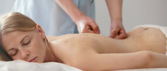
- classic massage
Recommendations and prevention
The main prevention of most diseases of the nervous system is maintaining a healthy and active lifestyle, giving up bad habits, timely and adequate treatment of infectious and non-infectious diseases.
If any neurological symptoms occur, do not delay contacting a doctor. Early diagnosis and timely treatment will help prevent the development of complications, prolongation of treatment and the consequences of uncontrolled use of medications. The most complete program of preventive measures is drawn up by a neurologist for each individual patient.
Diagnostic methods in neurosurgery
Radiography
Direct radiographs provide information about the normal anatomy and diseases of the spine, and can also detect pathologies such as fractures, cracks and curvatures in the neck, back, waist, tailbone and hip joints. The presence and degree of curvature of the back or waist can be determined using x-rays of scoliosis.
Computed tomography (CT)
Computed tomography is often used to examine and diagnose the spine. It allows you to determine the quality of bone tissue, its rarefaction, defects, osteoporosis, osteomyelitis, osteophytes at the initial stage of development, cartilage proliferation, degeneration of intervertebral discs (osteochondrosis, deforming spondylosis, intervertebral hernia, arthritis).
Magnetic resonance imaging (MRI)
Detailed images of extra and intracranial structures are obtained using MRI. This is the ability to make a cross-section (sagittal, coronal, axial, oblique) without changing the patient's position. With the help of MRI technology, the entire spinal column, spinal canal and spinal cord can be clearly visible from different angles. Superior to CT in the study of spinal pathologies.
Electromyography/Nerve Conduction Study (ENMG)
In the practice of neurosurgery, they are indispensable methods for diagnosing cuts of peripheral nerves, neuropathy, plexus lesions, disc herniation and other spinal cord lesions caused by other reasons, intramedullary pathology of the spinal cord.
Biopsy
A biopsy is performed using open surgery or a stereotactic method for accurate diagnosis and treatment planning for tumor, infectious, viral, and degenerative diseases of the central nervous system.
FAQ
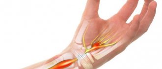
Three fingers on my right hand are numb and painful. What to do?
It is necessary to consult a neurologist to exclude carpal tunnel syndrome and treatment.
I had an attack of trigeminal neuralgia for a long time. Should I take something preventative to prevent another one?
It is imperative to see a neurologist. The doctor will select medications for you that need to be taken on a regular basis and prescribe preventive courses of injections (if necessary, drips in a day hospital) and tablet medications.
Severe burning pain appeared in the right half of the chest and some bubbles appeared. Which doctor should I see?
Similar manifestations, as you describe, can be observed with herpetic ganglioneuritis, the so-called. herpes zoster. This disease is treated by a neurologist.
After about 2 months, my legs began to go numb and weak. Why could this be?
Weakness and sensory disturbances in the limbs can be observed with polyneuropathies of various natures. For example, as a result of diabetes or intoxication. To establish the cause and select adequate therapy, you need to consult a neurologist. There may be a need for additional laboratory and instrumental examinations.
I developed weakness in my right hand after sleeping. Is it from the spine?
The cause of sudden weakness in the arm may also be diseases of the spine. However, a vascular cause or compression of the nerve trunk (nerve plexus) cannot be excluded. To find out the cause and prescribe treatment, you should consult a neurologist.
Can alcoholic neuropathy be cured?
Toxic and dysmetabolic polyneuropathies, incl. alcoholic diseases are most often chronic diseases. With adequate therapy, it is possible to achieve long-term and stable remission
Areas of expertise of a spine neurosurgeon
The expertise of a neurosurgeon includes chronic back pain that is poorly responsive to conservative treatment. The vast majority of patients are treated for a long time, sometimes unsuccessfully, by doctors of various specialties with spinal osteochondrosis.
A spinal neurosurgeon is engaged in finding the cause and eliminating back pain.
Deforming spondylosis, herniated intervertebral discs, segmental instability of the vertebrae, the development of weakness in the muscles of the limbs - all this requires consultation with a neurosurgeon.
Prevention of the development of complications of these diseases consists of a competent, timely examination, including highly informative methods (computer and magnetic resonance imaging). A neurosurgeon, having examined a patient with chronic back pain, determines a list of necessary examinations, and then the tactics of treating the patient - conservative, including drug therapy, physical therapy, massage and physiotherapy, or surgical.
In recent years, minimally invasive interventions on the spine for osteochondrosis have been widely used, including the use of endoscopes, medical lasers, ultrasound and high-frequency energy generators, carried out under X-ray control and local anesthesia by inserting a needle into the affected disc and restoring it. The result of these operations, when prescribed and carried out correctly, is in most cases quick and successful; in addition, patients spend only a few days in the clinic.
It should be noted that in some cases, spinal diseases require serious interventions, special structures and implants. After surgery, all patients undergo a course of rehabilitation, which allows them to consolidate the achieved results and significantly improve their quality of life.
There are other diseases of the spine and spinal cord: spinal hemangiomas, spinal cord tumors, metastases, spondylodiscitis, abscesses, myelitis, syringomyelia, etc., which are treated by neurosurgeons.
Another reason for turning to neurosurgeons is such a common symptom as dizziness. It can be quite difficult to figure out the cause of dizziness. The vestibular apparatus is located inside the temporal bone, connected to the inner ear, and the nerve leaving the ear is closely connected to the brain stem, has several nuclei and many connections with different parts of the cerebral cortex and cerebellum. These connections, as well as the vestibular system and nerve itself, can be damaged by swelling, inflammation, or improper blood supply. Neurosurgeons are assisted in identifying the cause of dizziness by ENT doctors and otoneurologists. Blood flow in the vessels of not only the brain, but also the spine, as well as the condition of the discs and ligaments around the vertebral arteries, is often examined to exclude instability of the cervical segments and arterial spasm.
In addition to the above diseases, neurosurgeons are involved in the treatment of acute injuries to the nervous system (damage to the spine and spinal cord, injuries to peripheral nerves and tendons of the extremities) and various consequences of these injuries.
Among other things, an important section of this specialty is the so-called functional neurosurgery - the provision of surgical and medicinal care to patients with various pain phenomena (pain due to neuralgia, neuritis, causalgia, phantom pain and others), including various types of therapeutic blockades, the use of electrical stimulation, the use of medical lasers , implantation of systems for analgesic electrical stimulation, operations to decompress nerve trunks in narrow canals (tunnel syndromes).
Some patients who have already had spinal surgery may require a consultation with a neurosurgeon as part of planning for “corrective” surgery to straighten the spine and correct overall balance that may have been compromised by previous interventions.
An important component in any branch of surgery is the rehabilitation of operated patients, especially in neurosurgery. The most important thing here is not to miss the time during which you can restore lost body functions. Therefore, with the participation of neurosurgeons, a plan of rehabilitation measures is developed and implemented, including therapeutic exercises, massage, the use of medical orthoses (corsets), the appointment of courses of physiotherapeutic procedures, and sanatorium-resort activities. Thanks to this, there is an earlier return to normal life in patients who have suffered even serious illnesses and injuries.
Treatment stories
Case No. 1
Patient Yu., 40 years old, noticed sudden facial asymmetry, lacrimation from the right eye and incomplete closure of the eyelids. I contacted a neurologist at the EXPERT Clinic. The patient was diagnosed with acute neuritis of the facial nerve and prescribed treatment and examination. The patient completed a course of intramuscular and intravenous drip injections in the day hospital of the EXPERT Clinic, courses of physical therapy and acupressure self-massage. Movements in the facial muscles have been completely restored. During the examination, the oncological nature of the disease was excluded, but its herpetic nature was revealed against the background of a secondary immunodeficiency state. After consulting an immunologist, patient Yu was prescribed a course of immunomodulatory therapy.
When is a consultation with a neurosurgeon required?
Postponing surgery for up to 2-3 months does not have a significant effect on the outcome of treatment. However, a period of 4 to 6 weeks is a certain limit for conservative approaches. If drug therapy for 10 days turns out to be ineffective, and there is no improvement even at week 6 (or pain has decreased, but the limbs and feet have become noticeably weaker, or muscle atrophy has begun), neuroimaging should be performed and a neurosurgeon should be consulted.
If the patient’s complaints coincide with his neurological condition, and CT and MR scans reveal a disc herniation at the appropriate level, surgery is performed. It may not give the desired result in cases if it is performed too late, that is, when there are already disturbances in the functioning of the internal organs of the small pelvis and - or persistent weakness of the legs (feet) or even paralysis has developed.
So, when is a consultation with a neurosurgeon necessary in the near future? When the following list of clinical signs develops:
- Numbness of the limbs, accompanied by pain in the lower back, radiating along the limb;
- No effect of treatment > 6 weeks;
- Recently had an injury;
- The pain is not associated with movement and does not go away at night or with rest;
- Urinary incontinence (retention), weakness in the legs or arms, and numbness of the perineum appeared.
What should you bring with you to your consultation?
As a rule, people come to see a neurosurgeon after consulting doctors of other specialties. First of all, in the direction of a neurologist.
When planning an appointment with a neurosurgeon, prepare in advance the medical documentation you already have on hand:
- conclusion of a neurologist and other specialists (ophthalmologist, otolaryngologist, etc.);
- MRI (magnetic resonance imaging) and/or CT (computed tomography) data, EMG. Be sure to take not only the conclusion, but also the result on film and disk.
After seeing a neurosurgeon, you may need to repeat MRI, CT and other instrumental studies. If a tumor process is suspected, it will be necessary to undergo laboratory diagnostics, in particular, determination of tumor markers in the blood).
At your initial appointment with a neurosurgery specialist, all treatment options are discussed. If the neurological disorder is not critical, patients may be prescribed drug therapy (painkillers or anti-inflammatory drugs), physical treatments (physiotherapy, manual therapy, reflexology, massage) or pain-relieving injections, blockades. If these so-called conservative treatment methods do not relieve pain, surgical treatment may be necessary, which is discussed in detail with patients and their relatives.