History of the study
The corpus callosum has long remained a mystery of human anatomy. Scientists could not determine exactly what function this part of the brain has. By the way, in 1981, the scientist who discovered the corpus callosum received the Nobel Prize for this. His name was Roger Sperry.
The first operations on the corpus callosum were aimed at treating epilepsy. Thus, by disrupting the connection between the hemispheres, doctors actually cured many patients from epileptic seizures. But over time, scientists drew attention to the occurrence of specific side effects in such patients - behavioral reactions and abilities changed. Thus, as a result of experiments, it was found that after an operation affecting the corpus callosum, a person could write exclusively with his right hand, and draw only with his left. So the corpus callosum, the functions of which were still unknown to scientists, was no longer dissected in surgery to treat epilepsy.
A few years later, scientists discovered a connection between the focus of the corpus callosum and the development of multiple sclerosis.
Localization
Spatially, this part of the brain is located under the hemispheres along the midline. From anterior to posterior, the corpus callosum can be divided into several different zones: genu, midsection, body, posterior end, and splenium. The knee, curving downwards, forms the beak, as well as the rostral plate. On top, the corpus callosum is covered with a thin layer of gray matter.
Another structure of this part of the brain is radiance. Strands of fan-shaped neurons extend to the frontal, parietal, temporal and occipital lobes of the cerebral hemispheres.
Agenesis of the corpus callosum
With agenesis, the corpus callosum of the brain is completely or partially absent. This brain abnormality can be caused by a number of different factors, including chromosomal mutations, genetic inheritance, intrauterine infections, as well as other reasons that have not yet been fully studied by scientists. Individuals with agenesis of the corpus callosum may experience cognitive and communication impairments. They also have difficulty understanding spoken language and social cues.
But, given the functions that the corpus callosum of the brain performs, how can people who do not have it from birth even live? How do they interact between the right and left hemispheres of the brain? Scientists have found that at rest, the brain activity of a healthy person is practically no different from that of a person diagnosed with agenesis of the corpus callosum. This fact indicates that the brain is rebuilt under these conditions, and the functions of the absent corpus callosum are performed by other healthy areas. How exactly and through what structures this process is carried out, scientists have not yet figured out.
Neuropsychological analysis of corpus callosum pathology
Chapter 1. Formation of the corpus callosum and interhemispheric interaction in ontogenesis
Deep in the longitudinal fissure of the brain, both hemispheres are connected to each other by a thick horizontal plate consisting of nerve fibers running transversely from one hemisphere to the other. The plate is called the corpus callosum (CC)
or
corpus callosum (CC)
.
According to some data, the area of MT fibers is 663–664 mm2 ( Jaenke
et al., 1997).
This is the largest commissure of the brain, 7–9 cm long. The MT is divided into anterior, middle and posterior sections. The anterior section forms the knee of the MT (genu corpus callosi). It, curving downwards, sharpens and forms a keel or beak (rostrum), which passes into a thin plate of the lamina rostralis, which in turn continues into the lamina terminalis, which is located in front and below the anterior commissure of the brain (commissural anterior). The middle section, the MT trunk (truncus corpus callosi), forms a convexity in the longitudinal direction and is the longest part of the large commissure of the brain. The posterior section forms a thickening, the MT cushion (splenium corpis callosi). In the white matter of the hemispheres, the MT fibers diverge fan-shaped, forming the MT radiation (radiatio corpus callosi), which in front passes into the frontal (small) forceps, and behind into the occipital (large) forceps. On the upper surface of the MT there is gray matter (gray vesture), which forms four small longitudinal thickenings: two medial longitudinal stripes, which anteriorly pass into the beak area and the periterminal gyrus, as well as two lateral longitudinal stripes, which connect to the dentate gyrus of the hippocampus. The narrowed part of the cingulate gyrus of the cerebral cortex behind the MT splenium, which passes into the parahippocampal gyrus, is called the isthmus (isthmus) ( Bolycheva
, 2004).
Rice. 1 a
.
Radiation of the corpus callosum ( Bolycheva
, 2004): 1. anterior commissure; 2. corpus callosum; 3. radiance of the corpus callosum; 4. forehead forceps; 5. nuchal forceps
Rice. 1 b
.
Parts of the corpus callosum ( Bolycheva
, 2004): 1. splenium; 2. trunk; 3. knee; 4. beak; 5. gray vestments; 6. medial longitudinal stripe; 7. lateral longitudinal stripe
Commissural fibers running in the beak and partially in the knee of the MT connect similar areas of the cortex of the left and right frontal lobes. The knee of the MT contains fibers connecting similar areas of the cortex of the central gyri, parietal and temporal lobes of both hemispheres. The MT roll consists of commissural fibers connecting the cortex of the occipital and posterior parietal parts of the left and right hemispheres.
The MT begins to form at 10–11 weeks of gestation and first develops rostrally in the shape of a knee. Other parts of the MT, the beak and the ridge, are formed after the trunk appears. At the 16th week of the embryonic period, the shape of the MT becomes recognizable, like that of an adult. In its early development, the knee grows faster than the roll, which shows rapid growth only after birth. Thus, similar development of the splenium and posterior region of the MT may explain why they are susceptible to damage in the third trimester of perinatal pregnancy ( Nosarti
et al., 2004).
MT is formed in the direction from the frontal parts of the brain to the occipital and consists of myelinated fibers, a significant part of which are the axons of pyramidal neurons of layers 3–5 of the cortex ( Innocenti
, 1986).
In a study of rhesus monkeys, it was found that the number of MT axons increases before birth but decreases thereafter ( Lamantia and Rakic
, 1990). It is likely that the increase in MT in childhood is most likely due to myelination rather than an increase in the number of axons. Interhemispheric transmission time in humans for the shortest callosal axons is 100–200 ms; for axons forming the longest paths, almost 1/3 of a second.
A significant body of evidence reviewed by Innocenti (1986, 1994) shows that MT fibers occupy their positions as a result of anatomical competition for synapses, which in turn are controlled by the external environment and the internal environment of the body. Synapses in the right hemisphere are associated with the expansion and complexity of a person’s internal and external perceptual fields, and in the left hemisphere - primarily with the processes of speech development ( Thatsher
et al., 1987;
Semenovich
, 2001). The hemispheres develop relatively independently, constantly competing both anatomically and functionally. And hemispheric specialization arises from long-term integration of the multisynaptic process, which is achieved in the human brain through interhemispheric connections.
Morphological and electrophysiological studies described in a collective monograph ( Mosidze
et al., 1977), and modern work shows that MT fibers connect not only symmetrical areas of the cortex (homotopic connections), but there is a large array of heterotopic connections in MT (
Clarke S.
, 2003). Callosal fibers originating in the superficial layers of the cortex of one hemisphere end in the superficial layers of the opposite hemisphere, and fibers originating in the deep layers end in the deep layers of the opposite hemisphere. It has been established that callosal fibers originating in different areas of the cerebral hemispheres can have both excitatory and inhibitory effects on the same neuron.
Callosal neurons are located in different layers of the cortex, and their axonal branches come into contact in the ipsilateral hemisphere with both cortical and subcortical neurons through pyramidal tract fibers.
The diameter of callosal fibers ranges from 0.3–6.9 μm. The vast majority of fibers are less than 3 microns in diameter, and 2/3 of them are less than 1 microns. Fibers with a diameter of more than 4 microns are rare. Fibers with a diameter of 0.3 to 1 µm communicate with the slow-conducting neurons of the pyramidal tract, and fibers with a diameter of more than 1 µm communicate with a group of fast-conducting pyramidal neurons.
MT, being formed prenatally, matures and develops quite late in ontogenesis, later than other commissures of the brain. When studying the brains of infants, children and adults, it was found that myelination of the anterior commissure and MT ends only by adolescence, by 12 years ( Yakovlev, LeCours
, 1967;
Joseph
et al., 1984).
Studies of rabbit MT have shown that 45% of callosal axons are unmyelinated ( Mosidze
et al., 1977).
Most modern MRI studies in humans have confirmed that MT matures towards the end of the second decade of life ( Barkovich and Maroldo
, 1993;
Giedd
et al., 1996;
Thompson
et al., 2000).
According to other data, MT size continues to increase until the mid-twenties ( Pujol
et al., 1993).
There are studies showing that the ratio of MT size to total brain size increases during the first two decades of life ( Rauch, Jinkins
, 1994).
A significant increase in MT size on sagittal MRI images was found in children between 4 and 18 years of age ( Geers
et al., 1999).
Similar studies describe a rostral-caudal pathway of MT myelination during childhood, with an increase in the frontal region between 3 and 6 years and predominant development of the posterior part of MT between 15 and 16 years ( Thompson
et al., 2000).
Subsequent longitudinal studies showed a nonlinear increase in posterior MT by age 18 ( Geers
et al., 1999).
Considering the formation of the brain organization of mental processes in ontogenesis, A. V. Semenovich also touches on the formation of pair work of the cerebral hemispheres ( Semenovich
, 2001). The proposed model reflects three main vectors for the formation of the brain organization of mental processes: from subcortical formations to the cerebral cortex (bottom up); from the right hemisphere of the brain to the left; from the back of the brain to the front. Speaking about the formation of interhemispheric interaction associated with the maturation of brain commissures in ontogenesis, the author of the model identifies several stages. At the first stage, including the prenatal period and the first 2–3 years of life, transcortical connections at the stem level are formed, which occur within the hypothalamic-diencephalic regions and basal ganglia. In accordance with the theory of three functional blocks of the brain by A. R. Luria, at this level, interhemispheric support for the work of the first functional block of the brain occurs, which underlies the somatic, affective and cognitive status of the child. The imprinting mechanism is associated with the formation of these areas. This period ends with the fact that during the period of adaptation to speech (2–3 years), selective lateralized brainstem activation occurs, which is the key and basis for consolidating stable prerequisites for the functional lateralization of the cerebral hemispheres and the formation of hemispheric control at the following stages.
At the second stage (from 2 to 7 years), the hypocampal commissure matures, which underlies the provision of multisensory intermodal integration and memory. Interhippocampal structures play the role of initiator and stabilizer of relationships between the right and left hemispheres. In this they fundamentally differ from the commissures of the subcortical level, the main prerogative of which is the initiation of the dynamics and vector (vertical and horizontal) of interhemispheric interaction. The most important function of interhippocampal connections is the interhemispheric organization and stabilization of mnestic processes, which in this age period bear the main responsibility for ontogenesis as a whole. When these connections are disrupted, amnestic syndrome occurs. In parallel, the formation of hemispheric dominance in speech and hand occurs.
In the period from 7 to 12–15 years (third stage), a complex of transcallosal connections is formed. According to other data, the maturation of the corpus callosum occurs up to 25 years of age (see above). MT increases its controlling function and influences the underlying commissural levels, ensuring the stability of connections and functions already achieved during ontogenesis. MT makes it possible to form an interhemispheric organization of mental processes at a regulatory, indirect, voluntary level of their occurrence (that is, it provides an interhemispheric basis for the activity of the third functional block of the brain), which allows an individual to successfully adapt socially and form individual cognitive styles. “Thanks to interhemispheric interactions at this level, it is possible to consolidate the functional priority of the frontal parts of the left hemisphere...” ( Semenovich
, 2001, p. 102). This level of interhemispheric interaction is the youngest and late maturing both in ontogenesis and phylogenesis, and therefore the most vulnerable to various negative influences that can manifest themselves during brain development.
One of the important ideas in the model proposed by A. V. Semenovich, from our point of view, is the idea of the commissures of the brain as a commissural system. Speaking about consolidating the leading role of the frontal lobes of the left hemisphere at the third stage of the formation of interhemispheric interaction, the author does not specify what the “leading” is for? Maybe they are talking about the leading role of the left hemisphere in general? In this case, we return to the concept of left hemisphere dominance, even if in a weak version. In our opinion, in the context of the problem of interhemispheric interaction, the leading role of the left hemisphere of the brain (and especially its anterior sections) can be discussed in relation to the processes of developing and consolidating skills and adaptive stereotypical forms of behavior. Only in this case do the “right to left” vector and the controlling function of MT become clear.
Symptoms of agenesis of the corpus callosum
Despite the extremely low occurrence of this diagnosis, scientists have studied its symptoms well. Some of the most common manifestations of agenesis of the corpus callosum are:
- Atrophy (complete or partial) of the auditory and (or) optic nerve.
- Cystic formations in the brain tissue (porencephaly).
- Connective tissue tumors – lipomas.
- A rare disorder of intrauterine development of the fetus, schizencephaly is a cleft of the brain.
- A significant decrease in the size of the brain and skull as a whole is microencephaly.
- Multiple pathologies of the digestive system.
- Spina bifida.
- Retinal structure disorders (Ecardi syndrome).
- Early puberty.
- Delay in psychomotor development.
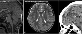
These and many other disorders are in one way or another closely related to the absence of the corpus callosum. As a rule, they allow a diagnosis to be made in the first 1-2 years of a child’s life. The final confirmation of the diagnosis is an MRI of the brain.
Corpus callosum in men and women
Male and female brains develop differently: from the prenatal formation of the neural tube according to sexual characteristics and ending with the lifelong action of hormones. Recently, you can often hear that the female body is no different from the male body. However, this is not true: neurophysiology, psychophysiology and neuropsychology provide a lot of experimental data in favor of the differences between the male and female brains.
This also applies to the corpus callosum, namely: the number of nerve fibers corresponding to the structure is greater in women than in men. This study suggests that the female sex operates better with speech concepts. Possessing a larger apparatus for exchanging information, a woman thus balances between the hemispheres, when the male brain “specializes” in one of them. However, in contrast to this statement, there are many reproaches.
Hypoplasia of the corpus callosum
Hypoplasia is a serious, but fortunately quite rare diagnosis. In essence, this, like agenesis, is a violation of the intrauterine development of brain tissue. If during agenesis the corpus callosum of the brain is completely absent, then with hopoplasia it is underdeveloped. Of course, treating this disease with modern medicine is impossible. Therapy involves a set of measures that minimize deviations in the patient’s development. Neuropsychologists recommend that patients regularly perform a specially designed set of physical exercises that help restore connections between the hemispheres, as well as information wave therapy.
Sexual dimorphism
A number of Russian and foreign scientists believe that the difference in thinking and behavioral reactions between men and women is associated with the different structure and size of the corpus callosum. Thus, Newsweek published an article explaining the nature of women's intuition: women have a slightly wider corpus callosum than men. This fact, according to the same scientists, also explains the fact that women, unlike men, are able to cope with several different tasks at the same time.
After some time, a group of French scientists reported that, as a percentage of brain size, men have a larger corpus callosum than women, but scientists did not draw any clear conclusions. Be that as it may, all scientists only agree that the corpus callosum is one of the most important structural components that performs a number of vital functions.
What is the corpus callosum responsible for?
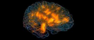
Possessing a colossal number of axons (structures responsible for transmitting electrical impulses to nerve cells), the corpus callosum literally connects the two hemispheres of the brain. Its fibers connect similar areas of the cortex (for example: the parietal cortex of the left hemisphere connects to that of the right). Thus, the fibrous cluster is responsible for the coordination and joint work of both parts of the brain. An exception is the temporal cortex, since the structure adjacent to the corpus callosum, the anterior commissure, is responsible for its connection.
The corpus callosum allows one hemisphere to “share” information with the other: when conducting experiments on higher mammals, it turned out that, by cutting the optic tract, the corpus callosum transmits information from the visual cortex of the left hemisphere to the right.
The functions of this structure also include maintaining human intellectual activity: by synthesizing information from two parts of the brain, the corpus callosum provides a deeper understanding of data received from outside. An experiment supports this position (all neurophysiology is based on experimental data): by dissecting and extracting a cluster of connecting nerve fibers, scientists noticed that the subjects were finding it difficult to understand written and spoken speech.
The most interesting and mysterious functions include the unity of consciousness and emotional response to a stimulus. When the corpus callosum was removed, people tended to show an ambivalent attitude towards the phenomenon or object (ambivalence). That is, they observed the presence of two diametrically opposed thoughts or emotions at the same time, such as: hatred and love, fear and pleasure, disgust and interest. A similar phenomenon is observed in the psychopathology of schizophrenia, when patients, without realizing it, showed love and hateful enmity towards something. This is not about the alternate manifestation of opposite feelings: emotions are located on parallel lines and in the same period of time.
