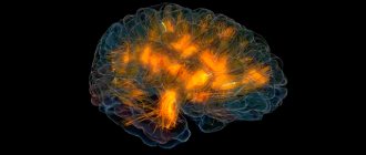
Anatomically realistic 3D brain model depicting localized activity in real time. Each color represents the source of bioelectrical activity and connectivity in a different frequency band (theta, alpha, beta, gamma); gold lines represent anatomical white matter fiber tracts. The supposed transfer of information between brain regions is visualized as pulses of light passing along fiber tracts connecting these regions
Bioelectric activity of the brain
(BEA) is the electrical activity of neurons associated with their excitation, i.e. with a shift in membrane potential.
The bioelectric activity of the brain is based on transmembrane ionic currents, which give rise to the phenomena of action potential and impulse activity of neurons. In this way, neural transmission of signals is carried out in the form of short electrical impulses, each of which represents the main information unit of the brain (quantum of information).12
Mechanism
The bioelectrical activity of the brain is explained by the physiology of the nervous tissue. Each neuron is covered by a membrane. Protein pumps built into the membrane create a difference in electrical potentials inside and outside the cell by pumping charged ions of sodium, potassium, chlorine, etc.
The excitation of a neuron is associated with the opening of ion channels and the passage of ions through the membrane, which leads to a change in voltage, which is recorded by various methods in the form of bioelectrical activity.
An individual neuron is in a continuous process of bioelectrogenesis. Excitation of a nerve cell can occur when it is irritated, i.e. when an impulse arrives from other neurons, and in some cases spontaneously. There are no other sources of the bioelectric field, except neurons, in the brain.
➥ Main article: Bioelectric potentials
Vertical integration of spatial scales from the molecular level (nanometer scale) to the entire brain (centimeter scale)
Levels of brain organization
Conventionally, there are three levels of brain organization at which different forms of bioelectrical activity can be studied: microlevel (activity of one neuron), mesolevel (activity of a local group of neurons) and macrolevel (activity of different areas of the brain).
➥ In more detail: Functional systems, structure and organization of the brain
Microscale is the lowest level of brain organization, reflecting the activity of single neurons due to synaptic and molecular mechanisms.
Mesoscale (English mesoscale) is a level located between the micro and macro levels, at which interactions between large neural ensembles occur.
Macroscale - used to define brain regions and large-scale connections between them.
Methods for recording bioelectrical activity of the brain
Registration of BEA at different levels of organization
(A) Scalp electrode (green), multicontact macroelectrode (red) and microelectrode (blue) (resolution: volume
The study of bioelectrical activity is carried out using electrophysiological methods based on recording fluctuations in the potential difference between two points in the medium, for example, between two electrodes located on the scalp.
Electroencephalography (EEG, English electroencephalography, EEG) is a non-invasive method that allows you to record the bioelectrical activity of the brain from the surface of the scalp.
➥ Main article: Electroencephalography
Magnetoencephalography (MEG, English magnetoencephalography, MEG) is a method based on recording a magnetic field that is formed due to the electrical activity of neurons.
Electrocorticography (ECoG, English electrocorticography
, ECoG) is an invasive method of recording bioelectrical activity using electrodes located on the surface of the cerebral cortex.
➥ Main article: Electrocorticography
Microelectrode methods are a set of methods that use microelectrodes for intra- or extracellular recording of biopotentials. These include recording the activity of a single neuron, multi-unit activity, local field potential (LFP), as well as patch clamp and dynamic clamp methods.
➥
Types of bioelectric activity
Bioelectrical activity, depending on the scale (from a single neuron to the coordinated activity of large neural ensembles), manifests itself in different forms.
➥ Main article: Functional systems, structure and organization of the brain
| Micro level of the brain | Meso level of the brain | Macro level of the brain | |
| Regular bioelectrical activity | spike trains; Regular pattern of subthreshold membrane potential oscillations (SMPO) | Oscillations of neural populations | Brain rhythms or waves generated by the largest neural ensembles |
| Irregular bioelectrical activity | thermal noise; Channel noise; burst noise; Synaptic noise; Spike | 1/f noise | Disorganized EEG 1/f noise |
| Transient bioelectrical activity | Irregular subthreshold membrane potential oscillations; Spindle-like subthreshold membrane potential oscillations | Non-epileptiform transient activity (SWR); Epileptiform transient activity (interictal epileptiform activity, IED) | Non-epileptiform (eng. nonepileptiform) transient activity; Epileptiform transient activity (eng. epileptiform discharges); Event-related potential (ERP) |
Rhythmic activity
Regular subthreshold oscillations of membrane potential
➥ Main article: Rhythmic activity of the brain
Rhythmic or regular bioelectrical activity is a repeating frequency pattern in the central nervous system. In other words, these are the same type or self-similar oscillatory events that repeat at equal intervals.
Neural oscillations generated by the bioelectrical activity of the brain are observed throughout the central nervous system at all levels of its organization and include spike trains/bursting, a regular pattern of subthreshold oscillations of the membrane potential, high-amplitude oscillations of the local field potential and large-scale oscillations (brain waves).
EEG is one of the most accessible electrophysiological methods for recording rhythmic activity. Since the electric field created by a single neuron or a collection of closely located neurons is so small that it cannot be recorded from the surface of the head, EEG is a method that reflects the total activity of a large pool of neurons, the magnitude of which is sufficient to generate a potential difference on the surface of the scalp. It should be emphasized that the electrical activity must be as powerful as is required for the signal to pass through the bones of the skull and skin, which have noticeable resistance to electric current. Again, based on the above necessary and sufficient conditions: the activity recorded from the scalp is due to the relatively powerful total activity of the neuronal pool, it logically follows that the total EEG is primarily due to the activity of cortical neurons as those closest to the skin surface heads - place of registration.
Rhythmic processes play a key role in the functional activity of the brain.
Irrhythmic activity
Irrhythmic or irregular activity refers to chaotic or stochastic changes in the bioelectrical activity of the brain.
At the micro level
Irregular brain activity can be divided into molecular, synaptic and 1/f noise,3 and spiking activity by mechanisms.
Spikes
(English spikes) can also be extremely irregular, both during constant, spontaneous activity, and during evoked activity at a high frequency of impulse emission. There is uncertainty about the source of this irregularity, ranging from intrinsic sources of noise in neurons to collective effects in large-scale cortical networks. Cortical interneurons show highly irregular spike timing in response to direct current injection in vitro. This is in sharp contrast to cortical pyramidal cells, which exhibit irregular bioelectrical activity in vivo but regular activity in vitro. In vitro recordings and computational models have shown that this is associated with the rapid activation kinetics of interneuronal K ion currents. In this case, an arrhythmic spike can contribute to the irregular activity of the entire cortical network.45
Above is a normal EEG, below is an EEG of a patient with hypsarrhythmia.
At higher levels of organization, nonrhythmic activity can manifest itself in the form of disorganization of the bioelectrical activity of the brain. Types of disorganization:
- Disorganization of bioelectrical activity with a predominance of alpha activity. On the EEG, alpha activity is the main one, but it is not regular enough or is completely irregular in frequency. This more or less disorganized alpha rhythm has an insufficiently high amplitude and may even dominate in all areas of the brain. Beta activity is also often enhanced, often represented by low-frequency oscillations of increased amplitude. Along with this, theta and delta waves with a fairly high amplitude may be present in the EEG.
- Disorganization of bioelectrical activity with a predominance of theta and delta activity. The structure of this type is characterized by a weak representation of alpha activity. Fluctuations in biopotentials in the alpha, beta, theta and delta ranges are recorded without any clear sequence. This non-dominant type of curve can have both medium and high amplitude levels.
- Hypsarrhythmia - manifests itself as disorganization of the background bioelectrical activity of the brain with irregular slow waves of high voltage; multifocal peaks and polyspikes can be superimposed during sleep.6
Paroxysmal activity
➥ Main article: Paroxysmal activity
Paroxysms or transients are short intervals (10 to 1000 ms)7 during which the signal changes abruptly and becomes atypical or relatively unpredictable.
(A) Spindle-shaped subthreshold oscillations of membrane potential; (B) Irregular subthreshold fluctuations in membrane potential
Transient activity reflects the functional integration of brain structures and is a fundamental element of neural interaction. Transients generated jointly by different neural populations indicate the connection of these populations. This communication may be synchronous or asynchronous.8
Examples of such activity include spontaneous synchronous synaptic transients generated by Ca2+ ion currents between adjacent cortical neurons.9 Spikes and sharp waves that constitute seizure or interictal activity in individuals with epilepsy or a predisposition to it, or transients in the form of vertices and sleep spindles , which are the norm. Irregular subthreshold membrane potential oscillations and spindle-shaped subthreshold membrane potential oscillations can also be recorded using in vivo intracellular recording.
Event related potential
➥Main article:ERP
Event related potentials
(EPS, English
event-related potential
- ERP) are electrical voltage fluctuations recorded from the scalp, generated in brain structures in response to certain events or stimuli.
Also refers to transient brain activity. These are EEG changes time-locked to sensory, motor or cognitive events that provide a safe and non-invasive approach to studying the psychophysiological correlates of mental processes. They are believed to reflect the total bioelectrical activity of postsynaptic potentials that occur when a large number of similarly oriented cortical neurons (on the order of thousands or millions) fire synchronously during information processing. ERP is measured using electroencephalography (EEG) and magnetoencephalography (MEG). The magnetoencephalographic (MEG) equivalent of ERP is the ERF or event -related
. 10 ERP subtypes include evoked and evoked potentials.
Evoked potentials
➥ Main article: Evoked potentials
Cognitive evoked potentials (EP, EP)
represent event-related activity that occurs as an electrical response from the brain to various types of sensory stimulation of neural tissue; auditory and visual stimulation are most often used. Recording of such electrical potentials is a non-invasive objective test that provides information, for example, about sensory pathway disorders, the location of lesions affecting sensory pathways, and disorders related to language and speech. Evoked potentials are recorded from the scalp using EEG. Potentials typically appear as a transient waveform, the morphology of which depends on the type and strength of the stimulus and the location of the electrodes on the scalp. The mental state of the subject, such as attention, wakefulness, and anticipation, also influences the morphology of the waveform.
Induced activity
Along with evoked activity, stimulus-related neural activity can lead to induced activity
. The induced response is associated with processes in the brain that are not directly related to the stimulus, but are caused indirectly by the nonlinear interaction of neurons after the stimulus. The functional component of the induced response is likely to represent corticothalamic feedback (top-down) processes linking external stimuli to the internal cortical model of the environment, i.e., combining externally controlled sensory information with internal brain activity.1112 The induced response may be related with cognitive brain functions such as perception, attention and learning, which can be considered higher order processes. A well-studied type of induced activity is a change in the amplitude of oscillatory activity. For example, gamma activity often increases during times of increased mental activity, such as during the presentation of an object. Since induced responses are not phase related and will therefore be excluded during averaging, they can only be obtained using time-frequency analysis. Induced activity typically reflects the activity of multiple neurons: amplitude changes in oscillatory activity are thought to result from synchronization of neuronal activity, such as spike timing or fluctuations in the membrane potential of individual neurons.
Alpha Rhythm
HE. Kirillovskikh, V.S. Myakotnykh, E.V. Sorokova, K.V. Myakotnykh, T.A. Borovkova Ural State Medical Academy, 620905 Ekaterinburg, st. Soboleva, 25
89 elderly and senile patients with various forms of epilepsy . The general characteristic features of the bioelectrical activity of the brain of elderly patients suffering from epilepsy were determined, and the features of the electroencephalographic picture were identified depending on the form of the disease, the age of its onset, and the severity of cerebrovascular pathology. It has been shown that dynamic EEG monitoring helps in selecting adequate antiepileptic therapy and avoids aggravation of cognitive deficit.
Key words: epilepsy, elderly and senile age, EEG, cerebrovascular pathology, cognitive deficit.
Relevance of the problem.
In recent years, the problem of epilepsy in the elderly and senile, the differential diagnosis of epileptic seizures and non-epileptic paroxysmal conditions of other origins, as well as the possibilities of treating epilepsy in the elderly suffering from multiple pathologies has received increasing attention [1,3,8]. It is believed that epilepsy is a disease that develops on the basis of a genetically determined predisposition of the brain in combination with exogenous factors that determine its actualization in the clinical form [4]. It has been proven that this genetically determined phenotypic precondition for the development of epilepsy in children and adolescents is high-amplitude synchronization of neuron activity in all frequency ranges. But in the process of natural age-related transformation of the bioelectrical activity of the brain, the main cortical rhythms slow down, primarily the alpha rhythm , which acts as a biological clock during aging. In addition, there is a decrease in the overall amplitude level of the EEG and an increase in the proportion of beta activity due to an increase in nonspecific activating influences of the mid-brain structures [4,7]. Also, with age, the number of slow waves (SW) increases, predominantly in the theta range, arising both regionally, mainly in the frontotemporal regions, and in the form of a generalized slowdown of the main cortical rhythms. However, despite the ongoing age-related changes in the bioelectrical activity of the brain and the entrenched opinion that epilepsy is a disease predominantly of young people, the incidence of epilepsy in older age groups continues to increase [7]. Therefore, we found it interesting to determine the characteristics of bioelectrical activity both in elderly patients with a long history of epilepsy and in patients with the onset of epilepsy over the age of 60 years.
Purpose of the study
Using electroencephalographic (EEG) studies, determine the characteristics of the bioelectrical activity of the brain in epilepsy in elderly and senile people.
Materials and methods.
For 5 years, from 2006 to 2010. A prospective study of the clinical and neurophysiological features of epilepsy and the possibilities of its treatment was conducted in 89 patients aged 63-96 years (m=75.5±6.87 years). The diagnosis of epilepsy in all cases was established based on the observation of at least two unprovoked epileptic seizures . The exclusion criterion was epileptic syndrome due to a brain tumor, Alzheimer's disease. The control group was represented by 30 patients aged 65-85 years (m=75±5.66 years), who did not suffer from epilepsy, but had a similar range of concomitant pathologies, mainly cardiovascular. Depending on the time of onset of epilepsy, all patients of the main observation group were divided into two compared groups: 1st – patients with the onset of epilepsy in old age, mainly of post-stroke origin (n=34); 2nd – patients with a long history of epilepsy with its onset before the age of 60 (n=55). Separately, from the representatives of group 2, subgroup 2A was identified - 18 elderly patients, over 80 years old, who suffered severe traumatic brain injury (TBI) during the Great Patriotic War and suffered from post-traumatic epilepsy. All patients in the study groups underwent EEG studies at intervals of 4-6 per year. We used EEG registration with visual assessment and calculation of indices for standard frequency ranges on a computer electroencephalograph "Encephalan-131-01" (Russia, Taganrog). In the process of selecting the optimal combination of antiepileptic drugs, EEG was recorded over time 5, 14 and 30 days after changing the treatment regimen. In the absence of epileptiform activity on a routine EEG or with questionable results, patients underwent EEG with sleep deprivation, daytime ambulatory EEG monitoring, EEG sleep monitoring using the Nicolet-one device. When conducting an EEG with partial sleep deprivation, which is better tolerated by the elderly, the patient was woken up at 4 a.m. the day before and an EEG recording was performed at 9 a.m. Recording was carried out with bipolar and monopolar installation of electrodes using 18-21 standard leads according to the 10-20 scheme; the following functional tests were used: opening and closing the eyes, rhythmic photostimulation with a frequency of 3, 5, 10 and 15 Hz, a block with a continuous increase in frequency photostimulation from 3 to 27 Hz and hyperventilation for 3 minutes. The EEG results were assessed using modern interpretation methods - computer processing with calculation of the index and amplitude of the main EEG rhythms, but the main method of EEG assessment was visual. The primary assessment of the EEG variant was carried out using the classification of E.A. Zhirmunskaya [2], a more detailed description of pathological changes - using the Classification of the American Association of Neurophysiologists [6].
Results and its discussion.
A study of the EEG of patients in both selected groups revealed some common features that distinguish the bioelectrical activity of the brain of elderly and senile patients with epilepsy from that of people of the same age, but not suffering from epileptic seizures (Table 1). This is, first of all, a higher amplitude level of the main EEG rhythms: if in the comparison group the EEG amplitude, as a rule, did not exceed 60 μV, then in patients with epilepsy the average amplitude level was twice as high, amounting to 120-150 μV. No significant differences were found between representatives of the main group and the control group only in the frequency of occurrence of interhemispheric asymmetry, bursts of beta activity and diffuse epileptiform activity. For other parameters, the differences identified are clear and significant. There is also a noticeable tendency towards synchronization of the main bioelectrical activity due to dysfunction of the mid-stem structures of the brain. Thus, when studying routine EEG, 38 (42.7%) patients showed a high-amplitude EEG variant for a given age with a tendency to synchronize the main cortical rhythms, in contrast to 2 (6.7%) patients in the control group (R
Table 1. Comparative characteristics of the main EEG variants and types of pathological activity.
| Main characteristics of EEG | Main group (n=89) | Comparison group (n=30) | |
| Hypersynchronous high-amplitude variant (sharp-looking) | 38 (42,7%) | 2 (6,7%) | |
| Desynchronous low-amplitude variant | 0 | 14 (46,7%) | |
| Disorganized hypersynchronous variant | 50 (56,2%) | 2 (6,7%) | |
| Disorganized desynchronous variant | 1 (1,1%) | 12 (40%) | |
| Interhemispheric asymmetry | 28 (31,5%) | 4 (13,3%) | |
| Increase in beta index > 40% | 7 (7,9%) | 18 (60%) | |
| Flashes of beta activity (excessive fast) | 15 (16,9%) | 3 (10%) | |
| Slowing down underlying background activity | I degree | 24 (27%) | 4 (13,3%) |
| II degree | 8 (9%) | 0 | |
| III degree | 3 (3,4%) | 0 | |
| Periodic regional slowdown | in the frontal leads | 23 (25,8%) | 5 (16,7%) |
| in the temporal leads | 41 (46,1%) | 0 | |
| Focal epileptiform activity | while awake | 36 (40,4%) | 0 |
| in a state of sleep | 14 (15,7%) | 0 | |
| Diffuse epileptiform activity | 5 (5,6%) | 0 | |
| Slow wave activity index (%) | 39,5±6,5 | 29,9±3,1 | |
Against the background of disorganization of background bioelectrical activity, hypersynchronization of the main cortical rhythms was also observed in 50 (56.2%) patients of the main group; in the control group there were only 2 (6.7%) observations of this kind; R
Among patients with epilepsy, the average index of slow wave activity reached 39.5±6.5%, in the control group – 29.9±3.1% (P
In the group of patients with epilepsy, in 24 (27%) cases, a slowdown in the main activity of the first degree (7 Hz and below) was noted, in the control group - only in 4 (13.3%) cases. Deceleration of the main activity of the second degree (6 Hz and below) was also significantly more often (P = 0.009) noted among people suffering from epilepsy (Table 1). This is consistent with the fact that a slowdown in basal activity compared to the age norm is always a sign of serious brain pathology [6]. Of course, in elderly patients, the slowdown in the main activity of the first degree can be considered a conditionally normal phenomenon, since after 60 years there is a gradual physiological decrease in the frequency of the alpha rhythm by approximately 1 Hz every 10 years [2,3,4,5]. Deceleration of the main activity of the II-III degree in elderly patients is a marker of severe cortical atrophy of the brain [6].
In the group of patients with epilepsy, almost all types of paroxysmal activity were epileptiform in nature; in 64 (71.9%) patients, high-amplitude pointed waves of the theta and delta range were more often localized in the frontal and temporal regions and were the most common type of conditioned epileptiform activity. Complexes “sharp-slow wave”, “spike-slow wave”, classified as true epileptiform activity, were less common - in 36 (40.4%) patients and were detected in a state of wakefulness. In 14 (15.7%) patients, epileptiform activity was detected only during sleep during EEG monitoring .
Thus, on the EEG of elderly patients with epilepsy, in addition to epileptiform activity itself, the following phenomena are more common: a) hypersynchronous alpha activity with an amplitude of over 100 μV, sharp-looking in shape - “sharp-looking alpha waves”; b) hypersynchronous beta activity (excessive fast) - beta activity with an amplitude of over 30 μV, often in the form of spindles extending beyond the normal fronto-central localization; c) periodic regional slowing of the main activity in the form of bursts of bilaterally synchronous or local high-amplitude pointed theta and delta waves; d) an increase in the average EEG amplitude and an increase in power during computer analysis in all spectral ranges; e) increase in the index of slow wave activity by more than 30%. The presence of these signs allows, even in the absence of true epileptiform activity, to draw a conclusion about excessive epileptic synchronization of brain neurons, and, therefore, confirm the diagnosis of epilepsy if the history and clinical picture are consistent.
A comparative analysis of the nature of EEG changes in various selected groups and subgroups of patients with epilepsy revealed the following changes (Table 2).
Table 2. Comparative characteristics of the main variants of diffuse changes in the EEG and types of pathological activity in patients of the main study group and the control group.
| Main characteristics of EEG | Main group (n=89) | |||
| Group 1 (n=34) | Group 2 without subgroup 2A (n=37) | 2A subgroup (n=18) | ||
| Disorganized hypersynchronous variant | 33 (97,1%) | 37 (100%) | 18 (100%) | |
| Desynchronous low-amplitude variant | 1 (2,9%) | 0 | 0 | |
| Interhemispheric asymmetry | 20 (58,8%) | 5 (13,5%)* | 3 (16,7%) | |
| Flashes of beta activity (excensive fast) | 4 (11,8%) | 6 (16,2%) | 5 (27,8%) | |
| Slow down main activity in background recording | I degree | 11 (32,4%) | 7 (18,9%) | 6 (33,3%) |
| II degree | 2 (5,9%) | 0 | 6 (33,3%) | |
| III degree | 0 | 0 | 3 (16,7%) | |
| Periodic regional slowdown | in the frontal leads | 8 (23,5%) | 9 (24,3%) | 0 |
| in the temporal leads | 15 (44,1%) | 21 (56,8%) | 5 (27,8%) | |
| Focal epileptiform activity | wakefulness | 16 (47,1%) | 15 (40,5%) | 5 (27,8%) |
| dream | 6 (17,6%) | 8 (21,6%) | 0 | |
| Average amplitude (µV) | 98±6,7 | 116±4,8 | 93±4.9 | |
| Slow wave activity index (%) | 39,4±4,4* | 35,4±4,34* | 48,4±4,59 | |
Note: * – p
All patients in the study groups with different types of epilepsy had statistically significant differences (p
Amplitude and frequency interhemispheric asymmetry significantly prevailed in the group of patients with late onset of epilepsy - in 20 (58.8%) patients (p
Rice. 1. Regional epileptiform activity “acute-slow wave” in the right temporo-parietal region in patient V., 72 years old, against the background of pronounced interhemispheric asymmetry and relative preservation of the background EEG in the intact left hemisphere of the brain. Recording against the background of sleep deprivation.
On routine EEG, in 16 (47.1%) patients with late onset of epilepsy , a lateralized focus of slow-wave activity was detected in the form of pointed delta waves with a frequency of 1.5-3 Hz, “sharp-slow wave” complexes, and less often “spike-waves”. slow wave”, in 6 (17.6%) patients epileptiform activity was detected on EEG sleep monitoring, in 2 (5.9%) diffuse (bilateral) epileptiform activity was detected. The preference for detecting epileptiform changes on the EEG and localizing post-stroke scar changes coincided in 70.6% (P = 0.016). In 10 (29.4%) patients, no focus of epileptiform activity was detected on routine EEG; These were mainly patients with small, less than 15 mm, foci of softening that formed as a result of a stroke.
EEG recording during sleep was possible only in 6 patients, since insomnia is a common concomitant symptom in this category of patients. The EEG picture of slow-wave sleep in representatives of group 1 was characterized by disorganization with prolongation of the first phase of slow-wave sleep. At the same time, sleep was superficial, with frequent awakenings for 8-12 seconds and motion artifacts; Specific sleep patterns - K-complexes, vertex potentials and sleep spindles - were not clearly expressed, delta sleep was shortened, and the amplitude of delta activity was reduced. The “rapid eye movement” stage was not recorded in any patient of group 1, which largely reflects the dysfunction of central somnogenic mechanisms in patients with cerebral vascular pathology. Against a disorganized background, in all 6 patients, mainly in the second stage of slow-wave sleep, focal epileptiform activity of the “sharp-slow wave” type was recorded, localized in the frontotemporal leads, on the side of the post-stroke focus of softening (Fig. 2).
Fig 2. EEG of patient M., 73 years old, diagnosed with: Consequences of ischemic stroke in the left internal carotid artery; symptomatic epilepsy, simple partial seizures with secondary generalization. In stages I-II of slow-wave sleep - epileptiform complexes “acute-slow wave” in the left frontotemporal region.
Thus, analyzing the structure of bioelectrical activity in patients with late onset of epilepsy, it can be assumed that initially, before the stroke, patients had a tendency of cortical neurons to paroxysmal forms of response in the form of a tendency to high-amplitude synchronization. However, these changes remained latent throughout life and did not lead to clinical manifestations of epilepsy . Ischemic stroke with localization of cerebral infarction in the cortical and cortical-subcortical areas was the triggering moment that led to the formation of epileptiform activity and the clinical manifestation of epilepsy.
When studying the EEG in patients with a long history of epilepsy (2nd group of observations), a significantly higher amplitude level of the EEG was observed (rEEG sleep monitoring (Fig. 3). The index of slow wave activity in patients of the 1st group, with early onset of epilepsy, significantly higher than in the control group, but lower than in patients of group 1 - with late onset of epilepsy (p
Rice. 3. EEG of patient K., 77 years old, diagnosed with: Symptomatic epilepsy, complex partial seizures with secondary generalization. In the second phase of slow-wave sleep there is a short discharge of diffuse epileptiform activity “spike-slow wave” with initiation in the left frontotemporal region and the phenomenon of secondary bilateral synchronization.
Thus, the bioelectrical activity in the group of patients with an early onset of epilepsy and a long history of it was distinguished by signs characteristic of the bioelectrical activity of young patients suffering from epilepsy, but in elderly patients these pathological signs were less common. Aging processes in the bioelectrical activity of the brain of patients with epilepsy are manifested, first of all, by an increase in the slow-wave activity index, a slight decrease in the overall amplitude level of the EEG and a decrease in the tendency to generalize epileptiform discharges, which is clinically reflected in a decrease in the proportion of secondary generalized epileptic seizures in elderly patients. It can be assumed that the genetically determined characteristics of the bioelectrical activity of the brain, which are the cause of the clinical manifestation of epilepsy, persist throughout life.
The characteristic features of the EEG in the subgroup of elderly patients (over 80 years old) with a history of combat TBI are due to the age of the patients and pronounced atrophic changes in the cerebral cortex. The main distinguishing feature of the EEG of representatives of this subgroup 2A was an increase in the index of slow wave activity (48.4±4.59), which is significantly higher (P
Rice. 4. EEG of patient Ch., 84 years old, with a diagnosis of Encephalopathy of complex origin (post-traumatic, cerebrovascular), severe cognitive and static-coordinating impairments. Symptomatic epilepsy, complex partial seizures of moderate frequency. The EEG shows diffuse slow-wave activity in the form of pointed high-amplitude theta-delta waves.
Regional epileptiform activity in elderly people was detected in a small percentage of cases - in 5 (27.8%) out of 18, however, the frequency of detection of regional epileptiform activity was higher than in the comparison group (P
Slow-wave activity in the EEG of patients in group 2A no longer reflects the degree of epileptiform activity, but rather the level of pathological morphological and functional changes in neurons due to traumatic and vascular factors. The index of slow wave activity in these patients turned out to be directly proportional to the degree of atherosclerotic lesions of cerebral vessels and inversely proportional to the number of points scored in the study of cognitive functions using the well-known MMSE scale. It is interesting that the index of slow wave activity is not a static value; it can decrease after a course of vascular therapy and, conversely, increase with the use of some anticonvulsants, primarily barbiturates. This is confirmed by EEG analysis in 3 (16.7%) representatives of the 2A subgroup who have been taking barbiturates for more than 60 years; In all cases, the EEG showed gross cerebral changes of an organic type, continued slow-wave activity of varying amplitude and degree of synchronization, but true epileptiform activity was not detected in these patients (Fig. 5).
Rice. 5. EEG of patient M., 85 years old, diagnosed with vascular atrophic disease of the brain, dementia, symptomatic post-traumatic epilepsy, rare generalized seizures. Constant intake of benzonal at a dose of 100 mg 3 times a day + phenobarbital 100 mg at night for 50 years. The EEG shows diffuse slow-wave activity with synchronization in the fronto-central regions. Characterized by a complete absence of physiological EEG rhythms. True epileptiform activity is not detected.
Thus, an increase in the index of slow-wave activity on the EEG during treatment with antiepileptic drugs is an unfavorable prognostic sign of worsening cognitive deficits, which indicates the need to replace the drug with a more modern one or a drug of a different group.
Conclusions.
- The frequency of detecting distinct epileptiform activity on the EEG in elderly and senile patients suffering from epilepsy is 40.4%, conditional epileptiform activity is even higher, up to 72%. In some cases, epileptiform activity can be detected during ambulatory EEG sleep monitoring. There are individual differences in EEG characteristics depending on the etiology of epilepsy, duration of the disease, and age of the patients.
- There are the following main distinctive neurophysiological features of the EEG of elderly and senile patients with epilepsy:
- an increase in the spectral power of the EEG in all frequency ranges – a high-amplitude version of the EEG;
- strengthening the synchronizing influences of mid-stem structures and, as a consequence, smoothing out zonal differences;
- decrease in the proportion of beta activity;
- an increase in the index of slow wave activity to 30% or higher, which includes both a slowdown of the main cortical rhythms in the background recording and periodic regional slowdown in the form of bilaterally synchronous bursts of theta-delta waves;
- Regional epileptiform activity is detected in approximately 40% during wakefulness, and in 16% during EEG sleep monitoring.
Literature.
- Gekht, A.B., Burd G.S., Selikhova M.V. Clinical and neurophysiological features of motor disorders in patients with post-stroke epilepsy // Journal of Neurology and Psychiatry. – 1998. – No. 7. – P. 4–8.
- Zhirmunskaya, E.A. In search of an explanation of EEG phenomena. - M.: NFP Biola, 1996. - 117 p.
- Zenkov L.R., Elkin M.N., Medvedev G.A. Clinical neurophysiology of neurogeriatric disorders // Advances in neurogeriatrics. – M., 1995. – P. 167-173.
- Zenkov, L.R. Clinical epileptology: 2nd ed., revised. and additional - M.: Medical Information Agency, 2010.- P. 123–129.
- Zenkov, L.R. Non-paroxysmal epileptic disorders. – M.: Medpress-inform, 2007. – 278 p.
- Mukhin K.Yu., Petrukhin A.S., Glukhova L.Yu. Epilepsy: Atlas of electroclinical diagnostics. – M.: Alvarez Publishing, 2004. – 440 p.
- Myakotnykh V.S., Galperina E.E. Electroencephalographic features in elderly and senile people with various cerebral pathologies: Educational and methodological recommendations. – Ekaterinburg: Publishing house. UGMA. – 30 s.
- Blalock, E.M., Chen K.C., Sharrow K. Gene microarrays in hippocampal aging: statistical profiling identifies novel processes correlated with cognitive impairment // J. Neurosci. – 2003.- Vol. 23. – R. 3807–3819.
- Carlson CE, St Louis ED, Granner MA Yield of video EEG monitoring in patients over the age of 50 // Program and abstract of the American Epilepsy Society 58th Annual Meeting. – 2004, 3 – 7 December. – Absract 2.- R. 226.
- Van Cott, AC Epilepsy and EEG in the Elderly // Epilepsia. –2002. –Vol. 43, suppl. 3. – P. 94–102.
Changes in bioelectrical activity of the brain
➥ Similar article: EEG changes
The frequency of bioelectrical activity can indicate various functional states of the brain: sleep, consciousness, cognition and some mental disorders. Decreased bioelectrical activity may appear on the EEG as slow waves, which are most often observed in conditions such as sleep, coma,13 brain death, depression,14 autism,15 brain tumors, obsessive-compulsive disorder (OCD),16 attention deficit disorder and hyperactivity (ADHD),17 encephalitis.18
In contrast, when there is increased bioelectrical activity in the brain, rapid waves are observed on the EEG, in conditions such as epilepsy,19 anxiety, post-traumatic stress disorder (PTSD) and substance use.20
It should be noted that the use of the terms “decrease” and “increase” of bioelectrical activity is incorrect, since the bioelectrical activity of the brain at different levels of organization manifests itself differently, and in the context of the whole brain one can only evaluate parameters (amplitude, power, index ) signals obtained during EEG recording. Then, based on numerous experimental data, one can already interpret changes in one or another parameter and talk about any changes in the functional activity of the brain.
Classification of BEA changes
Based on localization, changes in bioelectrical activity can be:21
- Focal – limited area (up to 3 leads);
- Regional – zone of the brain lobe (3 or more leads);
- Lateralized - pathological activity detected above the hemisphere;
- Generalized - general pathological activity, affecting the entire brain, recorded in all leads according to the mechanisms of primary and secondary generalization;
- Diffuse (unspecified) – cannot be classified into the above groups. According to the degree of severity, diffuse changes in bioelectrical activity can be divided into mild, moderate and pronounced.
Diffuse EEG changes
A. Flat EEG with burst suppression at intervals of 5–10 s.
B. Continuous generalized acute and slow complexes. C. Diffuse delta activity with peaks in the frontotemporal areas, spreading and predominant in the left hemisphere. A characteristic feature of pathological activity with diffuse changes in bioelectrical activity is the lack of locality, instability of spatial distribution (mosaicity), and violation of bilateral synchrony.
Diffuse brain damage is often associated with the development of encephalopathy, that is, widespread small-focal lesions. Due to the polymorphism of small focal changes and their prevalence, pathological changes are very diverse and do not form any organized activity, which is manifested by mosaic changes in the bioelectrical activity of the brain.
EEG disorganization
When the brain is damaged, pathological diffuse changes in bioelectrical activity are characterized by disorganization and the absence of regular dominant activity (alpha rhythm). Diffuse pathological activity can manifest itself in different ways (for example, in the form of slow-wave rhythms or epileptiform activity is detected), and also have different amplitudes (low-amplitude, medium-amplitude or high-amplitude).
Recording video EEG with single-channel ECG
The EEG demonstrates "slow" and "flat" phases during cardiac asystole, followed by a second "slow" phase after cardiac sinus rhythm resumes. Myoclonic seizures (MS) occur during asystole and after resumption of sinus rhythm. Tonic posture (tone) occurs during asystole. Pay attention to the artifact of tonic muscle contraction recorded on the ECG channel
Disorganization of bioelectrical activity is usually associated with a decrease in EEG amplitude and absence of alpha rhythm, which is described in more detail in the article alpha rhythm depression. Dysrhythmic brain activity can manifest itself in the form of a flat EEG (desynchronous type III, considered as a borderline norm), as well as with a predominance of alpha activity (disorganized type IV) and with a predominance of θ- and δ-activity (disorganized EEG type V). More details: classification of EEG disorders by type.
Slowing brain activity
A slowdown in the bioelectrical activity of the brain (when low frequencies predominate) does not always indicate pathological changes and may be associated with drowsiness. Slowing of the background rhythm in children is observed depending on the age group:
- 1 year – fundamental frequency less than 5 Hz
- 4 years – fundamental frequency less than 6 Hz
- 5 years – fundamental frequency less than 7 Hz
- over 8 years old – fundamental frequency less than 8 Hz
Slowing of cortical rhythms often occurs against the background of diffuse dysfunction of cortical and subcortical structures, due to vascular, metabolic or toxic lesions of the brain, and in children it can be a consequence of perinatal pathology.
Generalized slowdown
FIRDA pattern with slightly slowed background theta EEG in an elderly man with metabolic encephalopathy. Periods of well-formed occipital alpha activity are also noticeable