Microcephaly is a congenital anomaly, which is characterized by a significant reduction in the size of the brain (and, as a result, the skull) as a result of disruption of the growth processes of brain tissue during the period of intrauterine development of the baby.
It can be an independent defect or a manifestation of any other genetic diseases (in this case we are talking about true microcephaly), and it can also develop as a consequence of various diseases the mother suffered during pregnancy (secondary, or false, microcephaly). However, in any case, this pathology is accompanied by a lag in the child’s mental development of varying degrees of severity and the presence of other neurological symptoms.
Statistical information
The incidence of the defect ranges from approximately 1:2000 to 1:10000 among all newborns. As an independent disease, true microcephaly is much less common and is detected in one out of 50 thousand children, both girls and boys. If this pathology occurs as a manifestation of another hereditary disease, then its frequency in the population is determined by the frequency of the underlying disease.
Forecasts
The prognosis for microcephaly is in most cases unfavorable .
Even if a child has the ability to learn or master basic skills, his quality of life and life cycle will be significantly reduced.
Experts can strongly recommend that parents place their child in a special boarding school from a very early age. This need arises due to the rapid development of the disease and the importance of constant medical supervision of a small patient.
With properly selected therapy, the child may experience the following improvements :
- normalization of emotional state;
- reduction in the frequency of epileptic seizures or convulsive conditions;
- some improvement in memory and concentration;
- expansion of vocabulary;
- increasing the body's performance;
- increase in life expectancy to 30 years.
Anatomy of the brain
The brain is the main organ of the human nervous system; its formation and development begin in the earliest stages of the embryonic period.
The first rudiments of nervous tissue are formed already on the 18th day of intrauterine development. They are a neural plate, which consists of a small number of neurons (nerve cells). As the embryo forms, their number increases, as a result of which the brain and spinal cord and other elements of the system are formed from the neural plate.
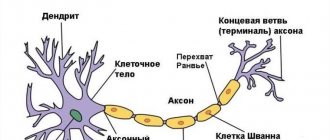
A neuron is a highly specialized cell that consists of a body and many processes through which it communicates with a huge number of other cells. Thanks to this structure, the main function of the nervous system is ensured, namely the reception and processing of information, including the regulation of the vital functions of the entire organism as a whole. Through some neurons (they are called afferent), information in the form of nerve impulses enters the brain, where it is processed and a response nerve impulse is formed, which is sent to organs and tissues through other neurons (they are called efferent).
The formation of nerve cells in the brain is completely completed only at the time of birth, on average reaching about 150 billion neurons. However, throughout a person’s life they are not able to reproduce (divide), and at the same time they gradually die due to so-called apoptosis (a genetically programmed process of death of malfunctioning or damaged cells).
The brain is anatomically divided into three sections.
- Large hemispheres of the brain (right and left). Being the higher parts of the brain, they consist of the cortex and subcortical structures (hypothalamus, thalamus, etc.). The cerebral cortex contains neurons that are responsible for the formation of personality, the processes of self-knowledge and learning (higher nervous activity of a person). Subcortical structures ensure the formation of instinctive and behavioral reactions (sexual, food, etc.). It is also worth noting that the cerebral cortex under normal conditions has an inhibitory effect on subcortical structures; in other words, a person’s consciousness can suppress his instinctive reactions.
- Brain stem. It includes the medulla oblongata, the pons, and the midbrain and diencephalon. They provide reflex activity of the human body, and also maintain communication between the brain and spinal cord. In addition, the trunk contains various nerve centers (for example, vasomotor, respiratory, etc.). They, in turn, ensure the performance of those physiological functions that are not controlled by consciousness (for example, a person breathes automatically, without thinking about it).
- The cerebellum is a small structural formation that is responsible for coordinating human movements, maintaining muscle tone and maintaining balance during complex movements (walking, running, etc.).
The human skull is divided into the brain and facial sections. The latter includes the bones of the face. The development of the skull comes from embryonic tissue called ectoderm. It surrounds the rudiments of the brain from the very beginning of their formation.
There are three stages in the formation of the skull bones:
- membranous - a layer of cells that surrounds the future brain, becomes soft membranous tissue, simultaneously increasing with the growth of brain tissue;
- cartilaginous - from about the sixth to seventh week of embryonic development, this tissue in the area of the base of the skull gradually turns into denser cartilaginous tissue;
- bone - already from the eighth to tenth week of the intrauterine period, ossification points are formed in various parts of the skull, which are zones of the beginning of the growth of bone tissue.
By the time of birth, some areas of membranous tissue (fontanelles) remain in the roof of the skull of a newborn child, which ensure bone mobility during childbirth or when the brain enlarges for any reason.
The large (anterior) fontanelle has a diamond shape and is located at the junction of the frontal and parietal bones. It completely ossifies and closes by the age of two years.
The small (posterior) fontanel has the shape of a triangle and is located in the occipital region at the junction of the parietal and occipital bones. As a rule, it becomes overgrown by the second month of life.
The lateral fontanels are paired and small in size, they are located on the side surface of the head, slightly in front and behind the ears, and are also completely overgrown by the second month of life.
The bones of the facial skull are formed from the so-called gill arches, which are derivatives of mesoderm (another embryonic tissue). Thus, the development of the brain and facial parts of the skull occurs independently of each other. In addition, the formation of the facial skull is practically independent of the development and growth of the brain; however, the size of the cerebral skull normally always corresponds to the volume of the cerebral hemispheres.
At the end of the 19th and beginning of the 20th centuries in Europe and North America, children with microcephaly were sold to the circus. There they took part in the so-called “freak show”.
Psychosocial adaptation
In the psychological and social adaptation of children with microcephaly, the main role is given to parents .
The child must take all prescribed medications in a timely manner, attend classes with speech therapists and other teachers, engage in physical therapy and receive a full-fledged massage.
Any interruptions in therapy can cause an exacerbation of the disease and accelerate its progression.
Recommendations for parents of children with this diagnosis:
- Raising children with microcephaly is accompanied by increased responsibility on the part of adults (parents must be aware of the scale of the problem).
- Psychological assistance should be provided not only to the child, but also to his parents.
- Slowing down the progression of the disease and the emergence of positive dynamics is only possible with constant training with the child and following the recommendations of doctors.
- Thanks to special classes, you can increase a child’s self-esteem and develop his individuality (such classes are in most cases conducted in a group form, children learn to communicate with each other, acquire basic skills and more easily adapt to external conditions).
- Children with a mild form of intellectual disability can attend remedial classes, but classes with a remedial teacher are mandatory.
Reasons for the development of the anomaly
Taking into account the causes and time of occurrence, primary microcephaly (true or hereditary) and secondary (syndromic) are distinguished.
Primary microcephaly in children is a manifestation of hereditary diseases; it accounts for about 7–34% of all types of this anomaly.
Primary microcephaly
The following diseases are independent forms.
Giacomini syndrome is a hereditary microcephaly, which is predominantly manifested by mental disorders in combination with the occurrence of convulsive conditions, as well as the progression of paralysis.
Payne's syndrome - this disease is also hereditary. It affects only boys and is manifested by constant convulsive conditions of the lower extremities, nervous disorders and abnormalities in the functioning of the heart.
Microcephaly can be a manifestation of the following pathologies:
- Down syndrome - the appearance of an extra chromosome in the XXI pair, clinically manifested by impaired mental development of the child, a decrease in the size of the brain and cerebellum, as well as other characteristic symptoms;
- Patau syndrome - the appearance of an extra chromosome in the XIII pair, characterized by low birth weight of the newborn, microcephaly and other anomalies of the skull and internal organs;
- Edwards syndrome - the appearance of an extra chromosome in the XVIII pair, while children have a small or long and narrow skull, low birth weight, underdeveloped brain and cerebellum, as well as developmental defects of the facial skeleton, legs (mainly feet) and internal organs;
- cat cry syndrome is a chromosome defect in the V pair, which is manifested by microcephaly, low body weight, retardation in physical and mental development, as well as a characteristic cry in a child, similar to the meowing of a cat (due to damage to the cartilage of the larynx);
- Prader-Willi syndrome - damage to chromosome XV, characterized by microcephaly, delayed psychomotor development and various anomalies of internal organs and skeletal bones;
- Miller-Dieker syndrome is a defect in the XVII chromosome, leading to disruption of the development of brain neurons in the embryo, as a result of which the brain of a newborn child is reduced and devoid of convolutions, in addition, there is severe muscle weakness, delayed physical and mental development and defects of internal organs;
- phenylketonuria is a hereditary disease characterized by impaired processing and use of one of the amino acids - phenylalanine; the resulting toxic substances have a damaging effect on the central nervous system of the pregnant woman, and also inhibit the development and growth of the fetal brain.
Secondary microcephaly develops due to the influence of unfavorable factors on the fetus in the embryonic period, when the formation and formation of all brain structures occurs. In this case, the genetic apparatus of the parents is not damaged.
Causes of secondary microcephaly
- viruses and infections (rubella, cytomegalovirus, measles, toxoplasmosis);
- alcohol abuse - ethanol almost freely penetrates from the mother’s blood through the placenta into the fetus’s body, the most severe manifestation is the so-called fetal alcohol syndrome, manifested by mental retardation and defects in the development of internal organs;
- drug abuse - they can also easily penetrate the placental barrier and adversely affect the fetus;
- taking certain medications that are contraindicated during pregnancy (for example, anticancer drugs such as colchicine or busulfan);
- intrauterine hypoxia - insufficient oxygen delivery to the tissues and organs of the fetus, which leads to a delay in its growth and development, as well as the occurrence of mental retardation and various anomalies;
- fasting - if the expectant mother fasts for a long time or does not take the required amount of protein food, then the fetal organs cannot form normally, which can be fraught with the development of congenital dystrophies and pathologies;
- mechanical injuries in the early stages of pregnancy can lead to disruption of the development of the brain and internal organs, often even incompatible with life;
- exposure to radiation - in the embryonic period, very intensive growth of all tissues occurs, therefore the damaging effects of radiation on the fetus lead to irreversible changes in the brain.
Microcephaly in children and basic facts about it
But in reality, this rare birth defect is not so easy to spot.
I know what I'm talking about: my six-year-old son, Blake, was diagnosed when he was a year and a half old. Our journey began with an inspection. At one and a half years old, Blake still couldn't sit up or walk on his own and didn't speak much. So such a slight developmental delay did not seem like something terrible to me. Some are fighting for a better place in life, some with their fears, some for a position at work. I'm fighting for my life. For the life of your child. For his health. And in this fight, my son and I will be winners! VIDEO ON THE TOPIC: Diagnosis. Headlong / June 7 21:40 / announcement
- Causes
- What should the treatment be?
- Symptoms of microcephaly in children
- Deformation of the skull in a newborn
- Psychosocial adaptation
- What is microcephaly
- The difference between baby teeth and molars
- How is microcephaly different from hydrocephalus?
- “My son has microcephaly!”: the story of one mother
- Types of disease
- Description
- Why was the child’s skull deformed?
- Causes of microcephaly, classification
Symptoms of microcephaly
In most cases, it appears immediately after the baby is born. If concomitant pathologies are compatible with life, then as the child grows older, the skull may undergo some changes and even increase slightly in size, but will always lag behind the age norm.
The usual appearance of such a newborn is characterized by the following features:
- noticeable predominance of the facial part of the skull over the brain;
- reduction in the vertical dimensions of the skull;
- prominent brow ridges;
- the presence of skin folds in the occipital region;
- the forehead is narrow and, in the case of a pronounced form, can be inclined at the back;
- wide and short nose;
- large, low-set ears.
The large fontanel and cranial sutures with this anomaly heal already in the first months of the baby’s life. In the future, children with microcephaly, as a rule, are lagging behind in weight and height, have a disproportionate physique, large sparse teeth and a narrow, high (Gothic) palate.
Neurological disorders can be represented by severe muscle weakness and loss of coordination, spastic paresis, convulsions and strabismus. Often such children may suffer from cerebral palsy (CP) and epilepsy. They begin to hold their heads up, crawl, sit and walk late. In addition, there is a noticeable delay in speech development, limited vocabulary, unclear articulation and impaired understanding of speech addressed to them.
With mild mental retardation, such patients can be quite trainable, capable of self-care, as well as performing simple tasks. But, in most cases, a microcephalic child requires care and supervision from adults.
Depending on their temperamental characteristics, these children can be classified into the so-called torpid or eretic group. Patients in the first case are characterized by lethargy, inactivity, and indifference to the environment; in the second case - hyperactivity, fussiness and unstable attention. The emotional sphere in patients with microcephaly is relatively preserved: children are good-natured and friendly, much less often they are emotionally unstable and have a tendency to affective outbursts.
Depending on the postnatal course of microcephaly, 4 types of its clinical manifestation are distinguished.
- Type I is characterized by the absence of an increase in symptoms of the disease. At the same time, the child retains completely normal motor activity, convulsions and spasms are not observed, and the IQ level is within 60;
- Type II is characterized by frequent manifestations and rapid progression of convulsive syndrome. The death of patients with this form of pathology, as a rule, occurs mainly from respiratory infections at the age of 10–12 years;
- Type III manifests itself with rapid progression; convulsive syndrome develops quite early, while its spastic component is sharply expressed. There is also a delay in psychomotor development;
- Type IV is an autosomal recessive pathology (unclassified microcephaly). It is characterized by the complete absence of convulsive and spastic syndrome.
How is microcephaly different from hydrocephalus?
hydrocephalus CSF there are four in total - two lateral, the third and fourth subarachnoid In congenital hydrocephalus the amount of CSF can reach several liters Comparative characteristics of microcephaly and hydrocephalus
| Criterion | Microcephaly | Hydrocephalus |
| Appearance time | During the intrauterine period. Always appears at the birth of a child. | In the prenatal period or after the birth of the child. It can also occur in adults. |
| Causes |
|
|
| Changing the shape of the head |
|
|
| CSF pressure | Normal (130 millimeters of water column) or reduced. | Always elevated. |
| Intellectual impairment | Always present (from mild severity to idiocy and inability to self-care). | Intelligence depends on the time of development, severity and duration of compression of brain structures. |
| Treatment | The disease is incurable. Symptomatic therapy and education in special schools are prescribed. Manifestations of mental retardation will persist throughout the child’s life. | Timely surgical treatment (consisting of restoring the patency of the anatomical foramina or removing the choroid plexus papilloma) can lead to a complete cure (if irreversible changes in the brain tissue have not developed). |
Consequences and quality of life
The quality of life of children with microcephaly is primarily determined by the severity of mental development disorders, which determines their future ability to self-care and learning.
Debility is a mild degree of mental retardation. In this case, children can be taught basic self-care skills, simple work, as well as speech, reading and writing. Such patients get along well in society, however, they cannot master the regular school curriculum, so they must go to special schools designed for children with mental disabilities. In this case, the prognosis for life is relatively favorable - these people can live up to 30 years, and in the most rare cases even into old age.
Imbecility is severe mental retardation. In such children, the ability to self-care is sometimes preserved, but often they still require constant care. Their intellectual abilities are extremely underdeveloped, and they are quite difficult to train. In this case, the prognosis for life is also less favorable - in quite rare cases, such patients even survive to adulthood. The cause of death, as a rule, is infectious diseases of the upper respiratory tract (pneumonia) or malformations of other internal organs, which are very often detected in children with microcephaly.
Idiocy - severe mental retardation. Such children are not able to care for themselves and cannot be taught. Their survival directly depends on the care of those around them. In this case, the prognosis is unfavorable - death occurs already in the first years of the child’s life due to disruption of the functioning of internal organs, concomitant developmental defects or infectious complications.
Description
Microcephaly is a developmental defect of the central nervous system, characterized by a decrease in brain mass and skull circumference.
Microcephaly is a cause of mental retardation and neurological abnormalities. It occurs with equal frequency among both boys and girls. It is believed that for every 10,000 newborns, there is 1 child with microcephaly. With microcephaly, a decrease in brain mass is detected (normally approximately 400 g), the circumference of the newborn’s head is reduced to 25–27 cm (normally 35–37 cm).
It is quite difficult to establish the exact cause of the formation of pathology. Possible factors are:
- alcohol, drug or nicotine addiction of the mother, leading to intoxication of the fetus;
- infectious diseases of the mother during pregnancy (for example, toxoplasmosis);
- serious toxic poisoning of the mother during pregnancy;
- exposure to radiation;
- taking toxic doses of certain medications (for example, some antibiotics that have a teratogenic effect);
- various genetic defects.
Unfortunately, there are no effective treatments for microcephaly. The main treatment is aimed at reducing the severity of symptoms of the pathology. For this purpose, nootropic, sedatives, anticonvulsants and decongestants, therapeutic exercises, and manual therapy are used. Methods of upbringing, training and development of the child’s personality develop elementary self-care abilities and motor skills. Often children with a similar diagnosis attend a special boarding school. The outcome of the disease is unfavorable, the pathology cannot be completely cured. The average life expectancy of a child is 15 years; in rare cases, patients live up to 30 years.
Diagnostics
Prenatal diagnosis of microcephaly includes ultrasound examination (ultrasound). During this procedure, the doctor compares the data obtained on the size of the fetus's head and torso with normal values. The most reliable information can only be obtained if you know exactly the duration of pregnancy.
Determining mutations of genes and chromosomes is the so-called invasive diagnostics. This method is carried out with a puncture of the fetal bladder, because the material for research is fetal villi, amniotic fluid, and also particles of epithelium.
Prenatal screening also includes a biochemical blood test. In addition, the pregnant woman is asked to fill out a special form, which, among other questions, contains a column about the timing of pregnancy. All information received and test results are processed in a special computer program that shows the likelihood of developing brain microcephaly.
A newborn baby is examined by a neonatologist in the delivery room in the very first minutes of his life through an external examination. If the diagnosis is confirmed, then an additional comprehensive examination is required.
Children with microcephaly must be consulted by a geneticist to identify concomitant hereditary diseases. In order to determine the degree and prognosis, it is also important to conduct a full instrumental examination: ultrasound of the brain (neurosonography), electroencephalography (EEG), radiography of the skull, as well as computed and magnetic resonance imaging (CT and MRI).
In addition, in order to identify defects in the development of other organs and systems, the child should receive consultations from specialized specialists: otolaryngologist (ENT), ophthalmologist, cardiologist, neurologist, neurosurgeon, orthopedist.
“My son has microcephaly!”: the story of one mother
Microcephaly is a neonatal malformation in which the baby's head is much smaller than that of other babies of the same age and gender. When combined with improper brain development, children with microcephaly may develop developmental disorders.
The severity of microcephaly ranges from mild to severe. Microcephaly is a rare condition. The estimated prevalence of microcephaly varies considerably due to different definitions and depending on the target populations.
Scientists are studying a potential, although unproven, link between rising rates of microcephaly and Zika virus infection. Microcephaly can sometimes be diagnosed using fetal ultrasound. The most appropriate period for diagnosis is the end of the second trimester around 28 weeks or the third trimester of pregnancy. Head circumference of newborns should be measured at least 24 hours after birth and compared with WHO standard child development indicators.
The results are interpreted based on the baby's gestational age, height and weight. If there are suspicions, the child is referred for examination to a pediatrician and for a brain scan, the circumference of his head is measured once a month in early infancy and the data obtained are compared with standard indicators.
Doctors should also test for known causes of microcephaly. Microcephaly has many potential causes, but often the cause remains unknown. The most common reasons include:. Many babies born with microcephaly may have no other symptoms at birth but may later develop epilepsy, cerebral palsy, learning disabilities, hearing loss and vision problems. Some children with microcephaly develop completely normally. There is no specific treatment for microcephaly.
A multidisciplinary team is required to assess and treat newborns and children with microcephaly. Early stimulation and play programs can have a positive impact on development. Family counseling and parental support are also very important. Since mid-year, WHO has been working closely with the Americas to investigate and respond to the outbreak.
The Strategic Response Program and Collaborative Action Plan outlines the steps WHO and partners are taking to address the Zika virus and its potential complications:. Children's growth standards. Microcephaly February 16 Key Facts Microcephaly is a condition in which a baby is born with a small head or the head stops growing after birth. One child in several thousand is born with microcephaly. The most reliable way to determine microcephaly in a child is to measure his head circumference 24 hours after birth, compare this value with the WHO standard indicators for child development, and then measure head growth in early infancy.
Children born with microcephaly may suffer from seizures as they grow, as well as physical disabilities and learning problems. There is no specific test to detect microcephaly in a fetus, but an ultrasound scan in the third trimester of pregnancy can sometimes reveal the problem.
Guillain-Barré syndrome October 31 Zika virus July 20
Treatment
Medical care for microcephaly comes down mainly to symptomatic support for patients, since it is impossible to restore normal brain activity, but there are ways to correct it. Treatment implies an integrated approach aimed at the physical and intellectual development of the child with the goal of maximizing his adaptation in society. It is carried out in three directions.
- Drug therapy. It is used to stimulate metabolic processes in the brain. For this, nootropic, sedative, anticonvulsant and dehydration drugs, as well as B vitamins, are prescribed.
- Physiotherapeutic procedures, physical therapy and massage.
- Therapeutic measures aimed at correcting the mental development of the child. In the event that mental retardation does not reach a severe degree (idiocy), then it is quite possible for such a patient to be taught basic self-care skills, performing simple work, and sometimes even speech and writing. For this purpose, long-term classes with specialists, special training programs and other necessary activities are held.
There is a so-called conductive pedagogy. Its essence is to create conditions that encourage the child to mental, motor and emotional activity, which creates the prerequisites for the further development of his intellect and psyche. During this treatment, the patient learns both simple and complex movements, learns to think and make decisions. At the beginning, the child develops motor behavioral stereotypes, which, as a result of long and hard work with teachers, gradually become meaningful and automated, that is, he does not just learn and repeat certain movements, but realizes the purpose for which he does this.
For each patient, an individual training program is drawn up, which in turn includes therapeutic exercises and physical education, exercises with various sports equipment, classes with a speech therapist, audiologist, psychotherapist and other specialists. This technique allows one to achieve fairly good results in children with various forms of mental retardation, including microcephaly.
Treatment of children with the primary form of pathology is sometimes successful. However, it should be remembered that even with timely and well-chosen therapy, the baby will never be completely healthy, but he will be able to lead a more simplified social life.
Why choose Israel for microcephaly treatment?
Every parent wants their child to be healthy and, at the slightest opportunity, chooses treatment only from the world's leading specialists. Israeli medicine has been at a high level of development for many years now, which attracts foreign patients. Here, everyone who turns to Israeli clinics can be sure that they will receive qualified and effective assistance. The private multidisciplinary medical center Assuta is one of the largest clinics in the country, where the most comfortable conditions for treating patients have been created. Comfortable diagnostic and operating rooms with all the necessary innovative equipment have been created here; the country's best specialists work here, who save people's lives every day. The main advantages of the medical center are the absence of queues, compared to public clinics, as well as the ability to choose the attending physician. In addition, diagnosis takes on average no more than 3 days, and with our help, patients can count on an individual approach and provision of an indicative program for the treatment of microcephaly in Israel even before arriving in the country. We will also take care of the patient’s accommodation in Israel, provide transfers to the hospital and be in touch with the client around the clock.
get a free consultation
How is microcephalic syndrome diagnosed?
There are several effective diagnostic methods that can accurately determine the presence of pathologies in a child’s brain.
Perinatal screening is part of medical monitoring of a woman's pregnancy. It allows you to timely identify all existing pathologies in the development of the fetus and, if necessary, interrupt it.
During observation, the pregnant woman's blood is taken for analysis, an ultrasound examination, a conversation and a visual examination are performed.
The results are entered into a special program, which indicates possible risks of microcephaly and other diseases in the unborn baby.
Throughout pregnancy, diagnosis is carried out using ultrasound. It allows timely diagnosis of brain pathology. The emphasis is on measuring the baby's head and body.
The results obtained are compared with normal values. It is also important to consider the size of the mother's belly, her height and body weight. The best and most reliable results are obtained by ultrasound examination only after the 27th week of pregnancy.
If microcephaly is suspected in a child, then invasive examination methods are performed. With their help, the presence of chromosomal and gene mutations is detected.
To do this, a sample of amniotic fluid is taken using a microscopic puncture, and a biopsy of fetal epithelial tissue is performed. In this case, tests will confirm or refute the diagnosis, and its causes will also be identified and studied.
It is mandatory to examine and diagnose a newborn immediately after birth. The size of the head, chest, height, and body weight are visually assessed. Parents are also examined by a geneticist, who determines the causes of the disease.
If the diagnosis in newborns is confirmed, it is recommended to conduct a comprehensive examination using MRI, neurosonography and x-ray examination of the child’s brain.
How long do children with microcephaly live?
Medical statistics state that on average one person is born with a diagnosis of microcephaly per 10,000 births without deviations from the norm. In some cases, doctors estimate that the child will only live for a few years.
Although the average life expectancy of patients is from 12 to 15 years.
There are cases where patients with microcephalic syndrome reached 36 years of age and continued to live. Often in such cases they were able to partially restore the process of brain development through hard work.
It is impossible to predict exactly how long a child with such a diagnosis will live. It all depends on its individual characteristics, the body’s hidden reserves, as well as the presence of other concomitant diseases.
It has been noted that children suffering from a congenital type of brain disease have a more positive prognosis.