MRI can be performed without a doctor's prescription
This is true.
The MRI machine does not have a harmful effect on human health, so the patient can apply for an MRI study himself if something bothers him, but he has not received a referral for the study from a doctor.
However, it is worth thinking about what is the point of conducting this research? To make a correct diagnosis, you need to accurately determine the area of study. For example, pain in the legs occurs for various reasons: diseases of the lumbar spine, hip joints, blood vessels, or diabetes mellitus.
Only the attending physician can determine the type and area of research after a careful examination and thorough analysis of the patient’s complaints. Without a doctor's direction, the patient may make a mistake and do the wrong test.
Before coming for a test without a doctor’s referral, we recommend weighing the pros and cons.
Pros:
- Most likely, nothing will be found out about you, and you will be able to calmly enjoy your life further.
- You will receive a research result that may be useful. If health problems arise in the future, taking pictures in a state of relative health will help doctors more accurately and quickly diagnose and prescribe treatment. Doctors will have the opportunity to compare images and track the dynamics of changes. For example, there are congenital characteristics that cannot always be accurately distinguished from diseases the first time. And if a doctor sees the same thing in a picture taken 10 years ago, then most likely these are not pathological changes, but congenital features.
- In very rare cases, the patient's concern is confirmed, and during the examination, serious diseases are found that require treatment.
Minuses:
- Most often, patients’ concerns are unfounded, and performing an MRI without a doctor’s referral makes no sense.
- Minor changes may be detected that are not clinically significant. But the very fact of their presence negatively affects the mood and sometimes the psyche of the patient. Instead of enjoying life, the patient falls into hypochondria, starts visiting all the doctors and taking unnecessary medications.
How to get an MRI if you have claustrophobia: 4 easy ways
Today, magnetic resonance and computed tomography are some of the most informative methods for examining patients.
Sometimes there is simply no alternative to them when it comes to non-invasively obtaining information.
There are several different options for this research method, for example, 3D modeling, which can either replace angiography or narrow down the search area for problems before this intervention.
Doctors have long been armed with tomographic examinations of the abdominal organs, brain and other areas. They are regarded as standard techniques. However, there are several contraindications to their implementation, which include:
- Artificial pacemaker - pacemaker;
- Installed all kinds of metal structures, magnetic implants;
- Claustrophobia.
It is important to understand that true claustrophobia, as a clear manifestation of the fear of closed spaces with obvious clinical manifestations, as a diagnosis established by a psychiatrist, is a very rare phenomenon.
That is, it is almost impossible to encounter such a pathology. But such a situation, when a person begins to have a panic attack when entering a confined space, is considered quite common.
In this case, varying degrees of severity may be noted.
Therefore, the occurrence of fear, discomfort, and tachycardia when entering the tube of the device are quite normal.
Here it is important to carry out psychological work on yourself in order to relax; you need to explain to yourself that you should not be afraid.
Hysteria will not lead to anything good, because the doctor will not receive the necessary data about your health status, and you will simply be ashamed. Under no circumstances should you imagine yourself in this apparatus as if you were in a coffin.
It should be noted that to obtain a high-quality picture on MRI, the patient must remain immobile for up to half an hour. Hysterical attacks can cause injury to the patient himself, as well as break this device, which costs a lot of money. Also, hysterical fits are not good for anyone around you. Most medical workers openly dislike such patients.
How to Perform an MRI
First of all, before undergoing an MRI, you should establish contact with the employees of the institution where the study will be conducted. It is important to indicate your preferences regarding the procedure:
- How bright he wants the lighting;
- Does it need a fan inside;
- Does he want to have his eyes open or closed?
The patient should also understand that inside the machine the doctor may ask him to perform certain actions, for example, hold his breath or try to move even less.
How to get an MRI if you have claustrophobia? In order for the procedure to be as successful as possible, there are several options for preparing the patient:
Psychological
It is considered the most preferable for all participants. The patient must notify the doctor that he has such a problem. Medical workers show that the device is not closed on all sides; on one of them it is completely open. That is, if there is strong fear, you can leave it on your own.
It must be said that during the manipulation with the patient there will be verbal contact through internal communication at all times. That is, nothing bad should happen, no one will forget about him.
Experienced tomographers can tell you how to deal with panic. Sometimes they resort to the help of Corvalol, which has some sedative effect. It is important for the patient to understand that it is quite acceptable to move around inside, and you can calmly look around inside the device.
If you have a headache, you should definitely do an MRI of the brain.
Most often, patients who are bothered by headaches come for MRI of the brain. But in 80% of cases it is a tension headache associated with stress, fatigue, neck muscle tension, scalp tension, etc. None of this can be seen on an MRI. Such patients turn out to be healthy from an organic point of view: they have no changes in the substance of the brain. And their headaches should be treated by neurologists and cephalologists. Using MRI, you can exclude possible tumors, strokes, developmental features, anomalies, etc., but finding and treating the cause of the headache is the task of other specialists.
How to prepare for an MRI?
You need to start preparing by searching for detailed information about the procedure. The more the patient is aware of the upcoming algorithm of actions, the calmer he will feel about the manipulations being carried out. The main thing you need to learn in order to overcome the fear of an MRI is how the examination will be carried out:
- without instrumental intervention, incisions, punctures, or radiation exposure;
- lying on your back;
- with a constantly working radio connection, through which you can communicate with staff;
- in a slowly moving and sound-producing apparatus that will not be in contact with the body;
- There is always an emergency stop button at hand, which can be pressed if severe panic occurs.
The attending specialist always refers you for a scan. Don't be shy to ask exciting questions. The doctor will always tell you about the nuances of the upcoming procedure. When going for screening, you need to remember that thousands of clients have already successfully undergone scanning and received only a useful result in the form of an effective diagnosis. Infants undergo the scan without experiencing any discomfort or discomfort.
An MRI machine is a confined space
Typically, patients are worried before the test because they are afraid of being in a confined space. In fact, an MRI machine is a kind of pipe where there is air, fans, lighting and communication with a doctor. Once the patient is there, he realizes that he is in an open space and his fear disappears.
There are certain groups of people, for example, combatants, with whose claustrophobia nothing can be done. For such patients, we offer another imaging method: computed tomography or open tomography.
How to undergo an MRI with claustrophobia without panic and anxiety
Computer methods for examining the body have significantly facilitated the diagnosis of serious diseases.
They are carried out without pain or discomfort for patients, allowing them to obtain a correct diagnosis in 30–60 minutes and identify hidden pathologies.
But the structure of the device makes it difficult to carry out MRI with claustrophobia: many scanners have a closed body, and therefore provoke a panic attack in people with severe mental disorders.
How tomography is performed
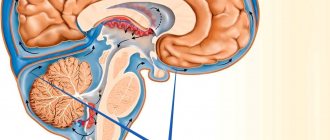
Magnetic resonance imaging is a unique examination method in which high-power radiation passes through the patient's body.
The patient is placed on a special table that enters the scanner through a tunnel. When turned on, a huge magnet is launched. It produces rays through the cavity of internal organs and soft tissues.
Sensitive sensors capture the movements of particles and ions, translating them into high-quality images.
MRI is often prescribed for diseases that require accurate diagnosis for final decision-making:
- oncological tumors;
- cerebrovascular disorders;
- abnormalities of the heart and coronary vessels;
- pathologies of internal organs (spleen, liver, kidneys);
- aneurysms;
- fistulas or dangerous abscesses on the mucous membrane of organs;
- distant metastases.
In some situations, MRI is the only way to correctly diagnose and assess the danger of the growth of a malignant tumor. But how do you get an MRI exam if you have claustrophobia? A specific disorder limits the capabilities of a person who cannot remain motionless in the tomograph tunnel.
What is fear of enclosed spaces?
A mental illness called “claustrophobia” is a condition in which a person experiences panic and anxiety when entering a confined space. For such people, a banal elevator ride or being in a small room becomes a problem.
At an advanced stage of the disease, the patient cannot get on a bus or get into a car.
Fear of enclosed spaces has characteristic features:
- a feeling of severe anxiety increases;
- pulse quickens, blood pressure rises;
- loss of body control;
- sweating;
- feeling of dryness in the throat;
- pain in the temples, dizziness.
Claustrophobia is often an acquired condition. A person faces a dangerous disease after suffering stress, disaster or physical trauma. Some patients manage to control themselves and can enter the elevator for a short time. But conducting an examination using tunnel-type devices takes at least 20–30 minutes, which is too long for a patient with a similar problem.
MRI doctors note that while in a tomograph, a panic attack sometimes begins in a person who has not previously shown signs of claustrophobia.
Perhaps the monotonous buzzing and flickering of sensors, new sounds and the need to remain motionless for half an hour have a strong impact on the psyche.
An aggravating factor is anxiety in anticipation of a diagnosis, fear of cancer, or poor health due to the underlying illness.
For such patients, it becomes a problem how to get an MRI if claustrophobia prevents them from being in the tomograph tunnel. In some situations, doctors try to choose a gentle method of examination, using radiography or ultrasound. If standard diagnostics do not give the desired result, different methods are used to correct the situation.
How to get an MRI if you have claustrophobia
Before conducting an MRI examination, the radiologist explains in detail to the patient the rules and features of the procedure. When a tunnel-type device operates, the table enters the scanner cavity, and the tomograph itself rotates around the body.
During this time, it is necessary not to move so that the sections are of high clarity without shadows or blurry areas. Sometimes you need to comply with the diagnostician’s request and hold your breath.
Claustrophobia and an MRI procedure are difficult concepts to combine. For patient comfort, modern tomographs have pleasant lighting inside: it can be easily adjusted according to the patient’s wishes. A small fan is responsible for the flow of oxygen. The diagnostician carefully monitors the person’s behavior and physical reactions and can stop the operation of the equipment at any time. If there are no contraindications, downloading and full diagnostics takes no more than 30 minutes.
If the diagnosis of claustrophobia is confirmed, it is recommended to undergo the procedure using an open-type MRI machine. These are relatively new tomograph models that do not have sides. They resemble an operating table with a small canopy. But the number of clinics using such scanners is limited. Therefore, diagnosticians try to use psychological methods to reduce the level of anxiety and prevent a panic attack:
- select a comfortable level of lighting inside the device;
- offer thick bandages to cover the eyes;
- remove the headrest to increase space;
- show the structure of the scanner, which is open from the head and legs.
Some radiologists recommend bringing a loved one with you to the MRI, whose voice and support can help cope with a panic attack. In addition, the patient must know that he is in control of the situation: when the panic button is pressed, the examination immediately stops.
How can you get an MRI if claustrophobia has not gone away?
When an injury or tumor threatens the patient's life, he has to undergo an MRI for claustrophobia. Radiologists recommend using weakened anesthesia, putting a person into a state of medical sleep. The dose is calculated depending on the expected operating time of the device. But the method is not suitable for cardiac dysfunction; it can complicate the course of kidney and liver dysfunction.
How to alleviate the patient's condition during the procedure
Doctors recommend to patients: if claustrophobia is confirmed, get an MRI using a sedative. Sometimes a light dose of an antidepressant is enough so that the patient can endure a complex examination with a reduced feeling of anxiety.
The following tips can help alleviate the condition:
- in the evening before the procedure, you should get enough sleep and take a relaxing bath;
- watch a video showing the principle of operation of the tomograph;
- visit a treating psychologist or psychiatrist;
- use self-hypnosis to normalize the condition.
In a number of patients, a panic state occurs only when a number of factors coincide. Therefore, diagnosticians sometimes suggest trying to undergo an MRI without anesthesia, trying to reduce the scanner operating time as much as possible.
MRI is harmful to health
The operating principle of the device is based on magnetic fields, and the research process is safe from the point of view of ionizing radiation - it is absent . There is simply no harmful radiation. Unpleasant sensations during the examination are caused by the need to lie still, sometimes for quite a long time. But for the sake of your health, it’s worth being patient, because MRI is one of the most informative methods for studying and diagnosing diseases of the central nervous system, human musculoskeletal and joint systems, etc., which means it helps to make an accurate diagnosis and prescribe the correct treatment.
How to overcome your fear of an MRI
Thousands of patients undergo hardware scans every day, and most of them experience wariness or emotional distress. This can be considered the norm, especially if the study is carried out using this method for the first time. Experiences are common to every person when faced with the unknown. The page contains information on how to overcome your fear of MRI. Many people unconsciously or purposefully put off visiting a doctor because they are afraid that a serious pathology will be discovered. You should not neglect the specialist’s recommendations about conducting a magnetic resonance scan, as delaying the examination can lead to critical health consequences. Most complex diseases are asymptomatic until the anomaly affects the functionality of vital organs. Diagnosis by MRI will not cause pain or discomfort, and will help identify the disease at the earliest stage of its occurrence.
The contrast agent has many side effects
Sometimes, for research, patients are given an intravenous drug containing the metal gadolinium. It helps to better visualize certain structures; by the accumulation of this drug in tissues, you can track the presence of tumors, their prevalence, etc. This drug is much safer than the one used in computed tomography. The risk of an allergic reaction is minimal.
The contrast agent, like all medications, has possible side effects, but in general it is hypoallergenic, well tolerated by patients, and most importantly, it increases the accuracy of diagnosis.
The administration of such a drug may be contraindicated only in patients with renal failure, who must notify the doctor about their disease. In such cases, the patient will first have to take a blood test, based on the results of which a decision will be made on whether to administer the drug.
The main reasons for fear of MRI: how to overcome nervousness?
The main cause of emotional distress is fear of the unknown. The patient does not know how the procedure is performed, whether it will be painful, embarrassing, uncomfortable, or whether the scan may cause radiation harm. In addition to the reasons listed, the following situations are common:
1. Noise effect from hardware equipment. The generation of a powerful magnetic field produces mechanical sound from the installation. Manufacturers of tomographic equipment are trying to minimize this feature of the machines, but the fact remains that the sound level is difficult to reduce, you just need to be mentally prepared for this. In addition, the staff always accommodates the client halfway and offers to use earplugs or headphones, which will play music during the session. You can take care of this in advance and bring your own player with your favorite tracks.
2. Limited space inside the device. Everyone experiences discomfort when immersed in a narrow room of a tomograph. However, such a true mental illness as claustrophobia is diagnosed extremely rarely. It is one of the contraindications to the procedure. During screening, you must be in a calm, motionless state. This is impossible to achieve during hysterical attacks. If the patient is afraid of diving, you can come to the selected diagnostic center in advance and ask for a short “excursion”, during which a specialist will show and talk about the structure of the equipment and demonstrate the ability to independently leave the tomograph during a panic attack. Modern machines are equipped with internal ventilation and light lighting, which is adjusted according to the wishes of the patient. If the fear grows and does not go away, it is recommended to use an open type unit with a ring rather than a cylindrical structure.
3. The myth about X-rays. Contrary to popular belief, no ionizing field is generated during MRI. The operating principle of the equipment is based on the production of magnetic flux, which does not accumulate in biological structures, does not damage cells, does not lead to mutations and does not affect nerve fibers and the immune system. Scanning using the described method is prescribed even for infants and pregnant women. Over the half-century history of the use of tomography, not a single case of harm to the health of subjects has been recorded. MR screening is not related to radiographic imaging modalities.
4. Contrast poisoning. In some cases, additional color enhancement is required to increase the contrast of images of the area being studied. For this purpose, the patient is injected with a contrast agent based on a rare earth metal. The risk of getting intoxicated after using the solution is minimal if the person does not suffer from chronic diseases of the urinary system. After administration, the substance quickly breaks down into its components and is excreted naturally through the kidneys within 24 hours after the session. You can speed up the process by drinking large amounts of liquid.
The presence of any metal implants is a contraindication for MRI
The only absolute contraindication to MRI is the presence of a pacemaker or cochlear implant. The presence in the patient's body of metal prostheses and implants, plates, surgical staples, vena cava filters, stents, dental pins, braces and other metal objects is not a contraindication to MRI.
Conducting research is possible. Difficulties may arise with visualizing tissues and organs around a metal object, but this does not pose a danger to the life and health of the patient. It is necessary to inform the doctor about the presence of metal in the body, but conducting a study is most likely possible.
How to get an MRI if you suffer from claustrophobia - fear of closed spaces is not a death sentence at all!
For patients with fear of closed spaces, diagnosis using a tomograph is not always acceptable. After all, how to do an MRI if claustrophobia does not allow you to stay inside the machine for a long time and provokes panic attacks.
Today, medicine offers a lot of methods for examining the body, but magnetic resonance imaging stands out among them for a number of reasons. This method is universal due to the fact that it is safe and highly informative. Despite a number of advantages, MRI still has its disadvantages.
Among them is that the patient is placed in a closed machine for scanning. This means that those who are afraid of confined spaces will find the procedure difficult. However, it is possible to do it!
How is tomography performed?
The scanning procedure is carried out in a special room, where the patient is placed horizontally on the table, laid flat and comfortable, if necessary, pads and bolsters are placed under the limbs, then the legs and arms are secured with belts, and the table slides into the machine.
A tomograph is a large capsule in which magnets are placed; under their influence, particles in the human body are aligned in a certain way, which makes it possible to read information about an organ in each layer, almost cell by cell, after which the computer converts the information into a picture, displaying a three-dimensional image on the diagnostician’s monitor. several projections. You can take pictures of blood vessels and nerve endings.
Despite the fact that the tomograph has good ventilation, fresh air supply and a comfortable temperature, it is still not an easy task to stay in a closed space for a long time. And for those suffering from claustrophobia it is simply real torture. In addition, examination rules require immobility throughout the entire tomography procedure.
If you are afraid of confined spaces, MRI is done under general anesthesia or using sedatives.
What is fear of confined spaces?
Claustrophobia is considered to be one of the most common phobias on the planet. Statistics show that about 40 million people on the planet suffer from it, and this is only data from recorded attacks among patients.
Fear of enclosed spaces is a real psychological problem. It occurs most often after a stressful situation in which a person has been.
It is noted that a number of patients did not even imagine the presence of such fear, but discovered it only when they found themselves in a closed room:
- stuck in an elevator;
- woke up in a closed ship cabin;
- traveled in a closed train carriage;
- were in a dark cinema;
- did not have access to the apartment doors.
Where is MRI performed in the capital?
If efforts to suppress unpleasant emotions do not work, the fear remains, it is recommended to use an open configuration scanner, without solid walls. In exceptional cases, only general medicated sleep can help. To search for a diagnostic center that provides the described services, located in any area of the city, there is a single city service 36go. Its pages provide a complete list of clinics where tomography is performed.
Medical organizations on the portal are equipped with general and contact information, reviews of clients who have undergone diagnostics, ratings, and are distributed according to the prices of the offers. You can sign up for a consultation or session using a single hotline number located at the top of the page. Service operators will provide complete information about the nearest clinics, prices, and diagnostic nuances. Consultation and time booking are free of charge.