An intracerebral hematoma (ICH) is an accumulation of blood in a limited area of the brain substance that occurs due to a violation of the integrity of a cerebral vessel. The pathological condition is provoked by weak or injured intracerebral arteries or veins, which, having ruptured, began to release blood into the intracranial space. The VMH cavity can be filled with liquid or coagulated blood, or a bloody substance mixed with brain detritus (crushed fragments of brain tissue).
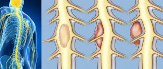
Schematic representation of a large hematoma.
Cerebral hematomas can be single or multiple, unilateral and bilateral, acute, subacute and chronic. They become chronic just 21 days after the hemorrhage occurs. Triggers of pathogenesis are often the consequences of arterial hypertension and atherosclerosis (70%-80% of cases), aneurysms and vascular malformations, mechanical intracranial damage due to TBI.
Intracerebral hematomas pose a serious threat to the health and life of the patient, since the structure-forming tissues of the brain at the site of the lesions are subject to compression, dislocation, edema and necrosis. Without adequate therapy, pathology can lead to severe morphological and functional brain damage, often irreversible. With ICH of any origin, the risks of disability and mortality are too high: disability affects up to 70% of surviving patients, the probability of death is 25%-50%.
Classification of intracerebral hematomas
Intracerebral hematomas in neurosurgery and neurology are classified by location and volume. The following types of VMH are diagnosed by location:
- medial (10%) – located medially from the internal capsule, determined in the zone of the thalamus and hypothalamus with possible spread to the midbrain and ventricles of the GM;
- lateral (the most common, accounting for about 50%-55% of all hematomas) – located outward from the internal capsule, localized mainly in the area of the shell, often extending to the level of the semioval centers and insular lobe;
- lobar (15%) – concentrated under the cerebral cortex in the subcortical white matter, usually within one lobe (occipital, temporal, frontal or parietal);
- cerebellar (10%) – located in the area of the right/left hemisphere of the cerebellum or worm-like structure (if the worm is damaged, a hematoma often breaks out with the contents entering the 4th ventricle, which causes the development of occlusive hydrocephalus);
- intra-stem (6%-9%) – formed in the brain stem, as a rule, more often such hematomas affect the structures of the pons by the type of hemorrhagic impregnation;
- mixed - simultaneously cover several intracerebral anatomical structures (for example, subcortical white matter, basal ganglia of the telencephalon and thalamus), or VMH is combined with subdural types of hematomas.
An important diagnostic indicator that will play a predetermining role in the choice of treatment algorithm is the size or volume of the formed hematoma. The following types of intracerebral hematomas are distinguished by volume:
- small – less than 40 cm³;
- medium – 40-60 cm³;
- large – 60-80 cm³;
- gigantic - more than 80 cm³.
Secondary complications in the form of increasing compression, deformation, swelling and displacement of the brain can occur with blood clots of not only large but also medium sizes. In some cases, especially if accompanied by a brain contusion, a hemorrhagic component of 30 cm³ may be sufficient to create a mass effect.
Predisposing factors
The formation of hematomas occurs after injuries, including pinching, blows, squeezing, and bruises. Subarachnoid hemorrhage does not fall into this category, since it does not appear due to trauma, but due to damage to an unchanged vessel. Often small hematomas appear due to eating large quantities of food or drinking alcoholic beverages. This is due to stretching of the gastrointestinal tract and the appearance of cracks.
The development of pathology is influenced by vascular weakness and problems with blood clotting. Often due to a weakened immune system due to infections or age-related changes, the likelihood of pus accumulating in the affected area increases.
Clinical signs of ICH
The clinical picture of intracerebral hematomas begins with the sudden onset of a crisis, characteristic of a cerebral hemorrhage. Hemorrhage is often preceded by high blood pressure or head trauma. During the acute period, typical symptoms are observed:
- sharp, intense headache;
- dizziness, loss of consciousness;
- nausea, vomiting;
- heavy wheezing breathing;
- abnormal heart rate;
- development of hemiparesis on the side opposite to the VMH with possible development into hemiplegia (usually in the leg, arm, facial muscles);
- coma.
The severity of symptoms depends on the size and location of the hematoma. As the pathology progresses, neurological deficits progress with a predominance of motor, sensory, speech, and thinking disorders. Patients with intracerebral hematomas are often bothered by epileptic-type convulsions, stiffness of the muscles of the back of the head and neck, impaired coordination and stunned consciousness syndrome of varying severity. Visual disturbances (mydriasis, loss of fields, unilateral hemianopsia, ptosis, etc.), bradycardia, memory problems, and mental disorders often occur.
To give a clear rationale for the symptoms occurring, it is necessary to conduct high-quality instrumental diagnostics of the brain using cerebral imaging methods. Without a normal examination of the brain, it is impossible to make a final conclusion on the diagnosis, its severity and location, it is impossible to choose a treatment method and form an idea about the prognosis of the outcome.
Symptoms
Symptoms of hemorrhages on the head may not be expressed, most often it manifests itself in the form of a headache. You may also experience an unpleasant sensation such as slight dizziness when changing your body position or moving vehicles in front of your eyes.
One of the main symptoms of the pathology is headache
Other possible signs of pathology:
- vomiting, nausea;
- drowsiness;
- confusion;
- loss of speech or slow speech;
- weakness in the limbs on one side of the body;
- difference in pupil size.
In the early stages of injury, an accurate diagnosis can often only be made through special studies. Therefore, in case of any head injury, it is recommended to immediately contact a neurologist or neurosurgeon.
Diagnosis of hematoma in brain structures
Neuroimaging is of clinical value in making a diagnosis. A computed tomography scan is prescribed as an initial examination of the brain. This inexpensive method allows you to quickly and easily determine the presence of blood in the GM substance, the location and volume of the clot. The information value of CT is highest when 2-3 weeks have passed since the hematoma appeared (maximum 5 weeks). Until this time, the VMH area has the highest density, which facilitates diagnosis and can be limited to one CT scan.
MRI.
As the lesion ages (on average, after 14-21 days), the density of the hemorrhagic mass decreases, it becomes isodense, that is, close to normal brain tissue. During this period and later, purely magnetic resonance imaging can provide qualitative information about the intracerebral hematoma and the state of the brain.
Additionally, the patient is often recommended to undergo cerebral angiography. This technique does not have the necessary potential for verification of VMG. But angiography of cerebral vessels determines the intensity and distribution of vasospasm and allows one to exclude or confirm the involvement of a vascular malformation or arterial aneurysm in the development of ICH.
Causes
Hemorrhage usually occurs after injury. This could be a bruise of the skin, internal organs, a concussion or bruise of the brain, an injection with thin (sharp) objects. Sometimes blood leaves the vessels and pours into the skin and internal organs as a result of infections, autoimmune diseases, and poisoning. The occurrence of hemorrhages and bruises is promoted by increased fragility of blood vessels, fasting, lack of vitamins in food, high blood pressure, and congenital bleeding disorders.
At CELT you can get a consultation with a traumatologist-orthopedic specialist.
- Initial consultation – 3,000
- Repeated consultation – 2,000
Make an appointment
Treatment methods for intracerebral hematoma
Conservative therapy or neurosurgery are used to treat cerebral hematomas. Recommendations for conservative elimination of hematoma are:
- small size of the lesion (≤ 40 cubic cm) without pronounced symptoms, without clinical signs of herniation and dislocation;
- old age of the patient (75 years and older);
- inappropriateness of surgery due to the high risk of unfavorable outcome (for example, large-scale hemorrhage in the dominant hemisphere or with extensive neurological damage);
- severe condition of the blood coagulation system, sepsis;
- diabetes mellitus in the stage of decompensation, uncontrolled hypertension.
The problem is solved conservatively by competent neurologists and neuroreanimatologists. The treatment plan is developed strictly individually. It may involve basic anti-ischemic, antihypertensive treatment, reduction of cerebral edema with osmodiuretics, therapy with hemostatics and neuroprotectors.
Surgical methods for removing VMG
Surgery is indicated in most cases. Its main goal is to save a person’s life and normalize the neurological status. The use of surgery is justified in the following conditions:
- hematomas of any size, accompanied by a pronounced mass effect, cerebral edema, dislocation of the central structures by more than 5 mm;
- lobar and lateral VMGs from 50 cm³ or more in volume;
- medial formations from 20 cm³ and above;
- cerebellar hematomas ≥ 15 cm³;
- progressive deterioration of the patient’s well-being (exception – stage 2-3 coma);
- young age of the patient;
- severe intracranial hypertension when it is impossible to correct ICP conservatively.
The options for surgical treatment are transcranial surgery with direct inspection under microscope control (the standard, most common technique), stereotactic aspiration, and endoscopic removal of the hematoma.
- Transcranial removal. The operation involves direct craniotomy (classical trepanation, often extended) in the projection of the lesion. Next, an encephalotomy is performed in the area of the closest apposition of the VMG to the cerebral cortex. Then they begin to remove the pathological formation, which is carried out by aspiration with washing out the cavity with a stream of saline solution. Heavily condensed elements are removed using special windowed tweezers. Hemostasis is carried out by bipolar coagulation of blood vessels, the use of antihemorrhagic agents (sponges, fibrin-thrombin glue, pads with hydrogen peroxide). A drainage is installed on the wound. The surgical session lasts approximately 3 hours, manipulations are performed under general anesthesia. Direct craniotomy is more common for lobar and cerebellar VMH.
- Stereotactic procedure. Stereotactic aspiration is considered a minimally invasive tactic. However, despite the gentle effect, recurrences of hematomas occur more often after such treatment than after craniotomy. This is explained by the inability to perform thorough hemostasis during stereotactic surgery. The basic type of anesthesia is neuroleptanalgesia. Intervention is performed more often for hematomas of medial and mixed types. The patient's head is pre-fixed in a special stereotactic frame. Next, after placing a small burr hole in the skull, a thin cannula (diameter about 5 mm) is inserted into the hematoma cavity. Through an installed cannula, to which an electric aspirator is connected, pathological contents are evacuated from the brain. Intraoperative control of cannula insertion and therapeutic manipulations is carried out using navigation systems and X-ray equipment. Session duration is 1-3 hours.
- Endoscopic surgery. This is a minimally aggressive neurosurgery tactic that involves removing blood accumulated in the brain tissue under endoscopic control. Anesthesia is usually used by intubation. Access is performed using a surgical device such as trephine, which serves to create a small burr hole in the skull of a round shape. A rigid endoscopic tube is placed into the formed trefination hole. The endoscope tube has a video system that transmits images of brain structures to a color intraoperative screen. The endoscope is carefully brought to the area of interest, then its tidal working system suctions the hematoma and rinses the cavity. Hemostasis is also carried out endoscopically, using monopolar coagulation and defocused laser irradiation. The duration of the procedure is 45-90 minutes.
Russian clinics offer direct craniotomy at a price of 60 thousand rubles or more, stereotactic surgery - 40-60 thousand rubles, endoscopic removal of a hematoma - from 50 thousand to 62 thousand rubles.
After any surgical intervention, a full course of rehabilitation is prescribed, including deep prevention of all possible postoperative complications (infections, thrombosis, pneumonia, etc.). In the majority of cases, after neurosurgery, preventive therapy to prevent epileptic seizures is advisable.
On average, it takes about 6 months for an adult patient to recover after removal of an intracranial hematoma. Depressed functions can be restored completely, but there is no absolute guarantee of this, since everything depends on the initial condition of the patient, age, and the nature and consequences of the intracerebral hematoma at the preoperative stage. In children, rehabilitation usually progresses at a faster pace, and much more often it ends in full recovery.
Where is the best treatment?
Intracerebral hematomas are a very serious problem, associated with high risks of mortality and complete disability. Patients with this diagnosis need to be observed and treated in medical institutions of the highest global level. Some of the best neurosurgical specialists in Europe practice in the Czech Republic.
Central Military Hospital of Prague.
Clinics in the Czech Republic follow only advanced diagnostic principles and cutting-edge technologies for the safe treatment of central nervous system lesions. In addition, the famous republic offers impeccable rehabilitation care for patients after brain surgery.
Czech neurosurgery means the quality of the services provided, as well as their affordable prices. In the Czech Republic, high-tech treatment programs for intracerebral hematomas cost 2-3 times lower than in Israel and Germany.