What is high intracranial pressure?
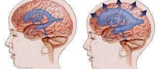
The pressure of intracranial contents (substance of the brain and spinal cord, blood and cerebrospinal fluid - cerebrospinal fluid) on the walls of the skull is called intracranial. Liquor circulates in the cerebrospinal fluid pathways: the subarachnoid space and the ventricles of the brain (Fig. 1). If his pressure rises above 10-12 mm Hg. Art., intracranial hypertension occurs. It can manifest itself as depression of consciousness, headache, nausea and can cause damage to brain tissue. If the pressure continues to rise, at 30-40 mm Hg. Art. a person most often falls into stupor (a sharply inhibited state), at 40-50 mm Hg. Art. - in a coma of varying depth.1
Figure 1. Cerebrospinal fluid circulation. Source: OpenStax / Wikipedia (Creative Commons Attribution 4.0 International license)
Intracranial hypertension occurs in children and adults. In infants under one year of age, it is almost always associated with hydrocephalus or birth trauma. In adults, common causes are head injuries and conditions that increase the volume of brain tissue or the cerebrospinal fluid and blood circulating inside the skull.
Causes
What can cause intracranial pressure to increase? In adults and children there are some differences in the etiology of the problem under consideration. What is common is the severity of the pathology. There are 2 groups of causes of intracranial hypertension.
- First group
- the presence of an additional formation that increases the volume of the brain (tumor growth, cysts, hematoma formation, cerebral aneurysm, development of an abscess);
- swelling of the brain substance that occurs against the background of encephalitis, traumatic brain injury, hypoxia, ischemic strokes, poisoning, and encephalopathy of hepatic etiology;
- swelling of the cerebral membranes – pachymeningitis, arachnoiditis;
- disorders of cerebrospinal fluid dynamics (hydrocephalus) - arising as a result of increased production, impaired absorption of cerebrospinal fluid or the presence of an obstacle to its outflow.
- Second group:
- increased blood flow to the brain during hyperthermia, hypercapnia (carbon dioxide poisoning), hypertension;
- difficulty in venous outflow from the cranial cavity (for example, dyscirculatory encephalopathy in elderly patients);
- constant increase in intrathoracic or intra-abdominal pressure.
In adult patients, cerebral hypertension most often occurs against the background of acquired encephalopathy of post-traumatic, vascular, toxic, discirculatory origin. In childhood, congenital factors predominate among the causes:
- various anomalies of the development of the central nervous system - microcephaly, a congenital form of hydrocephalus;
- birth injuries of the brain and their consequences - residual or residual encephalopathy with intracranial hypertension (manifests some time after injury and brain hypoxia during childbirth);
- intrauterine neuroinfections (meningitis, arachnoiditis, encephalitis);
- congenital brain tumors (craniopharyngioma).
According to the course, acute and chronic forms of ICH are distinguished. The first is usually a consequence of brain damage as a result of traumatic brain injury, stroke or infection, the second develops gradually against the background of slowly growing tumors, cystic formations or as vascular disorders increase. This includes residual encephalopathy in children and adults.
There is a pathology such as idiopathic or benign intracranial hypertension, the etiology of which is considered unknown. Most often it develops in overweight women. The role of endocrine disorders, chronic kidney disease, poisoning, treatment with corticosteroids and antibiotics is being studied. With this form of hypertension, no space-occupying lesions are detected, there is no thrombosis of the venous sinuses and there are no signs of infectious brain damage.
Causes of increased intracranial pressure
Intracranial pressure may increase due to the following reasons:
- Swelling of brain tissue due to injury, hypoglycemia, hyponatremia, the development of hypoxia as a result of a stroke, inflammation due to encephalitis or meningitis, poisoning.
- Increase in volume: when foreign bodies enter, tumors appear, hemorrhage (subdural or epidural hematoma, intracerebral or subarachnoid hemorrhage).
- Development of hydrocephalus: an increase in the volume of cerebrospinal fluid in the cranial cavity.
- Venous congestion: accompanies congestive heart failure and can develop with thrombosis of the cerebral veins.
- Increased cerebral blood flow during epileptic seizures, cerebral hyperperfusion (develops after ischemic strokes, surgical interventions, injuries).
Diagnostics
For diagnosis, the pressure inside the skull is measured by inserting a needle attached to a pressure gauge into the spinal canal or into the fluid cavities of the skull.
For staging, a number of signs are taken into account:
- It is determined by poor outflow of venous blood from the skull area.
- According to MRI (magnetic resonance imaging) and CT (computed tomography).
- Judged by the degree of rarefaction of the edges of the ventricles of the brain and the expansion of fluid cavities.
- According to the degree of dilation and blood filling of the veins of the eyeball.
- According to ultrasound data of cerebral vessels.
- According to the results of the encephalogram.
- If the ophthalmic veins are clearly visible and heavily filled with blood (red eyes), then we can indirectly say that there is an increase in pressure inside the skull.
In practice, in most cases, for a more accurate diagnosis and the degree of development of the disease, differentiation of the symptoms of clinical manifestations of hypertension is used in combination with the results of a hardware study of the brain.
Who is at risk?
The most common cause of increased intracranial pressure is considered to be traumatic brain injury. At risk are people for whom the risk of such injuries is increased (for example, due to professional activities or playing contact sports). Also, the likelihood of developing intracranial hypertension is higher for people with high blood pressure and the risk of ischemic stroke.
For children in the first year of life, the risk of high intracranial pressure is associated with hydrocephalus - water on the brain. It can be congenital and caused by disturbances during intrauterine development or acquired, resulting from birth trauma or infection.
Treatment and prognosis
Therapeutic measures are prescribed based on diagnostic results. First of all, it is important to get rid of the cause that provokes excessive fluid pressure on the skull. If this phenomenon is caused by traumatic brain injury, the patient is advised to rest completely, take gentle nutrition, and take anti-inflammatory drugs. In some cases, surgical intervention is necessary (in the presence of hematomas between the membranes of the brain, as well as in the presence of injuries that require surgical treatment). Surgery is also prescribed when various tumors are detected in brain tissue that is prone to rapid growth.
A separate set of measures is carried out when diagnosing hydrofecal in a child. To remove excess fluid, a shunt is installed, through which it flows into the abdominal cavity, and the pressure is normalized. The operation is repeated as the child grows, and the patient is constantly monitored. In some children, the need for artificial fluid removal gradually disappears.
Drug therapy for ICP pathologies is secondary. However, drugs are prescribed to eliminate symptoms and make the patient feel better. The following medications may be helpful:
- hormonal anti-inflammatory drugs;
- neuroprotectors and substances for stimulating blood circulation in the brain - the effectiveness of this group has not been proven, despite its widespread use;
- loop diuretics (diuretics) - medications that stimulate the excretion of excess fluid;
- osmodiuretics - including reducing the production of cerebrospinal fluid.
The Clinical Brain Institute specializes in the diagnosis and treatment of diseases of the central nervous system. Increased intracranial pressure is not a separate disease, but a symptom that indicates a number of pathologies. Experienced specialists will accurately determine the cause of such a violation, as well as its severity and possible consequences. It is worth understanding that only timely seeking medical help can guarantee successful treatment and a return to your normal lifestyle without headaches.
Clinical Brain Institute Rating: 4/5 — 56 votes
Share article on social networks
What are the risks of increased intracranial pressure? Complications and consequences
The brain is the highest energy system in the human body. It needs approximately five times more oxygen than the heart. Therefore, the quality of blood supply to the brain is a vital indicator that depends on two parameters - blood pressure and intracranial pressure. At constant blood pressure, the lower the intracranial pressure, the better the blood supply to the brain. If for some reason intracranial pressure increases, this proportionally worsens the blood supply to the brain, and ischemia develops. Therefore, high intracranial pressure is a dangerous condition that can cause the following complications:
- reduction and complete loss of vision;
- convulsions;
- acute cerebrovascular accident;
- depression and/or disorder of consciousness;
- depression of respiratory and circulatory function.
Important! With a rapid increase in intracranial pressure, there is a threat of death. If the symptoms are severe or appear sharply, suddenly and rapidly increase, you need to call an ambulance.
Symptoms
The formation of clinical hypertension syndrome and the nature of its manifestations depend on the localization of the pathological process, its prevalence and speed of development.
Intracranial hypertension syndrome manifests itself with the following symptoms in adults:
- Headache of increased frequency or severity (increasing headache), sometimes awakening from sleep, often forced positioning of the head, nausea, and repeated vomiting. It can be complicated by coughing, a painful urge to urinate and defecate, and actions similar to the Valsalva maneuver. Possible disturbances of consciousness and convulsive seizures. With prolonged existence, visual impairment occurs.
- History may include trauma, ischemia, meningitis, cerebrospinal fluid shunt, lead intoxication or metabolic disorder (Reye's syndrome, diabetic ketoacidosis). Newborns with cerebral ventricular hemorrhage or meningomyelocele are predisposed to intracranial hydrocephalus. Children with blue heart disease are predisposed to an abscess, and children with sickle cell disease may have a stroke leading to intracranial hypertension.
Objective signs of intracranial hypertension are swelling of the optic disc, increased cerebrospinal fluid pressure, increased osmotic pressure of the extremities, and typical x-ray changes in the bones of the skull. It should be borne in mind that these signs do not appear immediately, but after a long time (except for increased cerebrospinal fluid pressure).
There are also signs such as:
- loss of appetite, nausea, vomiting, headache, drowsiness;
- inattention, decreased ability to awaken;
- papilledema, upward gaze paresis;
- increased tone, positive Babinski reflex;
With a significant increase in intracranial pressure, disturbances of consciousness, convulsive attacks, and visceral-vegetative changes are possible. With dislocation and herniation of brain stem structures, bradycardia, respiratory failure occur, pupillary response to light decreases or disappears, and systemic blood pressure increases.
Symptoms indicating high intracranial pressure
One of the symptoms of increased intracranial pressure is morning headache.
Photo: AndrewLozovyi / Depositphotos Symptoms can vary greatly depending on how high the blood pressure is, as well as the causes of this condition, the speed of its development and concomitant diseases.
Main features:
- headache;
- nausea or vomiting;
- vision becomes blurred and there may be double vision;
- confusion;
- problems with movement or speech;
- restlessness, anxiety, fear or irritability.
With intracranial hypertension, only some of these symptoms may appear.
They can develop gradually or grow rapidly over several hours or minutes. If blood pressure rises quickly, a person may lose consciousness and fall into a coma (Table 1). Table 1. Symptoms of high intracranial pressure.
| Intracranial pressure (mmHg) | Symptoms |
| 20-30 | Headache, nausea and vomiting, drowsiness, anxiety, psychosyndrome (impaired memory, thinking and emotional reactions), high blood pressure, decreased heart rate, convulsions |
| 30-40 | Sopor |
| 40-50 | Coma of varying depths |
| >50 | Threat of brain death |
Headache with increased intracranial pressure has several features:
- It appears after waking up in the morning, worsens when lying down, and is localized in the back of the head, temples, and forehead.
- The pain is strong, pressing or bursting.
- Painkillers cannot relieve pain.
- Sneezing and coughing, physical activity or lying down provoke attacks of pain.
How to determine intracranial pressure in a child?
In children in the first year of life, intracranial hypertension may appear due to birth trauma, against the background of hydrocephalus (Fig. 3) or swelling of brain tissue.
In older age, traumatic brain injury is a more common cause. Figure 2. Hydrocephalus in children. Source: Journal of Neurosciences in Rural Practice / Open-i (Attribution-NonCommercial-ShareAlike 3.0 Unported (CC BY-NC-SA 3.0)
High intracranial pressure in children of the first year of life has several characteristic signs:
- The shape of the head changes; the bone plates that form the skull can move apart.
- The fontanel protrudes outward and pulsates.
- Increase in skull volume.
- Restless behavior, constant crying, or drowsiness and lethargy.
- Frequent regurgitation or severe vomiting.
- Trembling chin.
- Convulsions, loss of coordination of movements of the arms and legs.
- Visual impairment, limited range of rotation of the eyeballs, strabismus.
- Depression or loss of consciousness.
If these symptoms appear suddenly and increase quickly, you should immediately call emergency medical help.
Diagnosis of intracranial pressure
During diagnosis, the doctor will conduct a general examination, measure blood pressure, and check how the pupils react to light. He will need to talk about the existing symptoms, how long ago and how often they appear, as well as any concomitant diseases. After this, the neurologist will prescribe instrumental diagnostics. It is carried out in two directions:
- brain imaging;
- measurement of intracranial pressure.
Brain imaging is needed to obtain information about the cause of hypertension, how common this process is, and what complications there are. At this stage of diagnosis, a tumor, areas of impaired circulation of cerebrospinal fluid, and signs of strangulation can be identified. Computed tomography or magnetic resonance imaging is performed for imaging. When examining children under 1 year of age, an ultrasound scan of the brain is performed.
Measuring intracranial pressure is possible in one of three ways:
- intraparenchymal;
- intraventricular;
- extracerebral.
All three methods are invasive, require the installation of a special sensor, and are used only in a hospital setting. With such monitoring, external ventricular drainage is sometimes additionally provided, which helps control intracranial pressure (Fig. 3).
Figure 3. External ventricular drainage. Source: Children's Health Queensland, Dermatology reports / NCBI (Creative Commons Attribution 3.0 License (by-nc 3.0)
Noninvasive methods for measuring intracranial pressure do not provide accurate values, but they can be used to confirm hypertension. Among such methods are fundus examination and transcranial Doppler sonography. An ophthalmologist performs a fundus examination. He assesses the condition of the optic nerve and uses it to draw a conclusion about the level of intracranial pressure. Transcranial Doppler is an ultrasound examination that evaluates blood flow in the vessels running inside the skull.
Intracranial hypertension
Intracranial hypertension, or increased intracranial pressure, is a dangerous syndrome that can be fatal. A syndromic diagnosis is established by a neurologist using examination and additional research methods. Increased intracranial pressure requires immediate and adequate treatment.
Unfortunately, in our country, this diagnosis is often made without any reason by everyone who is in any way connected with the examination of the head: ophthalmologists, therapists, radiologists. Intracranial hypertension is diagnosed both in adults and children, right and left. In this case, treatment is prescribed (mainly vascular drugs), which, naturally, does not lead to a cure. Often at an appointment you can hear that the patient has been treated for intracranial hypertension unsuccessfully since childhood and has been taking courses of vascular medications all his life.
What is intracranial hypertension really?
The skull is a closed space in which the brain, blood vessels, as well as cerebrospinal fluid (cerebrospinal fluid) and other structures are located. All these organs create a certain pressure inside the skull, which can fluctuate normally. Due to the fact that the bones of the skull in an adult are normally motionlessly connected, any increase in volume in the cranial cavity leads to an increase in pressure inside it. For example, intracranial pressure increases slightly when straining, then returns to normal values.
Certain medical conditions can cause blood pressure to rise or fall significantly, causing damage to structures inside the skull. For example, intracranial pressure slowly and steadily increases as a brain tumor grows. Since the bones of the skull are motionless and the tumor continues to grow, the volume of the contents of the skull increases, the pressure inside increases, the brain and blood vessels are displaced or compressed. All this leads to irreversible consequences and even death.
Intracranial pressure can rise very quickly with a cerebral hemorrhage or severe meningitis. In this case, emergency measures are required. A sudden decrease in intracranial pressure is also dangerous, as it can cause the brain to wedge into the bony openings of the skull and cause rapid death.
There is another type of hypertension - benign intracranial hypertension. The peculiarity is that the cause of the increase in pressure cannot be found; the disease may go away on its own. However, benign intracranial hypertension is a fairly rare disease. It is known that women are more likely to suffer from it, especially during pregnancy or if they are overweight.
What symptoms may bother you?
If the pressure changes smoothly, the patient may have various complaints. Among them, the most common are headache and vomiting in the morning or after being in a horizontal position. Sometimes blurred vision and double vision occur. Infants may have a bulging or retracted fontanel. The remaining symptoms are nonspecific: weakness, irritability, in children - a sharp cry, etc.
If the pressure inside has changed sharply, then most likely the person will be unconscious in a serious condition.
Research methods
First of all, I would like to say that intracranial pressure can only be measured by opening the skull. To date, there are no other methods for accurately measuring intracranial pressure. A stable change in pressure (mostly an increase) can be assumed using the following methods:
- Examination by a doctor to assess the movements of the eyeballs
- the patient may have strabismus due to compression of the abducens nerve, double vision while checking the range of movements of the eyeballs. In children, head circumference may increase. Pathological neurological symptoms, associated, for example, with a tumor, may be observed. - Fundus examination
- there may be blurred optic disc, vascular congestion and other nonspecific signs. - X-ray of the skull
- so-called “finger impressions” and other nonspecific signs may be visible. - MRI and CT of the head
- “empty sella” syndrome, dilatation of the ventricles of the brain and other nonspecific signs. - Ultrasound of the vessels of the head and neck
- there may be a violation of the venous outflow from the cranial cavity.
All these data must be assessed in combination, because each of them separately does not allow establishing the syndrome of intracranial hypertension. To make a diagnosis, you must also visit at least a neurologist and an ophthalmologist.
Unfortunately, there are often cases when, based on an examination by one specialist or based on the results of a single study, intracranial hypertension syndrome is established and treatment is prescribed. In most cases, headaches and visual disturbances are caused by completely different reasons (tension headaches, migraines, cervicocranialgia, etc.).
Treatment of intracranial hypertension
Treatment of intracranial hypertension depends on the cause of the syndrome.
And remember that real intracranial hypertension cannot be treated with vascular, nootropic drugs, physiotherapy and massage! This is a serious condition. This often requires hospitalization in the intensive care unit and emergency assistance from a neurosurgeon.
Be healthy!
Maria Meshcherina
Photo istockphoto.com
Treatment of intracranial pressure
Intracranial hypertension is treated by reducing the pressure to 20 mmHg. Art. or lower and stabilizing the patient's clinical condition. To achieve this, therapy is carried out in several directions.
Conservative approach:
- Maintaining the correct position of the patient (lying position, body elevated by 30 degrees, head position straight).
- Hardware ventilation for breathing problems.
- Reducing body temperature if it is elevated.
- Reducing blood pressure with antihypertensive drugs.
- Reducing stress: stopping vomiting, coughing, eliminating factors that provoke a rise in blood pressure, including shivering from cold, careless transportation, pain and others.
After diagnosis, the cause of hypertension is eliminated. This may require removal of an aneurysm or tumor, bypass surgery, or other major interventions. If there is excess volume of cerebrospinal fluid, punctures are performed to reduce it. After the main stage of treatment, rehabilitation is carried out. The doctor will prescribe medications, diet therapy, and tell you how to adjust your lifestyle.
Prognosis and prevention
When driving at high speed, wearing a helmet is mandatory.
Photo: Paha_L / Depositphotos Increased intracranial pressure is most often associated with head injuries. To prevent them, you need to take precautions: wear a seat belt in a car, use a helmet when riding a bicycle, and reduce the risk of falls.
If there are signs of hypertension, you should see a doctor as soon as possible. The earlier treatment is started, the lower the risk of serious health consequences.
Bottom line
Thus, intracranial hypertension is a pathological condition that can occur in a variety of diseases of the brain and more. It requires mandatory treatment. Otherwise, a wide variety of outcomes are possible (including complete blindness and even death).
The earlier this pathology is diagnosed, the better results can be achieved with less effort. Therefore, you should not delay visiting a doctor if you suspect increased intracranial pressure.
Facts and myths about increased intracranial pressure
There are several misconceptions about intracranial hypertension:
- It does not need to be treated; the condition will go away on its own with age. In fact, both children and adults need treatment to avoid serious complications.
- The condition cannot be treated. False: Treatment results depend on the cause and severity of hypertension. Even if it cannot be completely cured, therapy greatly improves the condition and reduces the risk of health consequences.
- High intracranial pressure always causes developmental delays in children. This is a myth: with timely treatment, such consequences can be avoided.
It is important to know that:
- High intracranial pressure in children most often occurs due to hydrocephalus and birth injuries.
- Homeopathic or herbal preparations do not affect blood pressure in any way and do not help normalize it.
- If a person already has intracranial hypertension, he needs to be examined by a neurologist at least once every two years.
Treatment in adults
Therapy is carried out after determining the cause that led to the disease.
Cataract is a pathological condition of the lens, manifested by its clouding and deterioration of vision of varying degrees. Cataract is a disease that is more often diagnosed in older people (in 90% of cases). Read more in the article: “treatment of cataracts with folk remedies.”
Surgical removal of the cause
Indications for surgical intervention:
Advertising:
- hydrocephalus;
- ineffectiveness of puncture and medications;
- visual acuity decreases;
- open traumatic brain injury;
- brain tumors, hematoma, aneurysm.
Methods:
- If there is excessive secretion of cerebrospinal fluid due to hydrocephalus, shunting is performed. The method creates an additional pathway for fluid outflow and reduces intracranial pressure. A silicone catheter is installed into the hole in the skull. It is immersed in the ventricle of the brain. The cerebrospinal fluid is drained by a system of silicone tubes and valves, which is fixed in the abdominal cavity. X-rays are used to monitor the operation.
- Endoscopic perforation. Restores the physiological outflow of fluid. The risk of complications and trauma are minimal. The method is used for ICP after injury, if it is necessary to remove the shunt system, or for complications after shunting.
Other manipulations
- Ventricular puncture. Excess cerebrospinal fluid is drained from the ventricle using a long needle. In half of the cases complications arise and additional punctures may be required. The soft tissue is cut and a hole is made in the skull.
- Lumbar puncture. Excess fluid is removed from the spinal canal. The likelihood of brain damage is minimal. The needle is inserted between the lumbar vertebrae. The area is anesthetized with novocaine. A rubber tube connects the needle cannula to a sterile reservoir, which prevents bacteria from entering the spinal canal.
Medication correction
Advertising:
The following medications are used:
| Osmodiuretics | Mannitol | It will help reduce the amount of cerebrospinal fluid and lower ICP. Solution for intravenous administration. Has an anti-edematous effect. |
| Loop diuretics | Furosemide | A fast-acting diuretic that helps with pulmonary and cerebral edema. Has a diuretic effect. Available in tablets. Do not use with gentamicin, cephalosporins, or alcohol. |
| Hormonal agents | Dexamethasone | It has a pronounced anti-allergic, anti-shock, anti-inflammatory effect. Taken for a sharp drop in blood pressure, severe infections, and emergency allergic conditions. Regulates protein, carbohydrate, mineral metabolism. |
| Diakarb | A diuretic drug that has antiglaucoma and antiepileptic effects. The diuretic effect helps reduce moisture production in the structures of the central nervous system. Helps with edema syndrome, glaucoma, epilepsy, increased ICP. | |
| Neuroprotectors | Glycine | Improves brain metabolism in case of disorders, improves mental and physical abilities. Prescribed as a sedative for children and adults. Does not affect healthy tissue. Ineffective as a mental stimulant. |
Dosages, methods and duration of use of drugs are determined by the doctor!
In the absence of complications, they do without medications. They perform manual therapy, osteopathy, and gymnastics.
Diet
Proper nutrition is a necessary condition for normalizing intracranial pressure. Diet 10, 10a is used. They eat 6 times a day. Portions are small and do not burden the gastrointestinal tract.
Limit salt and liquid. The food is boiled, steamed, and consumed pureed. Fried, hot, cold foods, fatty meats and fish, smoked meats, caviar, hard-boiled eggs, fruits with coarse fiber and hard skin, coffee, soda, and kvass are excluded. You should not eat foods that stimulate the central nervous system and cardiovascular system or disrupt the functioning of the gastrointestinal tract.
Advertising:
Is the disease treated with herbs and tinctures? What is the effectiveness of such therapy?
ethnoscience
Folk remedies can activate and improve the functioning of the nervous system, improve blood circulation, and reduce the production of cerebrospinal fluid. Ineffective as a primary treatment.
- Mulberry branches are dried and chopped into pieces. Place two tablespoons in a saucepan, add a liter of cold water, bring to a boil, boil a little, leave for an hour. Strain and drink a glass three times a day. Headaches will become less intense after 10 days.
- Pour 100 ml of tincture of hawthorn, motherwort, valerian, 25 ml of mint tincture, eucalyptus into a dark glass container, mix, add 10 clove sticks, leave to infuse for two weeks. Stir 25 drops in a spoon of water and consume three times a day. The mixture will calm and eliminate spasm of the veins that are responsible for the absorption of cerebrospinal fluid.
- Cut a lemon, squeeze out the juice, add two tablespoons of honey, 100 ml of water. Mix and drink for 20 days.